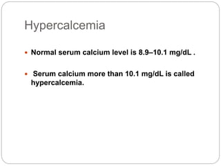
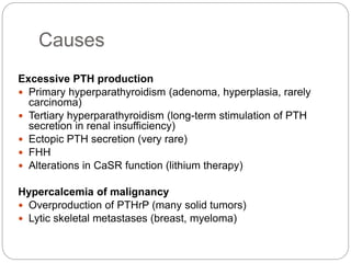
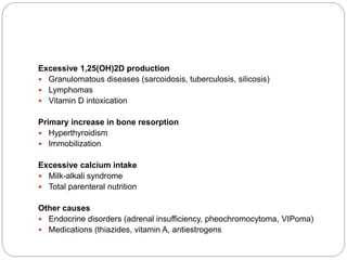
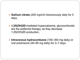
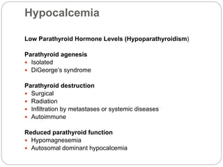
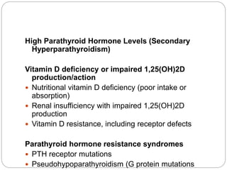
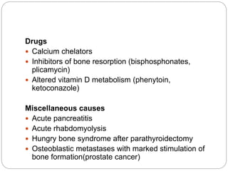
Dr Bikal Lamichhane
IM Resident(1st
year),NAMS
Hypercalcemia And Hypocalcemia
Hypercalcemia
 Normal serum calcium level is 8.9–10.1 mg/dL .
 Serum calcium more than 10.1 mg/dL is called
hypercalcemia.
Causes
Excessive PTH production
 Primary hyperparathyroidism (adenoma, hyperplasia, rarely
carcinoma)
 Tertiary hyperparathyroidism (long-term stimulation of PTH
secretion in renal insufficiency)
 Ectopic PTH secretion (very rare)
 FHH
 Alterations in CaSR function (lithium therapy)
Hypercalcemia of malignancy
 Overproduction of PTHrP (many solid tumors)
 Lytic skeletal metastases (breast, myeloma)
Excessive 1,25(OH)2D production
 Granulomatous diseases (sarcoidosis, tuberculosis, silicosis)
 Lymphomas
 Vitamin D intoxication
Primary increase in bone resorption
 Hyperthyroidism
 Immobilization
Excessive calcium intake
 Milk-alkali syndrome
 Total parenteral nutrition
Other causes
 Endocrine disorders (adrenal insufficiency, pheochromocytoma, VIPoma)
 Medications (thiazides, vitamin A, antiestrogens
Clinical Manifestations
.Mild hypercalcemia (up to 11–11.5 mg/dL) is usually
asymptomatic .
.Neuropsychiatric symptoms, including trouble
concentrating, personality changes, or depression.
.Peptic ulcer disease or nephrolithiasis, and fracture
risk.
 Gastrointestinal symptoms (nausea,anorexia,
constipation, or pancreatitis).
 Polyuria and polydipsia
ECG Changes
 Bradycardia
 AV block,
 Short QT interval; changes in serum calcium can be
monitored by following the QT interval.
Diagnostic Approach
 Alteration in calcium level due to abnormal albumin
concentration is corrected .
 When serum albumin concentrations are reduced, a
corrected calcium concentration is calculated by adding 0.2
mM (0.8 mg/dL) to the total calcium level for every
decrement in serum albumin of 1.0 g/dL below the reference
value of 4.1 g/dL for albumin.
 History regarding medication use,previous neck surgery and
symptoms suggestive of sarcoidosis or lymphoma.
 Once true calcium level established then should look for PTH.
.If the PTH level is increased (or “inappropriately
normal”) in the setting of elevated calcium and
low phosphorus, the diagnosis is almost always
primary hyperparathyroidism.
.A calcium/creatinine clearance ratio (calculated as
urine calcium/serum calcium divided by urine
creatinine/serum creatinine)of <0.01 is suggestive
of FHH, particularly when there is a family history
of mild, asymptomatic hypercalcemia.
 A suppressed PTH level in the face of hypercalcemia
is consistent with non-parathyroid-mediated
hypercalcemia, most often due to underlying
malignancy.
Treatment
 Mild and asymptomatic hypercalcemia doesnot
require immediate treatment.
 Initial therapy of significant hypercalcemia begins with
volume expansion because hypercalcemia invariably
leads to dehydration;
 4–6 L of intravenous saline may be required over the
first 24 h, keeping in mind that underlying
comorbidities .
 If there is increased calcium mobilization from bone
(as in malignancy or severe hyperparathyroidism),
drugs that inhibit bone resorption should be
considered.
 Zoledronic acid (e.g., 4 mg intravenously over~30
min), Pamidronate (e.g., 60–90 mg intravenously
over 2–4 h),and Ibandronate (2 mg intravenously
over 2 h) are bisphosphonates that are commonly
used for the treatment of hypercalcemia .
 Gallium nitrate (200 mg/m2 intravenously daily for 5
days.
 1,25(OH)2D-mediated hypercalcemia, glucocorticoids
are the preferred therapy, as they decrease
1,25(OH)2D production.
 Intravenous hydrocortisone (100–300 mg daily) or
oral prednisone (40–60 mg daily) for 3–7 days.
Hypocalcemia
Low Parathyroid Hormone Levels (Hypoparathyroidism)
Parathyroid agenesis
 Isolated
 DiGeorge’s syndrome
Parathyroid destruction
 Surgical
 Radiation
 Infiltration by metastases or systemic diseases
 Autoimmune
Reduced parathyroid function
 Hypomagnesemia
 Autosomal dominant hypocalcemia
High Parathyroid Hormone Levels (Secondary
Hyperparathyroidism)
Vitamin D deficiency or impaired 1,25(OH)2D
production/action
 Nutritional vitamin D deficiency (poor intake or
absorption)
 Renal insufficiency with impaired 1,25(OH)2D
production
 Vitamin D resistance, including receptor defects
Parathyroid hormone resistance syndromes
 PTH receptor mutations
 Pseudohypoparathyroidism (G protein mutations
Drugs
 Calcium chelators
 Inhibitors of bone resorption (bisphosphonates,
plicamycin)
 Altered vitamin D metabolism (phenytoin,
ketoconazole)
Miscellaneous causes
 Acute pancreatitis
 Acute rhabdomyolysis
 Hungry bone syndrome after parathyroidectomy
 Osteoblastic metastases with marked stimulation of
bone formation(prostate cancer)
Clinical features
.Mild or chronic hypocalcemia-asymtomatic.
.Moderate to severe hypocalcemia is associated with
paresthesias .
.Chvostek’s sign (twitching of the circumoral muscles in
response to gentle tapping of the facial nerve just
anterior to the ear) although it is also present in ~10%
of normal individuals.
.Carpal spasm may be induced by inflation of a blood
pressure cuff to 20 mmHg above the patient’s systolic
blood pressure for 3 min (Trousseau’s sign)
Diagnostic approach
 In addition to measuring serum calcium, it is useful to
determine albumin, phosphorus, and magnesium
levels
Treatment
 Acute, symptomatic hypocalcemia is initially managed
with calcium gluconate, 10 mL 10% wt/vol (90 mg or
2.2 mmol) intravenously, diluted in 50 mL of 5%
dextrose or 0.9% sodium chloride, given intravenously
over 5 min.
 Chronic hypocalcemia due to hypoparathyroidism is
treated with calcium supplements (1000–1500 mg/d
elemental calcium in divided doses) and either vitamin
D2 or D3 (25,000–100,000 U daily) or calcitriol
[1,25(OH)2D, 0.25–2 μg/d
 Vitamin D deficiency is best treated using vitamin D
supplementation.
 vitamin D deficiency due to malabsorption may
require much higher doses (100,000 U/d or more
Thank You

Hypercalcemia and hypocalcemia dr bikal

  • 1.
    Dr Bikal Lamichhane IMResident(1st year),NAMS Hypercalcemia And Hypocalcemia
  • 2.
    Hypercalcemia  Normal serumcalcium level is 8.9–10.1 mg/dL .  Serum calcium more than 10.1 mg/dL is called hypercalcemia.
  • 3.
    Causes Excessive PTH production Primary hyperparathyroidism (adenoma, hyperplasia, rarely carcinoma)  Tertiary hyperparathyroidism (long-term stimulation of PTH secretion in renal insufficiency)  Ectopic PTH secretion (very rare)  FHH  Alterations in CaSR function (lithium therapy) Hypercalcemia of malignancy  Overproduction of PTHrP (many solid tumors)  Lytic skeletal metastases (breast, myeloma)
  • 4.
    Excessive 1,25(OH)2D production Granulomatous diseases (sarcoidosis, tuberculosis, silicosis)  Lymphomas  Vitamin D intoxication Primary increase in bone resorption  Hyperthyroidism  Immobilization Excessive calcium intake  Milk-alkali syndrome  Total parenteral nutrition Other causes  Endocrine disorders (adrenal insufficiency, pheochromocytoma, VIPoma)  Medications (thiazides, vitamin A, antiestrogens
  • 5.
    Clinical Manifestations .Mild hypercalcemia(up to 11–11.5 mg/dL) is usually asymptomatic . .Neuropsychiatric symptoms, including trouble concentrating, personality changes, or depression. .Peptic ulcer disease or nephrolithiasis, and fracture risk.
  • 6.
     Gastrointestinal symptoms(nausea,anorexia, constipation, or pancreatitis).  Polyuria and polydipsia
  • 7.
    ECG Changes  Bradycardia AV block,  Short QT interval; changes in serum calcium can be monitored by following the QT interval.
  • 8.
    Diagnostic Approach  Alterationin calcium level due to abnormal albumin concentration is corrected .  When serum albumin concentrations are reduced, a corrected calcium concentration is calculated by adding 0.2 mM (0.8 mg/dL) to the total calcium level for every decrement in serum albumin of 1.0 g/dL below the reference value of 4.1 g/dL for albumin.  History regarding medication use,previous neck surgery and symptoms suggestive of sarcoidosis or lymphoma.  Once true calcium level established then should look for PTH.
  • 9.
    .If the PTHlevel is increased (or “inappropriately normal”) in the setting of elevated calcium and low phosphorus, the diagnosis is almost always primary hyperparathyroidism. .A calcium/creatinine clearance ratio (calculated as urine calcium/serum calcium divided by urine creatinine/serum creatinine)of <0.01 is suggestive of FHH, particularly when there is a family history of mild, asymptomatic hypercalcemia.
  • 10.
     A suppressedPTH level in the face of hypercalcemia is consistent with non-parathyroid-mediated hypercalcemia, most often due to underlying malignancy.
  • 11.
    Treatment  Mild andasymptomatic hypercalcemia doesnot require immediate treatment.  Initial therapy of significant hypercalcemia begins with volume expansion because hypercalcemia invariably leads to dehydration;  4–6 L of intravenous saline may be required over the first 24 h, keeping in mind that underlying comorbidities .
  • 12.
     If thereis increased calcium mobilization from bone (as in malignancy or severe hyperparathyroidism), drugs that inhibit bone resorption should be considered.  Zoledronic acid (e.g., 4 mg intravenously over~30 min), Pamidronate (e.g., 60–90 mg intravenously over 2–4 h),and Ibandronate (2 mg intravenously over 2 h) are bisphosphonates that are commonly used for the treatment of hypercalcemia .
  • 13.
     Gallium nitrate(200 mg/m2 intravenously daily for 5 days.  1,25(OH)2D-mediated hypercalcemia, glucocorticoids are the preferred therapy, as they decrease 1,25(OH)2D production.  Intravenous hydrocortisone (100–300 mg daily) or oral prednisone (40–60 mg daily) for 3–7 days.
  • 14.
    Hypocalcemia Low Parathyroid HormoneLevels (Hypoparathyroidism) Parathyroid agenesis  Isolated  DiGeorge’s syndrome Parathyroid destruction  Surgical  Radiation  Infiltration by metastases or systemic diseases  Autoimmune Reduced parathyroid function  Hypomagnesemia  Autosomal dominant hypocalcemia
  • 15.
    High Parathyroid HormoneLevels (Secondary Hyperparathyroidism) Vitamin D deficiency or impaired 1,25(OH)2D production/action  Nutritional vitamin D deficiency (poor intake or absorption)  Renal insufficiency with impaired 1,25(OH)2D production  Vitamin D resistance, including receptor defects Parathyroid hormone resistance syndromes  PTH receptor mutations  Pseudohypoparathyroidism (G protein mutations
  • 16.
    Drugs  Calcium chelators Inhibitors of bone resorption (bisphosphonates, plicamycin)  Altered vitamin D metabolism (phenytoin, ketoconazole) Miscellaneous causes  Acute pancreatitis  Acute rhabdomyolysis  Hungry bone syndrome after parathyroidectomy  Osteoblastic metastases with marked stimulation of bone formation(prostate cancer)
  • 17.
    Clinical features .Mild orchronic hypocalcemia-asymtomatic. .Moderate to severe hypocalcemia is associated with paresthesias . .Chvostek’s sign (twitching of the circumoral muscles in response to gentle tapping of the facial nerve just anterior to the ear) although it is also present in ~10% of normal individuals. .Carpal spasm may be induced by inflation of a blood pressure cuff to 20 mmHg above the patient’s systolic blood pressure for 3 min (Trousseau’s sign)
  • 18.
    Diagnostic approach  Inaddition to measuring serum calcium, it is useful to determine albumin, phosphorus, and magnesium levels
  • 19.
    Treatment  Acute, symptomatichypocalcemia is initially managed with calcium gluconate, 10 mL 10% wt/vol (90 mg or 2.2 mmol) intravenously, diluted in 50 mL of 5% dextrose or 0.9% sodium chloride, given intravenously over 5 min.  Chronic hypocalcemia due to hypoparathyroidism is treated with calcium supplements (1000–1500 mg/d elemental calcium in divided doses) and either vitamin D2 or D3 (25,000–100,000 U daily) or calcitriol [1,25(OH)2D, 0.25–2 μg/d
  • 20.
     Vitamin Ddeficiency is best treated using vitamin D supplementation.  vitamin D deficiency due to malabsorption may require much higher doses (100,000 U/d or more
  • 21.