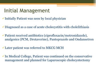
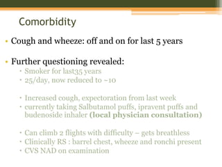
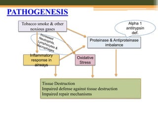
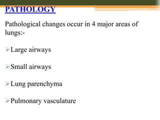
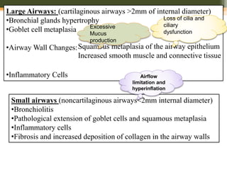
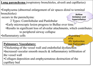
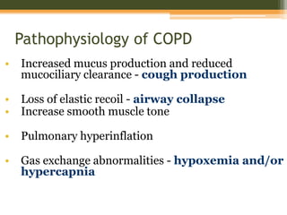
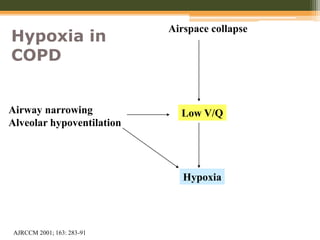
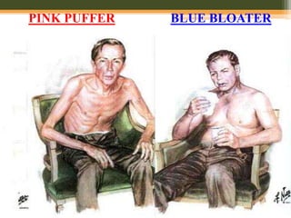
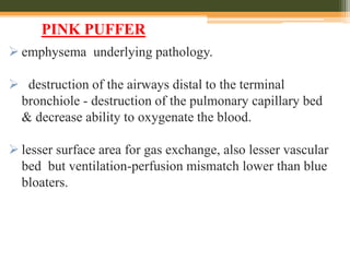
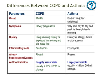
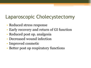
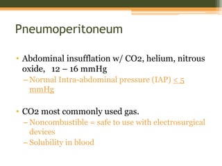
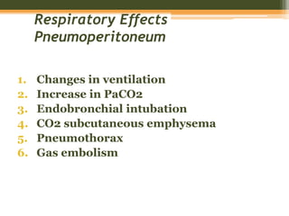
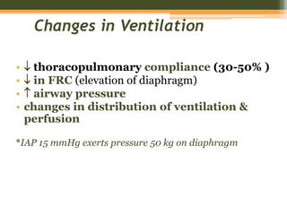
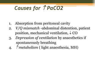
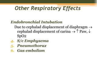
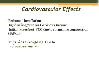
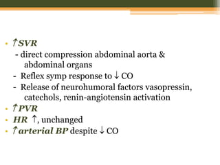
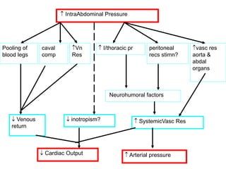
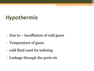
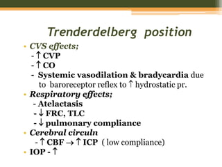
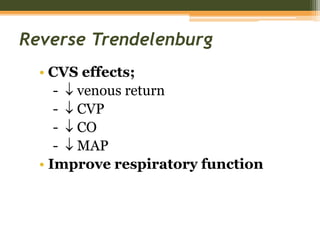
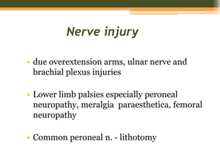
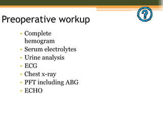
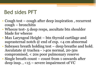
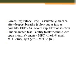
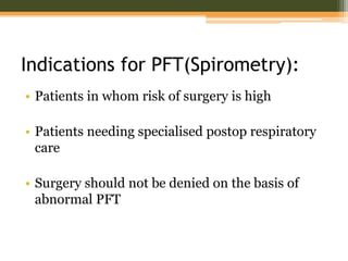
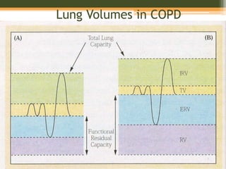
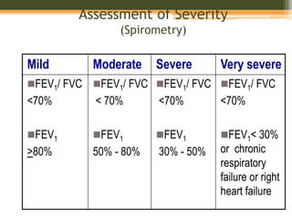
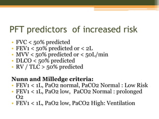
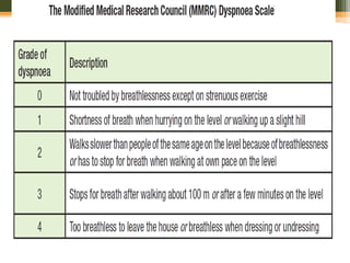
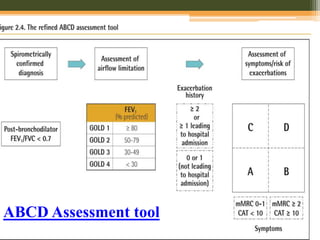
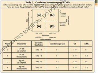
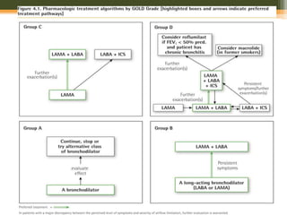
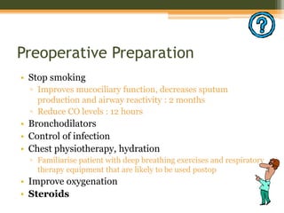
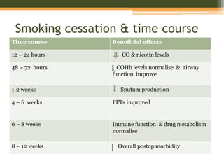
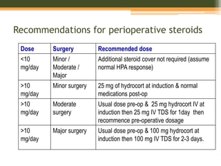
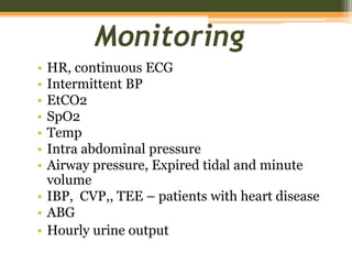
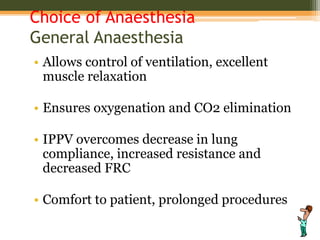
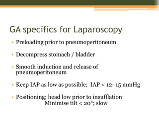
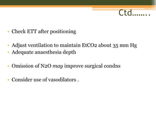
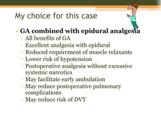
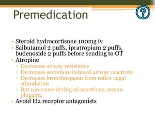
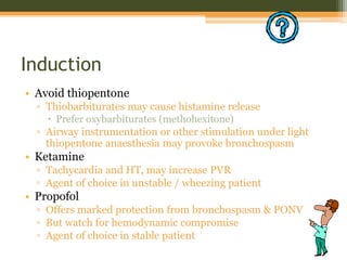
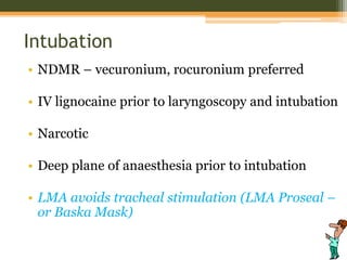
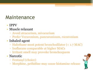
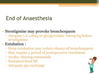
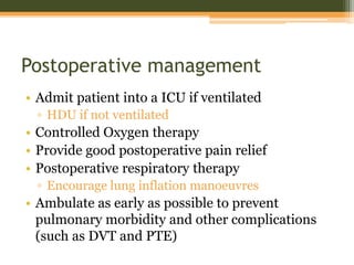
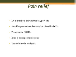
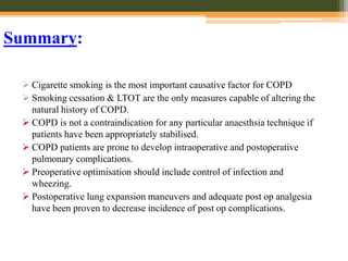
ANESTHETIC MANAGEMENT FOR LAPAROSCOPIC
CHOLECYSTECTOMY IN PATIENT WITH COPD
DR RAMKRISHNA BHUE
3RD YEAR PGT
MKCG MCH BERHAMPUR
Summary of the case
• NAME : Mr. Natraj Bisoi
• AGE/SEX : 56 YRS/ MALE
• Address : kukudakhandi , Ganjam
• Occupation : bus driver
• Education :Studied upto Class X
• DATE OF ADMISSION : 17/10/17
Chief complaints
• Pain abdomen off and on for last 3 months
• Fever for last 5 days
• 4 episodes of vomiting on the day of admission
Initial Management
Initially Patient was seen by local physician
Diagnosed as a case of acute cholecystitis with cholelithiasis
Patient received antibiotics (ciprofloxacin/metronidazole),
analgesics (PCM, Drotaverine), Pantoprazole and Ondansetron
Later patient was referred to MKCG MCH
In Medical College, Patient was continued on the conservative
management and planned for Laparoscopic cholecystectomy
Comorbidity
• Cough and wheeze: off and on for last 5 years
• Further questioning revealed:
 Smoker for last35 years
 25/day, now reduced to ~10
 Increased cough, expectoration from last week
 currently taking Salbutamol puffs, ipravent puffs and
budenoside inhaler (local physician consultation)
 Can climb 2 flights with difficulty – gets breathless
 Clinically RS : barrel chest, wheeze and ronchi present
 CVS NAD on examination
• B.P : 128/76mmHg
• PULSE : 78/min, regular, (arterial wall palpable)
• RESPIRATION : 22b/m
• TEMPREATURE : 36.6 c
• SpO2 : 93% (room air)
• Body wt :50 kg
Investigations available at PAC visit:
Blood:
Hb – 14gm/dl
TC:
RBC- 5.8* 10^6/ul
WBC- 16,000/ul
DC – 42(N), 38(L), 11(M), 9(E)
BILIRUBIN (TOTAL) 1.7 mg/ dL
BILIRUBIN (DIRECT) 1.0 mg/ dL
BILIRUBIN INDIRECT 0.7 mg/ dl
SGPT- 97, SGOT – 102,
Alk Phos – 205
FBS – 98 mg/dl
USG – Calculus Cholecystitis
ECG – NAD
Sr, Na – 141
Sr K – 4.7
Sr urea – 38
Sr creat. – 1.2
CXR:
• increased bronchovascular markings
• flattened hemidiaphragms(bilat)
Plan of management:
Patient of Acute Calculus Cholecystitis
with Acute on Chronic COPD
Posted for Laparoscopic Cholecystectomy
Discussion
• Chronic Obstructive Pulmonary Disease (COPD) is a
common, preventable and treatable disease that is
characterized by persistent respiratory symptoms and
airflow limitation that is due to airway and/or alveolar
abnormalities usually caused by significant exposure to
noxious particles or gases
• Includes:-
I. CHRONIC BRONCHITIS
II. EMPHYSEMA
III.SMALLAIRWAY DISEASE
Chronic Bronchitis: (Clinical Definition)
 Chronic productive cough for 3 months in each
of 2 successive years in a patient in whom other
causes of productive chronic cough have been
excluded.
Emphysema: (Pathological Definition)
 The presence of permanent enlargement of the
airspaces distal to the terminal bronchioles,
accompanied by destruction of their walls and
without obvious fibrosis
COPD
COPD: Risk factors
Host factors:
•Genetic factors: Eg. α1 Antitrypsin Deficiency
•Gender : Prevalence more in males.
?Females more susceptible
•Airway Hyperresponsiveness
Exposures:
•Smoking: Most Important Risk Factor
•Passive/second hand ,smoking exposure
• dust and smokes
•Environmental pollution
•Recurrent bronchopulmonary infections
PATHOGENESIS
Tobacco smoke & other
noxious gases
Inflammatory
response in
airways
Tissue Destruction
Impaired defense against tissue destruction
Impaired repair mechanisms
Proteinase & Antiproteinase
imbalance
Oxidative
Stress
Alpha 1
antitrypsin
def.
PATHOLOGY
Pathological changes occur in 4 major areas of
lungs:-
Large airways
Small airways
Lung parenchyma
Pulmonary vasculature
Large Airways: (cartilaginous airways >2mm of internal diameter)
•Bronchial glands hypertrophy
•Goblet cell metaplasia
•Airway Wall Changes:
•Inflammatory Cells
Squamous metaplasia of the airway epithelium
Increased smooth muscle and connective tissue
Small airways (noncartilaginous airways<2mm internal diameter)
•Bronchiolitis
•Pathological extension of goblet cells and squamous metaplasia
•Inflammatory cells
•Fibrosis and increased deposition of collagen in the airway walls
Excessive
Mucus
production
Loss of cilia and
ciliary
dysfunction
Airflow
limitation and
hyperinflation
Lung parenchyma (respiratory bronchioles, alveoli and capillaries)
•Emphysema (abnormal enlargement of air spaces distal to terminal
bronchioles)
occurs in the parenchyma:
2 Types: Centrilobular and Panlobular
•Early microscopic lesion progress to Bullae over time.
•Results in significant loss of alveolar attachments, which contributes
to peripheral airway collapse
•Inflammatory cells
Pulmonary Vasculature:
•Thickening of the vessel wall and endothelial dysfunction
•Increased vascular smooth muscle & inflammatory infiltration of
the vessel wall
•Collagen deposition and emphysematous destruction of the
capillary bed
Airflow
limitation and
hyperinflation
•Pulmonary
HTN
•RV dysfunction
(cor Pulmonale)
Pathophysiology of COPD
• Increased mucus production and reduced
mucociliary clearance - cough production
• Loss of elastic recoil - airway collapse
• Increase smooth muscle tone
• Pulmonary hyperinflation
• Gas exchange abnormalities - hypoxemia and/or
hypercapnia
Airspace collapse
Low V/Q
Hypoxia
Airway narrowing
Alveolar hypoventilation
Hypoxia in
COPD
AJRCCM 2001; 163: 283-91
Chronic hypoxia
Pulmonary vasoconstriction
Muscularization
Intimal
hyperplasia
Fibrosis
Obliteration
Pulmonary hypertension
Cor pulmonale
Death
Edema
Pulmonary Hypertension in COPD
Source: Peter J. Barnes, MD
PINK PUFFER BLUE BLOATER
 emphysema underlying pathology.
 destruction of the airways distal to the terminal
bronchiole - destruction of the pulmonary capillary bed
& decrease ability to oxygenate the blood.
 lesser surface area for gas exchange, also lesser vascular
bed but ventilation-perfusion mismatch lower than blue
bloaters.
PINK PUFFER
 compensate by hyperventilation (the "puffer" part)
so relatively normal ABGs.
 Eventually, low cardiac output, people develop
muscle wasting and weight loss.
 They actually have less hypoxemia and appear to
have a "pink" complexion and hence "pink puffer"
 primary lung pathology is chronic bronchitis.
 excessive mucus production with airway obstruction -
hyperplasia of mucus-producing glands, goblet cell
metaplasia, and chronic inflammation around bronchi.
BLUE BLOATER
 increased obstruction leads decrease in ventilation and
increasing cardiac output causing perfusion mismatch
leading to hypoxemia, hypercapnia and polycythemia.
 Because of increasing obstruction, their residual lung
volume gradually increases (the "bloating" part).
 They are hypoxemic/cyanotic because they actually have
worse hypoxemia than pink puffers and this manifests as
bluish lips and faces.
Comparative features of COPD
Differences Between COPD and Asthma
Parameters COPD Asthma
Onset Mid-life Early in life (often
childhood)
Symptoms Slowly progressive Vary from day to day and
peak in the night/early
morning
History Long smoking history or
exposure to smoking and
bio-mass fuel
History of allergy, rhinitis
and/or eczema.
Inflammatory cells Neutrophils Eosinophils
Airway
hyperresponsiveness
Absent Present
Airflow limitation Largely irreversible
usually < 15% or 200 ml
change
Largely reversible
usually > 15% or 200 ml
change.
Extrapulmonary comorbidities in COPD
• Commonly seen
▫ Weight loss
▫ Skeletal muscle wasting
• Increased risk of
▫ Myocardial infarction
▫ Angina
▫ Osteoporosis, bone
fractures
• Respiratory infection
• Depression
• Diabetes
• Sleep-disorders
• Anemia
• Glaucoma
 Common consequences
 RVH
 Cor pulmonale
Laparoscopic Cholecystectomy
• Reduced stress response
• Early recovery and return of GI function
• Reduced post op. analgesia
• Decreased wound infection
• Improved cosmetic
• Better post op respiratory functions
Pneumoperitoneum
• Abdominal insufflation w/ CO2, helium, nitrous
oxide, 12 – 16 mmHg
– Normal Intra-abdominal pressure (IAP) < 5
mmHg
• CO2 most commonly used gas.
– Noncombustible = safe to use with electrosurgical
devices
– Solubility in blood
Respiratory Effects
Pneumoperitoneum
1. Changes in ventilation
2. Increase in PaCO2
3. Endobronchial intubation
4. CO2 subcutaneous emphysema
5. Pneumothorax
6. Gas embolism
Changes in Ventilation
•  thoracopulmonary compliance (30-50% )
•  in FRC (elevation of diaphragm)
•  airway pressure
• changes in distribution of ventilation &
perfusion
*IAP 15 mmHg exerts pressure 50 kg on diaphragm
Causes for  PaCO2
1. Absorption from peritoneal cavity
2. V/Q mismatch -abdominal distention, patient
position, mechanical ventilation,  CO
3. Depression of ventilation by anaesthetics if
spontaneously breathing
4.  metabolism ( light anaesthesia, MH)
Other Respiratory Effects
Endobronchial Intubation
Due to cephalad displacement of diaphragm 
cephalad displacement of carina   Paw, 
SpO2
4. S/c Emphysema
5. Pneumothorax
6. Gas embolism
Cardiovascular Effects
• Peritoneal insufflations
Biphasic effect on Cardiac Output
Initial transient  CO due to splanchnic compression
(IAP<15)
Then  CO (10-30%) Due to
-  venous return
•  SVR
- direct compression abdominal aorta &
abdominal organs
- Reflex symp response to  CO
- Release of neurohumoral factors vasopressin,
catechols, renin-angiotensin activation
•  PVR
• HR , unchanged
•  arterial BP despite  CO
 IntraAbdominal Pressure
Pooling of
blood legs
caval
comp
Vn
Res
 I/thoracic pr peritoneal
recs stimn?
vasc res
aorta &
abdal
organs
Neurohumoral factors
 Venous
return
 inotropism?  SystemicVasc Res
 Cardiac Output  Arterial pressure
Cardiac arrhythmias
• Reflex ’s in vagal tone sudden peritoneal
stretch  bradycardia, arrhythmias, asystole
- Stop insuffln, atropine, deepen anaesthesia
•  PaCO2
• Use of halothane
• Pts with cardiac disease
• Gas embolism
• hypoxia
GIT Effects
• Due to  intra abdominal pressure  risk
of aspiration
• Head down position prevents regurgitated
material from entering the airway
Hypothermia
• Due to – insufflation of cold gases
• Temperature of gases
• cold fluid used for toileting
• Leakage through the ports etc
Trenderdelberg position
• CVS effects;
-  CVP
-  CO
- Systemic vasodilation & bradycardia due
to baroreceptor reflex to  hydrostatic pr.
• Respiratory effects;
- Atelactasis
-  FRC, TLC
-  pulmonary compliance
• Cerebral circuln
-  CBF   ICP ( low compliance)
• IOP - 
Reverse Trendelenburg
• CVS effects;
-  venous return
-  CVP
-  CO
-  MAP
• Improve respiratory function
Nerve injury
• due overextension arms, ulnar nerve and
brachial plexus injuries
• Lower limb palsies especially peroneal
neuropathy, meralgia paraesthetica, femoral
neuropathy
• Common peroneal n. - lithotomy
Anaesthetic management
• Calculus
cholecystitis
• COPD
Posted for
Laparoscopic
Cholecystectomy
Preoperative workup
• Complete
hemogram
• Serum electrolytes
• Urine analysis
• ECG
• Chest x-ray
• PFT including ABG
• ECHO
Bed sides PFT
• Cough test – cough after deep inspiration , recurrent
cough – bronchitis
• Wheeze test- 5 deep resps, ascultate btn shoulder
blade for wheeze
• Max Laryngeal Height – btn thyroid cartilage and
suprasternal notch @ end of exp. <4 cm abnormal
• Sabrasez breath holding test – deep breathe and hold.
Asculatate @ trachea - >40s normal, 20-30s
compromised, < 20s poor pulmonary reserve
• Single breath count – count from 1 onwards after
deep insp. , <15 – severe impairment of VC
• Forced Expiratory Time – ascultate @ trachea
after deepest breathe & blow out as fast as
possible- FET > 6s , severe exp. Flow obstuction
• Sniders match test – ability to blow candle with
open mouth @ 22cm – MBC >150l, @ 15cm
MBC <100l, @ 7.5cm – MBC < 50 L
•
Indications for PFT(Spirometry):
• Patients in whom risk of surgery is high
• Patients needing specialised postop respiratory
care
• Surgery should not be denied on the basis of
abnormal PFT
Lung Volumes in COPD
Assessment of Severity
(Spirometry)
Mild Moderate Severe Very severe
FEV1/ FVC
<70%
FEV1
>80%
FEV1/ FVC
< 70%
FEV1
50% - 80%
FEV1/ FVC
<70%
FEV1
30% - 50%
FEV1/ FVC
<70%
FEV1< 30%
or chronic
respiratory
failure or right
heart failure
PFT predictors of increased risk
• FVC < 50% predicted
• FEV1 < 50% predicted or < 2L
• MVV < 50% predicted or < 50L/min
• DLCO < 50% predicted
• RV / TLC > 50% predicted
Nunn and Milledge criteria:
• FEV1 < 1L, PaO2 normal, PaCO2 Normal : Low Risk
• FEV1 < 1L, PaO2 low, PaCO2 Normal : prolonged
O2
• FEV1 < 1L, PaO2 low, PaCO2 High: Ventilation
ABCD Assessment tool
Preoperative Preparation
• Stop smoking
▫ Improves mucociliary function, decreases sputum
production and airway reactivity : 2 months
▫ Reduce CO levels : 12 hours
• Bronchodilators
• Control of infection
• Chest physiotherapy, hydration
▫ Familiarise patient with deep breathing exercises and respiratory
therapy equipment that are likely to be used postop
• Improve oxygenation
• Steroids
Smoking cessation & time course
Time course Beneficial effects
12 – 24 hours CO & nicotin levels
48 – 72 hours COHb levels normalise & airway
function improve
1-2 weeks Sputum production
4 – 6 weeks PFTs improved
6 - 8 weeks Immune function & drug metabolism
normalise
8 – 12 weeks Overall postop morbidity
Recommendations for perioperative steroids
Dose Surgery Recommended dose
<10
mg/day
Minor /
Moderate /
Major
Additional steroid cover not required (assume
normal HPA response)
>10
mg/day
Minor surgery 25 mg of hydrocort at induction & normal
medications post-op
>10
mg/day
Moderate
surgery
Usual dose pre-op & 25 mg hydrocort IV at
induction then 25 mg IV TDS for 1day then
recommence pre-operative dosage
>10
mg/day
Major surgery Usual dose pre-op & 100 mg hydrocort at
induction then 100 mg IV TDS for 2-3 days.
Monitoring
• HR, continuous ECG
• Intermittent BP
• EtCO2
• SpO2
• Temp
• Intra abdominal pressure
• Airway pressure, Expired tidal and minute
volume
• IBP, CVP,, TEE – patients with heart disease
• ABG
• Hourly urine output
Choice of Anaesthesia
General Anaesthesia
• Allows control of ventilation, excellent
muscle relaxation
• Ensures oxygenation and CO2 elimination
• IPPV overcomes decrease in lung
compliance, increased resistance and
decreased FRC
• Comfort to patient, prolonged procedures
GA specifics for Laparoscopy
• Preloading prior to pneumoperitoneum
• Decompress stomach / bladder
• Smooth induction and release of
pneumoperitoneum
• Keep IAP as low as possible; IAP < 12- 15 mmHg
• Positioning; head low prior to insufflation
Minimise tilt < 20°; slow
Ctd……..
• Check ETT after positioning
• Adjust ventilation to maintain EtCO2 about 35 mm Hg
• Adequate anaesthesia depth
• Omission of N2O may improve surgical condns
• Consider use of vasodilators .
Regional anaesthesia
• Avoids risk of bronchospasm due to intubation
• Excellent intraoperative and postoperative analgesia
• Problems
▫ Spontaneous ventilation may lead to hypoventilation
▫ Hypercarbia and acidosis can increase PVR
▫ Inadequate muscle relaxation, coughing / bucking
▫ High levels of spinal / epidural block
 Increase parasympathetic tone and cause bronchospasm
 Decrease ERV by ~50%, detrimental for active
expiration
 Hypotension
▫ Prolonged procedure, patient discomfort, shivering
▫ Heavy sedation may required
My choice for this case
• GA combined with epidural analgesia
▫ All benefits of GA
▫ Excellent analgesia with epidural
▫ Reduced requirement of muscle relaxants
▫ Lower risk of hypotension
▫ Postoperative analgesia without excessive
systemic narcotics
▫ May facilitate early ambulation
▫ May reduce postoperative pulmonary
complications
▫ May reduce risk of DVT
Premedication
• Steroid hydrocortisone 100mg iv
• Salbutamol 2 puffs, ipratropium 2 puffs,
budenoside 2 puffs before sending to OT
• Atropine
▫ Decreases airway resistance
▫ Decreases secretion-induced airway reactivity
▫ Decreases bronchospasm from reflex vagal
stimulation
▫ But can cause drying of secretions, mucus
plugging
• Avoid H2 receptor antagonists
Induction
• Avoid thiopentone
▫ Thiobarbiturates may cause histamine release
 Prefer oxybarbiturates (methohexitone)
▫ Airway instrumentation or other stimulation under light
thiopentone anaesthesia may provoke bronchospasm
• Ketamine
▫ Tachycardia and HT, may increase PVR
▫ Agent of choice in unstable / wheezing patient
• Propofol
▫ Offers marked protection from bronchospasm & PONV
▫ But watch for hemodynamic compromise
▫ Agent of choice in stable patient
Intubation
• NDMR – vecuronium, rocuronium preferred
• IV lignocaine prior to laryngoscopy and intubation
• Narcotic
• Deep plane of anaesthesia prior to intubation
• LMA avoids tracheal stimulation (LMA Proseal –
or Baska Mask)
Maintenance
• IPPV
• Muscle relaxant
▫ Avoid atracurium, mivacurium
▫ Prefer Vecuronium, pancuronioum, rocuronium
• Inhaled agent
▫ Halothane most potent bronchodilator (< 1.7 MAC)
▫ Isoflurane comparable at higher MACs
▫ Irritant smell may provoke bronchospasm
• Narcotic
▫ Fentanyl (choice)
▫ Morphine, pethidine may cause histamine release
End of Anaesthesia
• Neostigmine may provoke bronchospasm
▫ Atropine 1.2-1.8mg or glyopyrrolate 10mcg/kg before
neostigmine
• Extubation :
▫ Deep extubation may reduce chance of bronchospasm
▫ May require a period of postoperative ventilation
▫ Awake, obeying commands
▫ Sustained head lift
▫ Adequate gas exchange
Postoperative management
• Admit patient into a ICU if ventilated
▫ HDU if not ventilated
• Controlled Oxygen therapy
• Provide good postoperative pain relief
• Postoperative respiratory therapy
▫ Encourage lung inflation manoeuvres
• Ambulate as early as possible to prevent
pulmonary morbidity and other complications
(such as DVT and PTE)
Pain relief
• LA infiltration- intraperitoneal, port site
• Shoulder pain - careful evacuation of residual CO2
• Preoperative NSAIDs
• Intra & post operative opioids
• Use multimodal analgesia
LONG TERM OXYGEN THERAPY
 Long-term oxygen therapy(home oxygen therapy) is
recommended if the Pao2 <55 mmHg, the hematocrit >55% or
there is evidence of cor pulmonale
 The goal of supplemental oxygen administration is to achieve a
Pao2 between 60 and 80 mmHg. This goal can usually be
accomplished by delivering oxygen through a nasal cannula at
2L/min. The flow rate of oxygen is titrated as neeeded according
to arterial blood gas or pulse oximetry measurements
Summary:
 Cigarette smoking is the most important causative factor for COPD
 Smoking cessation & LTOT are the only measures capable of altering the
natural history of COPD.
 COPD is not a contraindication for any particular anaesthsia technique if
patients have been appropriately stabilised.
 COPD patients are prone to develop intraoperative and postoperative
pulmonary complications.
 Preoperative optimisation should include control of infection and
wheezing.
 Postoperative lung expansion maneuvers and adequate post op analgesia
have been proven to decrease incidence of post op complications.
Thank You

ANESTHETIC MANAGEMENT LAP CHOLECYSTECTOMY WITH COPD

  • 1.
    ANESTHETIC MANAGEMENT FORLAPAROSCOPIC CHOLECYSTECTOMY IN PATIENT WITH COPD DR RAMKRISHNA BHUE 3RD YEAR PGT MKCG MCH BERHAMPUR
  • 2.
    Summary of thecase • NAME : Mr. Natraj Bisoi • AGE/SEX : 56 YRS/ MALE • Address : kukudakhandi , Ganjam • Occupation : bus driver • Education :Studied upto Class X • DATE OF ADMISSION : 17/10/17
  • 3.
    Chief complaints • Painabdomen off and on for last 3 months • Fever for last 5 days • 4 episodes of vomiting on the day of admission
  • 4.
    Initial Management Initially Patientwas seen by local physician Diagnosed as a case of acute cholecystitis with cholelithiasis Patient received antibiotics (ciprofloxacin/metronidazole), analgesics (PCM, Drotaverine), Pantoprazole and Ondansetron Later patient was referred to MKCG MCH In Medical College, Patient was continued on the conservative management and planned for Laparoscopic cholecystectomy
  • 5.
    Comorbidity • Cough andwheeze: off and on for last 5 years • Further questioning revealed:  Smoker for last35 years  25/day, now reduced to ~10  Increased cough, expectoration from last week  currently taking Salbutamol puffs, ipravent puffs and budenoside inhaler (local physician consultation)  Can climb 2 flights with difficulty – gets breathless  Clinically RS : barrel chest, wheeze and ronchi present  CVS NAD on examination
  • 6.
    • B.P :128/76mmHg • PULSE : 78/min, regular, (arterial wall palpable) • RESPIRATION : 22b/m • TEMPREATURE : 36.6 c • SpO2 : 93% (room air) • Body wt :50 kg
  • 7.
    Investigations available atPAC visit: Blood: Hb – 14gm/dl TC: RBC- 5.8* 10^6/ul WBC- 16,000/ul DC – 42(N), 38(L), 11(M), 9(E) BILIRUBIN (TOTAL) 1.7 mg/ dL BILIRUBIN (DIRECT) 1.0 mg/ dL BILIRUBIN INDIRECT 0.7 mg/ dl SGPT- 97, SGOT – 102, Alk Phos – 205 FBS – 98 mg/dl USG – Calculus Cholecystitis ECG – NAD Sr, Na – 141 Sr K – 4.7 Sr urea – 38 Sr creat. – 1.2 CXR: • increased bronchovascular markings • flattened hemidiaphragms(bilat)
  • 8.
    Plan of management: Patientof Acute Calculus Cholecystitis with Acute on Chronic COPD Posted for Laparoscopic Cholecystectomy
  • 9.
    Discussion • Chronic ObstructivePulmonary Disease (COPD) is a common, preventable and treatable disease that is characterized by persistent respiratory symptoms and airflow limitation that is due to airway and/or alveolar abnormalities usually caused by significant exposure to noxious particles or gases • Includes:- I. CHRONIC BRONCHITIS II. EMPHYSEMA III.SMALLAIRWAY DISEASE
  • 10.
    Chronic Bronchitis: (ClinicalDefinition)  Chronic productive cough for 3 months in each of 2 successive years in a patient in whom other causes of productive chronic cough have been excluded. Emphysema: (Pathological Definition)  The presence of permanent enlargement of the airspaces distal to the terminal bronchioles, accompanied by destruction of their walls and without obvious fibrosis COPD
  • 11.
    COPD: Risk factors Hostfactors: •Genetic factors: Eg. α1 Antitrypsin Deficiency •Gender : Prevalence more in males. ?Females more susceptible •Airway Hyperresponsiveness Exposures: •Smoking: Most Important Risk Factor •Passive/second hand ,smoking exposure • dust and smokes •Environmental pollution •Recurrent bronchopulmonary infections
  • 12.
    PATHOGENESIS Tobacco smoke &other noxious gases Inflammatory response in airways Tissue Destruction Impaired defense against tissue destruction Impaired repair mechanisms Proteinase & Antiproteinase imbalance Oxidative Stress Alpha 1 antitrypsin def.
  • 13.
    PATHOLOGY Pathological changes occurin 4 major areas of lungs:- Large airways Small airways Lung parenchyma Pulmonary vasculature
  • 14.
    Large Airways: (cartilaginousairways >2mm of internal diameter) •Bronchial glands hypertrophy •Goblet cell metaplasia •Airway Wall Changes: •Inflammatory Cells Squamous metaplasia of the airway epithelium Increased smooth muscle and connective tissue Small airways (noncartilaginous airways<2mm internal diameter) •Bronchiolitis •Pathological extension of goblet cells and squamous metaplasia •Inflammatory cells •Fibrosis and increased deposition of collagen in the airway walls Excessive Mucus production Loss of cilia and ciliary dysfunction Airflow limitation and hyperinflation
  • 15.
    Lung parenchyma (respiratorybronchioles, alveoli and capillaries) •Emphysema (abnormal enlargement of air spaces distal to terminal bronchioles) occurs in the parenchyma: 2 Types: Centrilobular and Panlobular •Early microscopic lesion progress to Bullae over time. •Results in significant loss of alveolar attachments, which contributes to peripheral airway collapse •Inflammatory cells Pulmonary Vasculature: •Thickening of the vessel wall and endothelial dysfunction •Increased vascular smooth muscle & inflammatory infiltration of the vessel wall •Collagen deposition and emphysematous destruction of the capillary bed Airflow limitation and hyperinflation •Pulmonary HTN •RV dysfunction (cor Pulmonale)
  • 16.
    Pathophysiology of COPD •Increased mucus production and reduced mucociliary clearance - cough production • Loss of elastic recoil - airway collapse • Increase smooth muscle tone • Pulmonary hyperinflation • Gas exchange abnormalities - hypoxemia and/or hypercapnia
  • 17.
    Airspace collapse Low V/Q Hypoxia Airwaynarrowing Alveolar hypoventilation Hypoxia in COPD AJRCCM 2001; 163: 283-91
  • 18.
    Chronic hypoxia Pulmonary vasoconstriction Muscularization Intimal hyperplasia Fibrosis Obliteration Pulmonaryhypertension Cor pulmonale Death Edema Pulmonary Hypertension in COPD Source: Peter J. Barnes, MD
  • 19.
  • 20.
     emphysema underlyingpathology.  destruction of the airways distal to the terminal bronchiole - destruction of the pulmonary capillary bed & decrease ability to oxygenate the blood.  lesser surface area for gas exchange, also lesser vascular bed but ventilation-perfusion mismatch lower than blue bloaters. PINK PUFFER
  • 21.
     compensate byhyperventilation (the "puffer" part) so relatively normal ABGs.  Eventually, low cardiac output, people develop muscle wasting and weight loss.  They actually have less hypoxemia and appear to have a "pink" complexion and hence "pink puffer"
  • 22.
     primary lungpathology is chronic bronchitis.  excessive mucus production with airway obstruction - hyperplasia of mucus-producing glands, goblet cell metaplasia, and chronic inflammation around bronchi. BLUE BLOATER
  • 23.
     increased obstructionleads decrease in ventilation and increasing cardiac output causing perfusion mismatch leading to hypoxemia, hypercapnia and polycythemia.  Because of increasing obstruction, their residual lung volume gradually increases (the "bloating" part).  They are hypoxemic/cyanotic because they actually have worse hypoxemia than pink puffers and this manifests as bluish lips and faces.
  • 24.
  • 25.
    Differences Between COPDand Asthma Parameters COPD Asthma Onset Mid-life Early in life (often childhood) Symptoms Slowly progressive Vary from day to day and peak in the night/early morning History Long smoking history or exposure to smoking and bio-mass fuel History of allergy, rhinitis and/or eczema. Inflammatory cells Neutrophils Eosinophils Airway hyperresponsiveness Absent Present Airflow limitation Largely irreversible usually < 15% or 200 ml change Largely reversible usually > 15% or 200 ml change.
  • 26.
    Extrapulmonary comorbidities inCOPD • Commonly seen ▫ Weight loss ▫ Skeletal muscle wasting • Increased risk of ▫ Myocardial infarction ▫ Angina ▫ Osteoporosis, bone fractures • Respiratory infection • Depression • Diabetes • Sleep-disorders • Anemia • Glaucoma  Common consequences  RVH  Cor pulmonale
  • 27.
    Laparoscopic Cholecystectomy • Reducedstress response • Early recovery and return of GI function • Reduced post op. analgesia • Decreased wound infection • Improved cosmetic • Better post op respiratory functions
  • 28.
    Pneumoperitoneum • Abdominal insufflationw/ CO2, helium, nitrous oxide, 12 – 16 mmHg – Normal Intra-abdominal pressure (IAP) < 5 mmHg • CO2 most commonly used gas. – Noncombustible = safe to use with electrosurgical devices – Solubility in blood
  • 29.
    Respiratory Effects Pneumoperitoneum 1. Changesin ventilation 2. Increase in PaCO2 3. Endobronchial intubation 4. CO2 subcutaneous emphysema 5. Pneumothorax 6. Gas embolism
  • 30.
    Changes in Ventilation • thoracopulmonary compliance (30-50% ) •  in FRC (elevation of diaphragm) •  airway pressure • changes in distribution of ventilation & perfusion *IAP 15 mmHg exerts pressure 50 kg on diaphragm
  • 31.
    Causes for PaCO2 1. Absorption from peritoneal cavity 2. V/Q mismatch -abdominal distention, patient position, mechanical ventilation,  CO 3. Depression of ventilation by anaesthetics if spontaneously breathing 4.  metabolism ( light anaesthesia, MH)
  • 32.
    Other Respiratory Effects EndobronchialIntubation Due to cephalad displacement of diaphragm  cephalad displacement of carina   Paw,  SpO2 4. S/c Emphysema 5. Pneumothorax 6. Gas embolism
  • 33.
    Cardiovascular Effects • Peritonealinsufflations Biphasic effect on Cardiac Output Initial transient  CO due to splanchnic compression (IAP<15) Then  CO (10-30%) Due to -  venous return
  • 34.
    •  SVR -direct compression abdominal aorta & abdominal organs - Reflex symp response to  CO - Release of neurohumoral factors vasopressin, catechols, renin-angiotensin activation •  PVR • HR , unchanged •  arterial BP despite  CO
  • 35.
     IntraAbdominal Pressure Poolingof blood legs caval comp Vn Res  I/thoracic pr peritoneal recs stimn? vasc res aorta & abdal organs Neurohumoral factors  Venous return  inotropism?  SystemicVasc Res  Cardiac Output  Arterial pressure
  • 36.
    Cardiac arrhythmias • Reflex’s in vagal tone sudden peritoneal stretch  bradycardia, arrhythmias, asystole - Stop insuffln, atropine, deepen anaesthesia •  PaCO2 • Use of halothane • Pts with cardiac disease • Gas embolism • hypoxia
  • 37.
    GIT Effects • Dueto  intra abdominal pressure  risk of aspiration • Head down position prevents regurgitated material from entering the airway
  • 38.
    Hypothermia • Due to– insufflation of cold gases • Temperature of gases • cold fluid used for toileting • Leakage through the ports etc
  • 39.
    Trenderdelberg position • CVSeffects; -  CVP -  CO - Systemic vasodilation & bradycardia due to baroreceptor reflex to  hydrostatic pr. • Respiratory effects; - Atelactasis -  FRC, TLC -  pulmonary compliance • Cerebral circuln -  CBF   ICP ( low compliance) • IOP - 
  • 40.
    Reverse Trendelenburg • CVSeffects; -  venous return -  CVP -  CO -  MAP • Improve respiratory function
  • 41.
    Nerve injury • dueoverextension arms, ulnar nerve and brachial plexus injuries • Lower limb palsies especially peroneal neuropathy, meralgia paraesthetica, femoral neuropathy • Common peroneal n. - lithotomy
  • 42.
    Anaesthetic management • Calculus cholecystitis •COPD Posted for Laparoscopic Cholecystectomy
  • 43.
    Preoperative workup • Complete hemogram •Serum electrolytes • Urine analysis • ECG • Chest x-ray • PFT including ABG • ECHO
  • 44.
    Bed sides PFT •Cough test – cough after deep inspiration , recurrent cough – bronchitis • Wheeze test- 5 deep resps, ascultate btn shoulder blade for wheeze • Max Laryngeal Height – btn thyroid cartilage and suprasternal notch @ end of exp. <4 cm abnormal • Sabrasez breath holding test – deep breathe and hold. Asculatate @ trachea - >40s normal, 20-30s compromised, < 20s poor pulmonary reserve • Single breath count – count from 1 onwards after deep insp. , <15 – severe impairment of VC
  • 45.
    • Forced ExpiratoryTime – ascultate @ trachea after deepest breathe & blow out as fast as possible- FET > 6s , severe exp. Flow obstuction • Sniders match test – ability to blow candle with open mouth @ 22cm – MBC >150l, @ 15cm MBC <100l, @ 7.5cm – MBC < 50 L •
  • 46.
    Indications for PFT(Spirometry): •Patients in whom risk of surgery is high • Patients needing specialised postop respiratory care • Surgery should not be denied on the basis of abnormal PFT
  • 47.
  • 48.
    Assessment of Severity (Spirometry) MildModerate Severe Very severe FEV1/ FVC <70% FEV1 >80% FEV1/ FVC < 70% FEV1 50% - 80% FEV1/ FVC <70% FEV1 30% - 50% FEV1/ FVC <70% FEV1< 30% or chronic respiratory failure or right heart failure
  • 49.
    PFT predictors ofincreased risk • FVC < 50% predicted • FEV1 < 50% predicted or < 2L • MVV < 50% predicted or < 50L/min • DLCO < 50% predicted • RV / TLC > 50% predicted Nunn and Milledge criteria: • FEV1 < 1L, PaO2 normal, PaCO2 Normal : Low Risk • FEV1 < 1L, PaO2 low, PaCO2 Normal : prolonged O2 • FEV1 < 1L, PaO2 low, PaCO2 High: Ventilation
  • 51.
  • 54.
    Preoperative Preparation • Stopsmoking ▫ Improves mucociliary function, decreases sputum production and airway reactivity : 2 months ▫ Reduce CO levels : 12 hours • Bronchodilators • Control of infection • Chest physiotherapy, hydration ▫ Familiarise patient with deep breathing exercises and respiratory therapy equipment that are likely to be used postop • Improve oxygenation • Steroids
  • 55.
    Smoking cessation &time course Time course Beneficial effects 12 – 24 hours CO & nicotin levels 48 – 72 hours COHb levels normalise & airway function improve 1-2 weeks Sputum production 4 – 6 weeks PFTs improved 6 - 8 weeks Immune function & drug metabolism normalise 8 – 12 weeks Overall postop morbidity
  • 56.
    Recommendations for perioperativesteroids Dose Surgery Recommended dose <10 mg/day Minor / Moderate / Major Additional steroid cover not required (assume normal HPA response) >10 mg/day Minor surgery 25 mg of hydrocort at induction & normal medications post-op >10 mg/day Moderate surgery Usual dose pre-op & 25 mg hydrocort IV at induction then 25 mg IV TDS for 1day then recommence pre-operative dosage >10 mg/day Major surgery Usual dose pre-op & 100 mg hydrocort at induction then 100 mg IV TDS for 2-3 days.
  • 57.
    Monitoring • HR, continuousECG • Intermittent BP • EtCO2 • SpO2 • Temp • Intra abdominal pressure • Airway pressure, Expired tidal and minute volume • IBP, CVP,, TEE – patients with heart disease • ABG • Hourly urine output
  • 58.
    Choice of Anaesthesia GeneralAnaesthesia • Allows control of ventilation, excellent muscle relaxation • Ensures oxygenation and CO2 elimination • IPPV overcomes decrease in lung compliance, increased resistance and decreased FRC • Comfort to patient, prolonged procedures
  • 59.
    GA specifics forLaparoscopy • Preloading prior to pneumoperitoneum • Decompress stomach / bladder • Smooth induction and release of pneumoperitoneum • Keep IAP as low as possible; IAP < 12- 15 mmHg • Positioning; head low prior to insufflation Minimise tilt < 20°; slow
  • 60.
    Ctd…….. • Check ETTafter positioning • Adjust ventilation to maintain EtCO2 about 35 mm Hg • Adequate anaesthesia depth • Omission of N2O may improve surgical condns • Consider use of vasodilators .
  • 61.
    Regional anaesthesia • Avoidsrisk of bronchospasm due to intubation • Excellent intraoperative and postoperative analgesia • Problems ▫ Spontaneous ventilation may lead to hypoventilation ▫ Hypercarbia and acidosis can increase PVR ▫ Inadequate muscle relaxation, coughing / bucking ▫ High levels of spinal / epidural block  Increase parasympathetic tone and cause bronchospasm  Decrease ERV by ~50%, detrimental for active expiration  Hypotension ▫ Prolonged procedure, patient discomfort, shivering ▫ Heavy sedation may required
  • 62.
    My choice forthis case • GA combined with epidural analgesia ▫ All benefits of GA ▫ Excellent analgesia with epidural ▫ Reduced requirement of muscle relaxants ▫ Lower risk of hypotension ▫ Postoperative analgesia without excessive systemic narcotics ▫ May facilitate early ambulation ▫ May reduce postoperative pulmonary complications ▫ May reduce risk of DVT
  • 63.
    Premedication • Steroid hydrocortisone100mg iv • Salbutamol 2 puffs, ipratropium 2 puffs, budenoside 2 puffs before sending to OT • Atropine ▫ Decreases airway resistance ▫ Decreases secretion-induced airway reactivity ▫ Decreases bronchospasm from reflex vagal stimulation ▫ But can cause drying of secretions, mucus plugging • Avoid H2 receptor antagonists
  • 64.
    Induction • Avoid thiopentone ▫Thiobarbiturates may cause histamine release  Prefer oxybarbiturates (methohexitone) ▫ Airway instrumentation or other stimulation under light thiopentone anaesthesia may provoke bronchospasm • Ketamine ▫ Tachycardia and HT, may increase PVR ▫ Agent of choice in unstable / wheezing patient • Propofol ▫ Offers marked protection from bronchospasm & PONV ▫ But watch for hemodynamic compromise ▫ Agent of choice in stable patient
  • 65.
    Intubation • NDMR –vecuronium, rocuronium preferred • IV lignocaine prior to laryngoscopy and intubation • Narcotic • Deep plane of anaesthesia prior to intubation • LMA avoids tracheal stimulation (LMA Proseal – or Baska Mask)
  • 66.
    Maintenance • IPPV • Musclerelaxant ▫ Avoid atracurium, mivacurium ▫ Prefer Vecuronium, pancuronioum, rocuronium • Inhaled agent ▫ Halothane most potent bronchodilator (< 1.7 MAC) ▫ Isoflurane comparable at higher MACs ▫ Irritant smell may provoke bronchospasm • Narcotic ▫ Fentanyl (choice) ▫ Morphine, pethidine may cause histamine release
  • 67.
    End of Anaesthesia •Neostigmine may provoke bronchospasm ▫ Atropine 1.2-1.8mg or glyopyrrolate 10mcg/kg before neostigmine • Extubation : ▫ Deep extubation may reduce chance of bronchospasm ▫ May require a period of postoperative ventilation ▫ Awake, obeying commands ▫ Sustained head lift ▫ Adequate gas exchange
  • 68.
    Postoperative management • Admitpatient into a ICU if ventilated ▫ HDU if not ventilated • Controlled Oxygen therapy • Provide good postoperative pain relief • Postoperative respiratory therapy ▫ Encourage lung inflation manoeuvres • Ambulate as early as possible to prevent pulmonary morbidity and other complications (such as DVT and PTE)
  • 69.
    Pain relief • LAinfiltration- intraperitoneal, port site • Shoulder pain - careful evacuation of residual CO2 • Preoperative NSAIDs • Intra & post operative opioids • Use multimodal analgesia
  • 70.
    LONG TERM OXYGENTHERAPY  Long-term oxygen therapy(home oxygen therapy) is recommended if the Pao2 <55 mmHg, the hematocrit >55% or there is evidence of cor pulmonale  The goal of supplemental oxygen administration is to achieve a Pao2 between 60 and 80 mmHg. This goal can usually be accomplished by delivering oxygen through a nasal cannula at 2L/min. The flow rate of oxygen is titrated as neeeded according to arterial blood gas or pulse oximetry measurements
  • 72.
    Summary:  Cigarette smokingis the most important causative factor for COPD  Smoking cessation & LTOT are the only measures capable of altering the natural history of COPD.  COPD is not a contraindication for any particular anaesthsia technique if patients have been appropriately stabilised.  COPD patients are prone to develop intraoperative and postoperative pulmonary complications.  Preoperative optimisation should include control of infection and wheezing.  Postoperative lung expansion maneuvers and adequate post op analgesia have been proven to decrease incidence of post op complications.
  • 73.