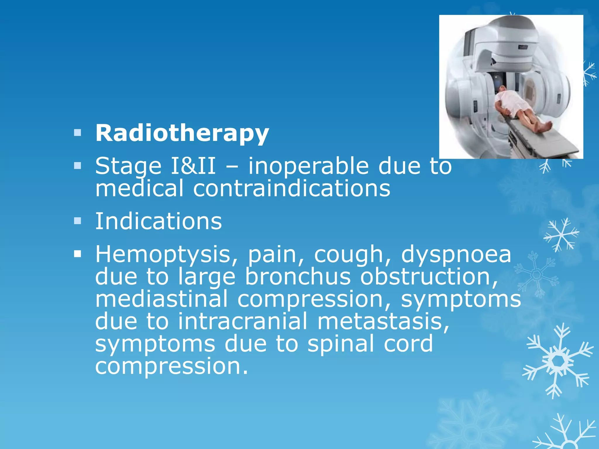
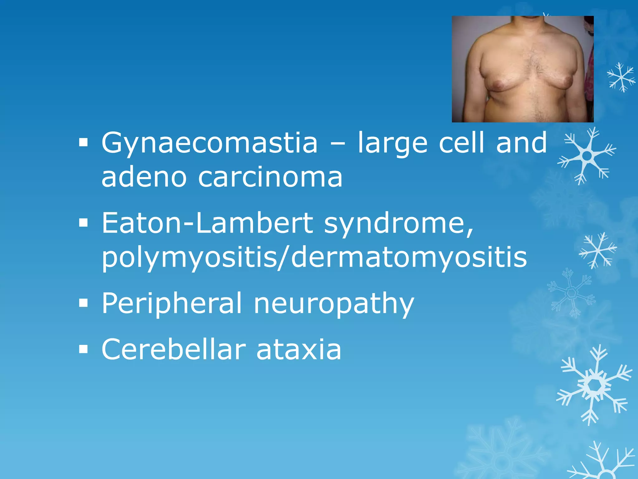
Lung and Mediastinal
Tumours
Dr. Manu Mohan. K
Associate Professor
Pulmonary Medicine
Epidemiology
Most common form of malignant
diseases
40,000 new patients per year
8% male deaths and 4% of all
female deaths
Men > women, middle age
Etiological factors
Tobacco smoking
Cigarette smokers are 8-20
times more likely to develop
lung cancer than life long non
smokers.
Squamous and small cell
carcinoma have clear
association with smoking.
Adenocarcinoma is commonest
histological type in a non
smoker
Atmospheric pollution
Controversial
Radon - radiation
Occupational factors
Asbestos – mining, processing,
usage.
Radioactivity – metal ore mining,
uranium mining.
Nickel – refining.
Chromium salt – extraction,
production, usage.
Arsenic – metal refining, chemical
industry, insecticides.
Pulmonary scarring
Localised areas of pulmonary scarring
Diffuse pulmonary fibrosis
Cryptogenic fibrosing alveolitis is
associated with adenocarcinoma
Tuberculosis – scar carcinoma,
adenocarcinoma
Bronchioloalveolar carcinoma also occur
in areas of scarring
Histological classification
1. Squamous cell carcinoma
(epidermoid carcinoma)
2. Small cell carcinoma
a. oat cell carcinoma
b. intermediate cell type
c. combined oat cell
carcinoma
3. Adenocarcinoma
a. acinar
b. papillary adenocarcinoma
c. bronchioloalveolar
d. solid carcinoma with mucous
4. Large cell carcinoma
5. Adenosquamous carcinoma
6. Carcinoid tumours
7. Bronchial gland carcinoma
a. adenoid cystic carcinoma
b. mucoepidermoid carcinoma
c. others
Growth factors
Polypeptides that take part in the
control of cell differentiation and
proliferation
Bombesin/gastrin releasing peptide
– growth factor for small cell
carcinoma
Non small cell carcinoma – few
growth factors are recognized, EGF,
TGF
Genetic abnormalities
Loss of short arm of
chromosome in small cell
carcinoma (p14, p23)
CDKN2 gene on chromosome 9
– Non small cell lung carcinoma
Oncogenes
myc genes – small cell lung
carcinoma
Kras – adenocarcinoma
Tumour markers
Substances produced by
tumour cells that are released
in to blood stream.
Neuron specific enolase,
creatinine phophokinase BB,
CEA
Modes of presentation
Worsening of preexisting respiratory
state.
No symptoms, detected by the chance
of finding an opacity.
Nonspecific symptoms of malignancy
like malaise, anorexia, and weight loss
Metastatic disease
Central tumours
Cough – most common symptom
New cough that persists longer than 2
weeks in a patient of 40 years who is a
smoker.
Hemoptysis – usually streaky
Breathlessness – due to central airway
narrowing, partial or total collapse of a
distal segment
Chest pain – deep chest discomfort,
due to peribronchial and perivascular
nerve involvement.
Peripheral tumours
Cough and hemoptyis
Bronchorrhoea
Dyspnoea
Chest pain
Distant spread
Skeletal metastasis – bone
pain, pathological fractures
Cerebral metastasis –
progressive neurological
symptoms
Clinical features
Frequently no abnormal
findings
Hoarseness
Bovine cough
Clubbing
HPOA
Lymphatic involvement –
scalene and supraclavicular
Axillary lymph nodes due to
chest invasion
Stridor, wheezes
Atelectasis
Pleural effusion
SVC obstruction
Diaphragm palsy
Enlarged liver
Raised intracranial pressure
Dysphagia
Investigations
Chest radiography
Nearly always abnormal
Collapse
Pleural effusion
Elevated hemi diaphragm
Widening of mediastinum
Lymphangitis carcinomatosa
Pneumonic shadow –
bronchioloalveolar carcinoma
Pancoast tumour
Solitary pulmonary nodule (SPN)
Opacity of less than 3 cm without
surrounding atelectasis and or
adenopathy.
Doubling time
Calcification
Solitary pulmonary nodule
Sputum cytology – more yield
in central tumours
60-70% positive yield in
experienced hands
Single sample – 40%
4 samples – 80%
Bronchoscopy – most useful
for central tumours
Tumours beyond bronchoscopic
view – Transbronchial needle
biopsy, blind brushing and
washing
Other investigations
Percutaneous needle biopsy
Aspiration of subcutaneous
swelling
Pleural fluid study
Thoracoscopic lung biopsy
Mediastinoscopy
Thoracotomy
Staging
Non small cell carcinoma
TNM staging
Primary tumour (T)
Tx, T0, T1s, T1, T2, T3, T4
Nodal involvement (N)
N0, N1, N2, N3.
Distant metastasis (M)
M0, M1
Staging
Small cell lung carcinoma
Limited
Extensive
Treatment
Non small cell lung carcinoma
 Surgery – best result, but only a small
minority
 Types of surgery
 Pneumonectomy
 Lobectomy
 VATS – segmentectomy
 5 year survival rate overall 35%
 Radiotherapy
 Stage I&II – inoperable due to
medical contraindications
 Indications
 Hemoptysis, pain, cough, dyspnoea
due to large bronchus obstruction,
mediastinal compression, symptoms
due to intracranial metastasis,
symptoms due to spinal cord
compression.
Endobronchial treatment
 Laser therapy
 Endobronchial radiotherapy
 Photodynamic therapy
Chemotherapy
 Poor response to chemotherapeutic agents
 Combined modalities
Small cell carcinoma
At presentation 70% have extensive
disease
Chemotherapy
More sensitive to chemotherapy
Combined therapy preferred than
monotherapy
Radiotherapy
Primary tumour control
Prophylatic cranial irradiation
Surgery
Paraneoplastic syndrome
 Non metastatic
metabolic/neuromuscular
manifestations
 Hypercalcemia
 SIADH
 Ectopic ACTH
 HPOA
 Gynaecomastia – large cell and
adeno carcinoma
 Eaton-Lambert syndrome,
polymyositis/dermatomyositis
 Peripheral neuropathy
 Cerebellar ataxia
Superior venacava obstruction
 Small cell carcinoma
 Diagnosis – swelling of face and upper
torso and distension of veins across the
chest, upper arms and neck.
 Treatment – chemotherapy,
radiotherapy and stenting
Superior sulcus tumour
Pancoast
Pain in lower part of shoulder and
inner aspect of the arm (C8, T1
and T2)
Sympathetic ganglion
involvement – stellate
Diagnosis
Treatment - radiotherapy and
surgery
Prevention
Primary prevention
Stop smoking
Mediastinum lies centrally
within the chest and spans the
region vertically from the
thoracic inlet to the
diaphragmatic hiatus,
transversally between the
parietal pleura, and coronally
between the sternum and
vertebral column.
Mediastinal Compartments
3 compartments
Anterior compartment
Middle compartment
Posterior compartment
Symptoms and Mechanisms
Symptoms Mechanisms
cough Airway narrowing,
compression
Chest pain Chest wall invasion,
neural invasion
Dyspnoea Airway compromise,
pericardial tamponade,
pleural effusions,
pulmonary stenosis,
heart failure.
Hemoptysis Bronchogenic
carcinoma, airway
invasion, pulmonary
stenosis, heart failure
Dysphagia Oesophageal
narrowing/obstructio
n, oesophageal
motor dysfunction
Hoarseness Vocal cord paralysis
Facial swelling Superior vena cava
syndrome
Incidence
Adults
65% in Anterosuperior, 10% in the
middle and 25% in the posterior
compartments
Children
28% Anterosuperior, 10% in middle,
62% in the posterior compartment
Investigations
• Noninvasive diagnostic procedures
• Computed tomography
• Magnetic resonance imaging
• Ultrasonography
• Radio nuclides
Biochemical Markers
 CEA
 AFB, HCG – nonseminomatous germ cell
tumour, Teratoma, Carcinoma
 Catecholamines,vanillylmandelic acid,
homovanillic acid – Pheochromocytoma
 Nor epinephrine, epinephrine –
paraganglioma,ganglioneuroma,
neuroblatoma
Invasive biopsy procedures
FNAB
Surgical procedures
Lesions masquerading as
mediastinal tumours
• Substernal Goiter
• Cystic Hygroma
• Lesions originating from thoracic
skeleton
• Vascular lesions
• Oesophageal lesions
• Pulmonary lesions
• Sub diaphragmatic Lesions
Paraneoplastic syndromes
associated with Thymoma
Well established
Myasthenia gravis
Pure red cell aplasia
Acquired
hypogammaglobulinemia
Non Thymic cancers
Less well established
Pancytopenia
Lambert-Eaton
Peripheral neuropathies
CNS changes
Multiple endocrine defects
Multiple rheumatologic disorders
Nephrotic syndrome
Thymoma is the most common
primary neoplasm of the mediastinum
15% of Thymic lesions
Equal frequency in male and female
40-60 years
75% in anterior mediastinum
More than 90% are visible on chest
radiograph
Surgical resection
Radiotherapy
Unresectable, recurrent or
metastatic Thymoma-
chemotherapy
Tumours of lymph nodes
Lymphomas
10-14% of mediastinal tumours
Rare in posterior mediastinum
 Hodgkin’s and Non Hodgkin’s
20-30% asymptomatic
60-70% symptoms of local
invasion
30-35% systemic symptoms
Non-Hodgkin’s lymphoma
5% with mediastinal involvement
Large irregular anterior and
superior mediastinal involvement
Radiation therapy effective in low
grade lymphoma
chemotherapy
Germ cell tumours
Benign and malignant
Benign germ cell tumours
(Teratoma)
Constitute 70% of the lesions in
children and 60% in adults.
Contain multiple tissues that
are foreign to the part of the
body in which they develop.
Symptomatic only when
infected
Malignant germ cell tumours
Malignant mediastinal teratoma
Mediastinal seminoma
Nonseminomatous tumours-
embryonal carcinoma,
choriocarcinoma, endodermal
sinus tumours, teratocarcinoma
Chemotherapy and
radiotherapy, surgery
Middle mediastinal tumours
Bronchogenic cysts
Mediastinal cysts form
20% of mediastinal
tumours
60% of mediastinal cysts
are bronchogenic cyst
Oesophageal cysts
 Neuroenteric cysts
Mesothelial cysts
Pericardial or
pleuropericardial cysts
Thoracic duct cyst
• Neurogenic tumours
• Most common malignancy in
children
• In children 50% malignant,
adults 10%
• Dumbbell tumours – intraspinal
extension
• CT, MRI, myelography
Posterior mediastinal tumours
Tumours of nerve sheath origin
Benign – neurilemoma or
neurofibroma
Malignant tumours-incidence
of malignancy more in von
Recklinghausen’s disease
Poor prognosis
Tumours of autonomic nervous
system
Neuroblatoma,
ganglioneuroblastoma rare in
adults
Endocrine tumours
Mediastinal pheochromocytoma
Parathyroid adenoma
Lung mediastinal tumors_mbbs

Lung mediastinal tumors_mbbs