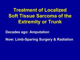
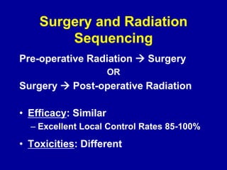
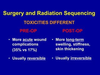
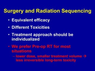
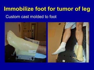
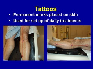
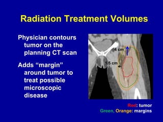
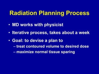
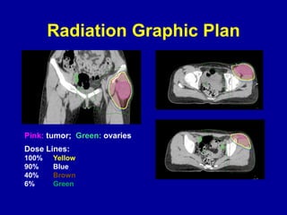
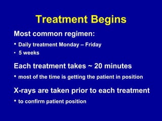
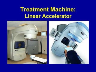
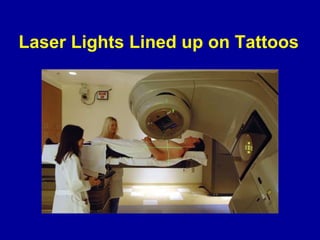
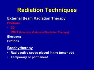
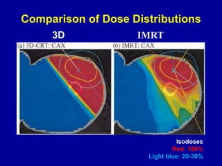
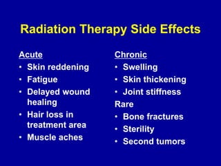
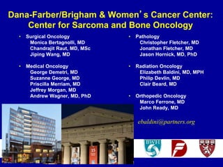
RADIATION THERAPY
SOFT TISSUE SARCOMA
Elizabeth H. Baldini MD, MPH
Radiation Oncology Director
Center for Sarcoma and Bone Oncology
Dana Farber Cancer Institute
Associate Professor of Radiation Oncology
Harvard Medical School
Soft Tissue Sarcoma
Management by Multimodality Team
Radiology
Pathology
Surgery: Oncologic/Orthopedic; Plastic
Radiation Oncology
Medical Oncology
Physical Therapy
Social Work
Case Review at Multi-disciplinary Tumor
Board
Management at Experienced Center
•Associated with better outcome
Guadagnolo, A J Clin Onc 2011
Gutierrez, Ann Surg 2007
Paszat, Sarcoma 2002
Clasby, Br J Surg 1997
Gustafson, Acta Orthop Scand 1994
Treatment
Localized Disease
• Surgery
• Radiation Therapy
Metastatic Disease (beyond local site)
• Systemic therapy
Treatment of Localized
Soft Tissue Sarcoma of the
Extremity or Trunk
Decades ago: Amputation
Now: Limb-Sparing Surgery & Radiation
Surgery and Radiation
Sequencing
Pre-operative Radiation  Surgery
OR
Surgery  Post-operative Radiation
• Efficacy: Similar
– Excellent Local Control Rates 85-100%
• Toxicities: Different
Surgery and Radiation Sequencing
PRE-OP RT
Lower Dose:
50 Gy
5 weeks
Smaller Irradiated
Volume: tumor
plus margins
POST-OP RT
Higher Dose:
60-66 Gy
6-7 weeks
Larger Irradiated
Volume: entire
operative bed +
margins
Surgery and Radiation Sequencing
PRE-OP
• More acute wound
complications
(35% vs 17%)
• Usually reversible
POST-OP
• More long-term
swelling, stiffness,
skin thickening
• Usually irreversible
TOXICITIES DIFFERENT
Surgery and Radiation Sequencing
• Equivalent efficacy
• Different Toxicities
• Treatment approach should be
individualized
• We prefer Pre-op RT for most
situations
– lower dose, smaller treatment volume 
less irreversible long-term toxicity
RADIATION PLANNING
PROCESS
SIMULATION
• Decide on patient position on
treatment table
• Make custom immobilization device
to reproduce position on a daily
basis for treatment
• Obtain CT scan in treatment position
Radiation Simulator
Treatment
couch
CT scan
Immobilize hand for tumor of arm
Custom cast molded to hand
•
Immobilize foot for tumor of leg
Custom cast molded to foot
Tattoos
• Permanent marks placed on skin
• Used for set up of daily treatments
Radiation Treatment Volumes
Physician contours
tumor on the
planning CT scan
Adds “margin”
around tumor to
treat possible
microscopic
disease
4 cm
1.5 cm
Red: tumor
Green, Orange: margins
Radiation Planning Process
• MD works with physicist
• Iterative process, takes about a week
• Goal: to devise a plan to
– treat contoured volume to desired dose
– maximize normal tissue sparing
Radiation Graphic Plan
Pink: tumor; Green: ovaries
Dose Lines:
100% Yellow
90% Blue
40% Brown
6% Green
Treatment Begins
Most common regimen:
• Daily treatment Monday – Friday
• 5 weeks
Each treatment takes ~ 20 minutes
• most of the time is getting the patient in position
X-rays are taken prior to each treatment
• to confirm patient position
Treatment Machine:
Linear Accelerator
Laser Lights Lined up on Tattoos
Or Lasers Lined up on Mask
Verification X-rays or CT scan
• Taken prior to
each treatment to
confirm patient is
set up correctly
• Adjustments are
made as necessary
and new films are
taken for set-up
confirmation
Radiation Techniques
External Beam Radiation Therapy
Photons
• 3D
• IMRT (Intensity Modulated Radiation Therapy)
Electrons
Protons
Brachytherapy
• Radioactive seeds placed in the tumor bed
• Temporary or permanent
Comparison of Dose Distributions
Isodoses
Red: 100%
Light blue: 20-30%
3D IMRT
Radiation Therapy Side Effects
Acute
• Skin reddening
• Fatigue
• Delayed wound
healing
• Hair loss in
treatment area
• Muscle aches
Chronic
• Swelling
• Skin thickening
• Joint stiffness
Rare
• Bone fractures
• Sterility
• Second tumors
Dana-Farber/Brigham & Women’s Cancer Center:
Center for Sarcoma and Bone Oncology
• Surgical Oncology
Monica Bertagnolli, MD
Chandrajit Raut, MD, MSc
Jiping Wang, MD
• Medical Oncology
George Demetri, MD
Suzanne George, MD
Priscilla Merriam, MD
Jeffrey Morgan, MD
Andrew Wagner, MD, PhD
• Pathology
Christopher Fletcher, MD
Jonathan Fletcher, MD
Jason Hornick, MD, PhD
• Radiation Oncology
Elizabeth Baldini, MD, MPH
Philip Devlin, MD
Clair Beard, MD
• Orthopedic Oncology
Marco Ferrone, MD
John Ready, MD
ebaldini@partners.org

How Radiation Therapy is Used to Treat Soft Tissue Sarcoma

  • 1.
    RADIATION THERAPY SOFT TISSUESARCOMA Elizabeth H. Baldini MD, MPH Radiation Oncology Director Center for Sarcoma and Bone Oncology Dana Farber Cancer Institute Associate Professor of Radiation Oncology Harvard Medical School
  • 2.
    Soft Tissue Sarcoma Managementby Multimodality Team Radiology Pathology Surgery: Oncologic/Orthopedic; Plastic Radiation Oncology Medical Oncology Physical Therapy Social Work Case Review at Multi-disciplinary Tumor Board
  • 3.
    Management at ExperiencedCenter •Associated with better outcome Guadagnolo, A J Clin Onc 2011 Gutierrez, Ann Surg 2007 Paszat, Sarcoma 2002 Clasby, Br J Surg 1997 Gustafson, Acta Orthop Scand 1994
  • 4.
    Treatment Localized Disease • Surgery •Radiation Therapy Metastatic Disease (beyond local site) • Systemic therapy
  • 5.
    Treatment of Localized SoftTissue Sarcoma of the Extremity or Trunk Decades ago: Amputation Now: Limb-Sparing Surgery & Radiation
  • 6.
    Surgery and Radiation Sequencing Pre-operativeRadiation  Surgery OR Surgery  Post-operative Radiation • Efficacy: Similar – Excellent Local Control Rates 85-100% • Toxicities: Different
  • 7.
    Surgery and RadiationSequencing PRE-OP RT Lower Dose: 50 Gy 5 weeks Smaller Irradiated Volume: tumor plus margins POST-OP RT Higher Dose: 60-66 Gy 6-7 weeks Larger Irradiated Volume: entire operative bed + margins
  • 8.
    Surgery and RadiationSequencing PRE-OP • More acute wound complications (35% vs 17%) • Usually reversible POST-OP • More long-term swelling, stiffness, skin thickening • Usually irreversible TOXICITIES DIFFERENT
  • 9.
    Surgery and RadiationSequencing • Equivalent efficacy • Different Toxicities • Treatment approach should be individualized • We prefer Pre-op RT for most situations – lower dose, smaller treatment volume  less irreversible long-term toxicity
  • 10.
  • 11.
    SIMULATION • Decide onpatient position on treatment table • Make custom immobilization device to reproduce position on a daily basis for treatment • Obtain CT scan in treatment position
  • 12.
  • 13.
    Immobilize hand fortumor of arm Custom cast molded to hand •
  • 14.
    Immobilize foot fortumor of leg Custom cast molded to foot
  • 15.
    Tattoos • Permanent marksplaced on skin • Used for set up of daily treatments
  • 16.
    Radiation Treatment Volumes Physiciancontours tumor on the planning CT scan Adds “margin” around tumor to treat possible microscopic disease 4 cm 1.5 cm Red: tumor Green, Orange: margins
  • 17.
    Radiation Planning Process •MD works with physicist • Iterative process, takes about a week • Goal: to devise a plan to – treat contoured volume to desired dose – maximize normal tissue sparing
  • 18.
    Radiation Graphic Plan Pink:tumor; Green: ovaries Dose Lines: 100% Yellow 90% Blue 40% Brown 6% Green
  • 19.
    Treatment Begins Most commonregimen: • Daily treatment Monday – Friday • 5 weeks Each treatment takes ~ 20 minutes • most of the time is getting the patient in position X-rays are taken prior to each treatment • to confirm patient position
  • 20.
  • 21.
    Laser Lights Linedup on Tattoos
  • 22.
    Or Lasers Linedup on Mask
  • 23.
    Verification X-rays orCT scan • Taken prior to each treatment to confirm patient is set up correctly • Adjustments are made as necessary and new films are taken for set-up confirmation
  • 24.
    Radiation Techniques External BeamRadiation Therapy Photons • 3D • IMRT (Intensity Modulated Radiation Therapy) Electrons Protons Brachytherapy • Radioactive seeds placed in the tumor bed • Temporary or permanent
  • 25.
    Comparison of DoseDistributions Isodoses Red: 100% Light blue: 20-30% 3D IMRT
  • 26.
    Radiation Therapy SideEffects Acute • Skin reddening • Fatigue • Delayed wound healing • Hair loss in treatment area • Muscle aches Chronic • Swelling • Skin thickening • Joint stiffness Rare • Bone fractures • Sterility • Second tumors
  • 27.
    Dana-Farber/Brigham & Women’sCancer Center: Center for Sarcoma and Bone Oncology • Surgical Oncology Monica Bertagnolli, MD Chandrajit Raut, MD, MSc Jiping Wang, MD • Medical Oncology George Demetri, MD Suzanne George, MD Priscilla Merriam, MD Jeffrey Morgan, MD Andrew Wagner, MD, PhD • Pathology Christopher Fletcher, MD Jonathan Fletcher, MD Jason Hornick, MD, PhD • Radiation Oncology Elizabeth Baldini, MD, MPH Philip Devlin, MD Clair Beard, MD • Orthopedic Oncology Marco Ferrone, MD John Ready, MD ebaldini@partners.org