Downloaded 144 times
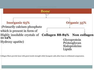
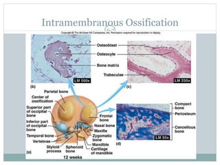
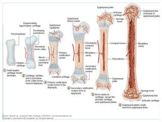
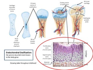
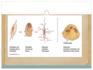
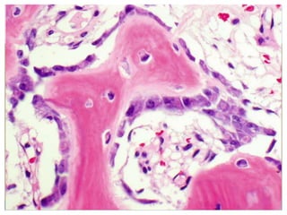
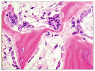
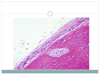
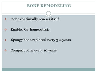
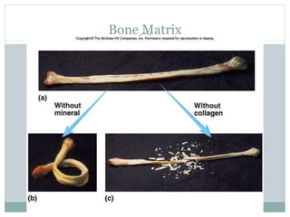


This document provides an overview of bone anatomy, physiology, and pathology. It discusses the following key points in 3 sentences or less: - Bone is composed of inorganic minerals (hydroxyapatite crystals) and organic collagen fibers, which provide strength and allow bone to withstand compression and tension. Bone develops through two processes: intramembranous and endochondral ossification. Bone remodeling is a continuous process where old bone is resorbed and new bone is formed, enabling calcium homeostasis and repair of microdamage.


