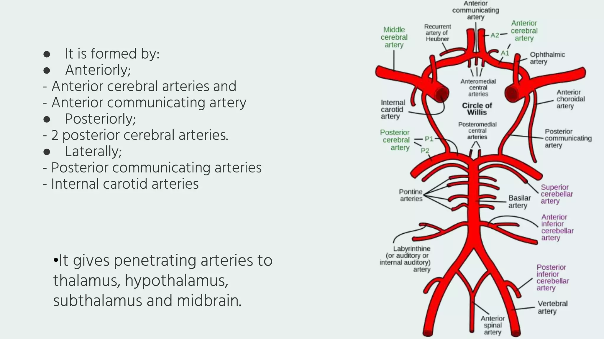
SlideShare a Scribd company logo
BLOODSUPPLYOF
THEBRAIN
TableofCONTENTS
• Arteries of the brain
• Circle of willis
• Veins of the brain
Name: Moustafa Gouda
Under supervision of Dr: Hadeel saed
blood supplyofthebrain &circle ofWillis
ArteriesoftheBrain
Twopairs oflarge
arteries:
1. Vertebral artery
2. Internal carotid artery
InternalCarotid Artery
• It pierces roof of cavernous sinus
•At medial margin of anterior clenoid process.
•It lies lateral to optic chiasma close to anterior
perforated substance.
Branchesofinternalcarotid arteries
1. Anterior cerebral artery
2. Middle cerebral artery
3. Posterior communicating artery
4. Ophthalmic artery
5. Anterior choroidal artery
6. Superior and inferior hypophyseal arteries
7. Hypothalamic artery
AnteriorCerebral Artery
● It passes above optic chiasma into
longitudinal fissure.
● It runs on medial surface above corpus
callosum.
● It ends at parieto- occipital sulcus.
● It is connected by its fellow of opposite side
by short anterior communicating artery
2. Central branches (striate arteries):
● Small arteries pierce ant. perforated substance.
● Supply a part of caudate nucleus and putamen.
● Supply anterior limb of internal capsule.
● Branches:
1. Cortical branches: supply
● Large part of medial surface
● Upper most one finger’s breadth of superolateral
surface as far as parieto- occipital sulcus
● Medial part of orbital surface of frontal lobe
MiddleCerebral Artery
● It runs laterally in stem of lateral sulcus.
● On cortex of insula it divides into cortical branches supplying
the superolateral surface.
1. Cortical branches
● They supply cortex of greater part of superolateral surface.
● Supply motor and sensory areas of trunk and face by:
 Rolandic artery
 Pre-Rolandic artery
 Post central artery
● Broca’s area.
● arteries.
2. Centralbranches:
● Medial and lateral lenticulo- striate arteries pierce A.P.S. to supply:
1. caudate nucleus
2. putamen and globus pallidus
3. Anterior and posterior limbs of internal capsule.
● OphthalmicArtery:
● Enters orbit via optic canal with optic nerve.
● Central artery of retina a branch of ophthalmic
artery is an end artery and its occlusion results in
blindness.
● Posterior Communicatingartery:
● Arises from I.C.A. before giving terminal branches
● It joins posterior cerebral artery.
● Supplies optic chiasma, tract, hypothalamus, subthalamus and
ant. half of thalamus by anterior thalamo- striate artery.
● Superiorandinferiorhypophysealarteries:
● They supply pituitary gland.
● Hypothalamicartery:supplies hypothalamus
Anteriorchoroidalartery:
● Supplies choroid plexus of inf. Horn of lateral ventricle.
● Supplies optic tract, L.G.B. and part of optic radiation.
● Supplies globus pallidus and part of post. Limb of internal
capsule.
● Supplies hippocampus and amygdaloid body.
VertebralArtery
● It is a branch of first part of subclavian artery.
● It enters cranial cavity through foramen magnum.
● Both arteries ascend on either side of medulla oblongata.
● They unite at lower border of pons to form basilar artery
BranchesofVertebral ArteryinCranial Cavity
1. Posterior spinal artery:
one on each side of spinal
cord.
2. Anterior spinal artery:
joins its fellow ant. To
medulla to form single
median artery.
3. Posterior inferior
cerebellar artery: supply
post. Part of inferior
surface of cerebellum and
forms choroid plexus of
4th ventricle.
4. Medullary branches
BasilarArtery
● It begins at lower border of pons by union of two
vertebral arteries.
● It ascends in median sulcus on ventral surface of
pons.
● It ends at upper border of pons by dividing into 2
posterior cerebral arteries.
Branchesofbasilarartery
1. Pontine branches.
2. Labyrinthine artery: runs laterally with
7th& 8th cranial nerves to internal
auditory meatus.
3. Anterior inferior cerebellar artery:
supply anterior part of inferior surface
of cerebellum.
4. Superior cerebellar artery: from upper
part of the artery to supply superior
surface of cerebellum.
5. Posterior cerebral artery: terminal
branch of basilar artery.
Posterior Cerebral Artery
• It runs backwards on lateral aspect of midbrain.
• It is separated from superior cerebellar artery by 3rd& 4th cranial nerves.
• It reaches lower and medial surfaces of occipital lobe where it divides into:
1. Parieto- occipital artery 2. Calcarine artery
● Cortical branches: supply
● The whole occipital lobe including visual cortex.
● Inferior surface of temporal lobe except its pole.
● Lower most one finger’s breadth of lat. surface of
temporal lobe.
Centralbranches:
1. Perforating arteries pierce post. Perforated
substance.
They supply crus cerebri of midbrain and medial and lateral
geniculate bodies.
2. Posterior thalamo- striate artery supply post. Half of
thalamus.
3. Lateral and medial posterior choroidal arteries supply dorsum
of thalamus, pineal body and choroid plexus of 3rd v. and lateral v.
CircleofWillis
● It lies on the base of the brain and is related to
inter- peduncular fossa.
● It is formed by:
● Anteriorly;
- Anterior cerebral arteries and
- Anterior communicating artery
● Posteriorly;
- 2 posterior cerebral arteries.
● Laterally;
- Posterior communicating arteries
- Internal carotid arteries
•It gives penetrating arteries to
thalamus, hypothalamus,
subthalamus and midbrain.
VeinsofTheBrain
1- Superior cerebral veins; end in SSS.
2- Inferior cerebral veins; end in superficial middle cerebral vein.
3- Superficial middle cerebral vein; ends in cavernous sinus and anastomoses
with SSS and transverse sinus.
4-Deep middle cerebral vein; lies deep to gyri of insula and ends in basal vein.
A-Superficial Cerebral
Veins:
● BasalVein
● Formed at anterior perforated
substance by:
1. Deep middle cerebral vein.
2. Anterior cerebral vein.
3. Striate vein.
● 2 basal veins encircle the brain
stem and drain into great
cerebral vein
● Receives tributaries from optic
chiasma,& tract,
hypothalamus, corpus
striatum, tectum and inferior
cerebral vein
B-DeepCerebral Veins
1. Internal cerebral veins:
● Each one starts at interventricular foramen by:
1. Thalamostriate vein
2. Superior choroid vein
● It runs backwards between two layers of tela
choroidea of 3rd ventricle.
● Below splenium of CC it units with its fellow to
form great cerebral vein
2. GreatCerebral Vein
● Located below splenium of
CC.
● It receives 2 internal cerebral
veins, 2 basal veins, posterior
callosal; vein and 2 internal
occipital veins.
● It joins inferior sagittal sinus
to form straight sinus
ThankYou

More Related Content

What's hot

Anatomy of brainstem
Anatomy of brainstemAnatomy of brainstem
Anatomy of brainstem
MBBS IMS MSU
 
Anatomy of cerebellum
Anatomy of cerebellumAnatomy of cerebellum
Anatomy of cerebellum
MBBS IMS MSU
 
Anatomy of meninges, ventricles, cerebrospinal fluid
Anatomy of meninges, ventricles, cerebrospinal fluidAnatomy of meninges, ventricles, cerebrospinal fluid
Anatomy of meninges, ventricles, cerebrospinal fluid
MBBS IMS MSU
 

What's hot (20)

Anatomy of brainstem
Anatomy of brainstemAnatomy of brainstem
Anatomy of brainstem
 
Anatomy of midbrain
Anatomy of midbrainAnatomy of midbrain
Anatomy of midbrain
 
Cerebrum
CerebrumCerebrum
Cerebrum
 
Cerebellum parts and functions
Cerebellum parts and functionsCerebellum parts and functions
Cerebellum parts and functions
 
Cerebellum-Connections and Functions
Cerebellum-Connections and FunctionsCerebellum-Connections and Functions
Cerebellum-Connections and Functions
 
Anatomy of cerebellum
Anatomy of cerebellumAnatomy of cerebellum
Anatomy of cerebellum
 
MIDBRAIN basic anatomy and applied aspects.
MIDBRAIN basic anatomy and applied aspects.MIDBRAIN basic anatomy and applied aspects.
MIDBRAIN basic anatomy and applied aspects.
 
Meninges
MeningesMeninges
Meninges
 
The blood supply of the brain and spinal cord
The blood supply of the brain and spinal cordThe blood supply of the brain and spinal cord
The blood supply of the brain and spinal cord
 
4th ventricle
4th ventricle4th ventricle
4th ventricle
 
Axillary artey ppt
Axillary artey pptAxillary artey ppt
Axillary artey ppt
 
Spinal cord
Spinal cordSpinal cord
Spinal cord
 
Spinal cord (2)
Spinal cord (2)Spinal cord (2)
Spinal cord (2)
 
Cerebrum
CerebrumCerebrum
Cerebrum
 
Anatomy of meninges, ventricles, cerebrospinal fluid
Anatomy of meninges, ventricles, cerebrospinal fluidAnatomy of meninges, ventricles, cerebrospinal fluid
Anatomy of meninges, ventricles, cerebrospinal fluid
 
Cubital fossa
Cubital fossaCubital fossa
Cubital fossa
 
Muscles and nerves in the thoracic wall
Muscles and nerves in the thoracic wallMuscles and nerves in the thoracic wall
Muscles and nerves in the thoracic wall
 
Spinal cord
Spinal cordSpinal cord
Spinal cord
 
Anatomy of Femoral nerve
Anatomy of Femoral nerve Anatomy of Femoral nerve
Anatomy of Femoral nerve
 
Third ventricle
Third ventricleThird ventricle
Third ventricle
 

Similar to circle of Willis.pptx

Lecture 16 - blood supply of cerebrum.ppt
Lecture 16 - blood supply of cerebrum.pptLecture 16 - blood supply of cerebrum.ppt
Lecture 16 - blood supply of cerebrum.ppt
besaryogya
 
‏لقطة شاشة ٢٠٢٣-١١-١٢ في ١١.٤٥.٤٨ ص.pdf
‏لقطة شاشة ٢٠٢٣-١١-١٢ في ١١.٤٥.٤٨ ص.pdf‏لقطة شاشة ٢٠٢٣-١١-١٢ في ١١.٤٥.٤٨ ص.pdf
‏لقطة شاشة ٢٠٢٣-١١-١٢ في ١١.٤٥.٤٨ ص.pdf
ahmad2100799
 
Blood supply of the brain (1).pptx
Blood supply of the brain (1).pptxBlood supply of the brain (1).pptx
Blood supply of the brain (1).pptx
GAFOCalaqqii
 

Similar to circle of Willis.pptx (20)

Blood supply of the brain.pptx
Blood supply of the brain.pptxBlood supply of the brain.pptx
Blood supply of the brain.pptx
 
Blood supply of brain
Blood supply of brainBlood supply of brain
Blood supply of brain
 
Blood supply of brain.pdf
Blood supply of brain.pdfBlood supply of brain.pdf
Blood supply of brain.pdf
 
Arterial supply of brain
Arterial supply of brainArterial supply of brain
Arterial supply of brain
 
Lecture 16 - blood supply of cerebrum.ppt
Lecture 16 - blood supply of cerebrum.pptLecture 16 - blood supply of cerebrum.ppt
Lecture 16 - blood supply of cerebrum.ppt
 
Blood supply to the brain
Blood supply to the brainBlood supply to the brain
Blood supply to the brain
 
Blood supply of nervous system
Blood supply of nervous  systemBlood supply of nervous  system
Blood supply of nervous system
 
Dural venous sinuses, Ankur Saxena
Dural venous sinuses, Ankur SaxenaDural venous sinuses, Ankur Saxena
Dural venous sinuses, Ankur Saxena
 
Cerebrovascular anatomy
Cerebrovascular anatomyCerebrovascular anatomy
Cerebrovascular anatomy
 
Venous drainge
Venous drainge Venous drainge
Venous drainge
 
Cardiovascular system
Cardiovascular systemCardiovascular system
Cardiovascular system
 
Blood supply of the brain & spinal cord by dr sarwar
Blood supply of the brain & spinal cord by dr sarwarBlood supply of the brain & spinal cord by dr sarwar
Blood supply of the brain & spinal cord by dr sarwar
 
Blood supply of the brain
Blood supply of the brainBlood supply of the brain
Blood supply of the brain
 
Blood supply of the brain
Blood supply of the brainBlood supply of the brain
Blood supply of the brain
 
Blood supply of brain
Blood supply of brainBlood supply of brain
Blood supply of brain
 
‏لقطة شاشة ٢٠٢٣-١١-١٢ في ١١.٤٥.٤٨ ص.pdf
‏لقطة شاشة ٢٠٢٣-١١-١٢ في ١١.٤٥.٤٨ ص.pdf‏لقطة شاشة ٢٠٢٣-١١-١٢ في ١١.٤٥.٤٨ ص.pdf
‏لقطة شاشة ٢٠٢٣-١١-١٢ في ١١.٤٥.٤٨ ص.pdf
 
Brain blood supply
Brain blood supplyBrain blood supply
Brain blood supply
 
Blood supply of the brain (1).pptx
Blood supply of the brain (1).pptxBlood supply of the brain (1).pptx
Blood supply of the brain (1).pptx
 
Blood supply of the brain
Blood supply of the brainBlood supply of the brain
Blood supply of the brain
 
NEUROLOGICAL VESSELS. ELABORATED BY ANATOMY
NEUROLOGICAL VESSELS. ELABORATED BY ANATOMYNEUROLOGICAL VESSELS. ELABORATED BY ANATOMY
NEUROLOGICAL VESSELS. ELABORATED BY ANATOMY
 

Recently uploaded

Additional Benefits for Employee Website.pdf
Additional Benefits for Employee Website.pdfAdditional Benefits for Employee Website.pdf
Additional Benefits for Employee Website.pdf
joachimlavalley1
 
The Roman Empire A Historical Colossus.pdf
The Roman Empire A Historical Colossus.pdfThe Roman Empire A Historical Colossus.pdf
The Roman Empire A Historical Colossus.pdf
kaushalkr1407
 
plant breeding methods in asexually or clonally propagated crops
plant breeding methods in asexually or clonally propagated cropsplant breeding methods in asexually or clonally propagated crops
plant breeding methods in asexually or clonally propagated crops
parmarsneha2
 

Recently uploaded (20)

Solid waste management & Types of Basic civil Engineering notes by DJ Sir.pptx
Solid waste management & Types of Basic civil Engineering notes by DJ Sir.pptxSolid waste management & Types of Basic civil Engineering notes by DJ Sir.pptx
Solid waste management & Types of Basic civil Engineering notes by DJ Sir.pptx
 
Additional Benefits for Employee Website.pdf
Additional Benefits for Employee Website.pdfAdditional Benefits for Employee Website.pdf
Additional Benefits for Employee Website.pdf
 
Supporting (UKRI) OA monographs at Salford.pptx
Supporting (UKRI) OA monographs at Salford.pptxSupporting (UKRI) OA monographs at Salford.pptx
Supporting (UKRI) OA monographs at Salford.pptx
 
GIÁO ÁN DẠY THÊM (KẾ HOẠCH BÀI BUỔI 2) - TIẾNG ANH 8 GLOBAL SUCCESS (2 CỘT) N...
GIÁO ÁN DẠY THÊM (KẾ HOẠCH BÀI BUỔI 2) - TIẾNG ANH 8 GLOBAL SUCCESS (2 CỘT) N...GIÁO ÁN DẠY THÊM (KẾ HOẠCH BÀI BUỔI 2) - TIẾNG ANH 8 GLOBAL SUCCESS (2 CỘT) N...
GIÁO ÁN DẠY THÊM (KẾ HOẠCH BÀI BUỔI 2) - TIẾNG ANH 8 GLOBAL SUCCESS (2 CỘT) N...
 
Matatag-Curriculum and the 21st Century Skills Presentation.pptx
Matatag-Curriculum and the 21st Century Skills Presentation.pptxMatatag-Curriculum and the 21st Century Skills Presentation.pptx
Matatag-Curriculum and the 21st Century Skills Presentation.pptx
 
Embracing GenAI - A Strategic Imperative
Embracing GenAI - A Strategic ImperativeEmbracing GenAI - A Strategic Imperative
Embracing GenAI - A Strategic Imperative
 
Synthetic Fiber Construction in lab .pptx
Synthetic Fiber Construction in lab .pptxSynthetic Fiber Construction in lab .pptx
Synthetic Fiber Construction in lab .pptx
 
Students, digital devices and success - Andreas Schleicher - 27 May 2024..pptx
Students, digital devices and success - Andreas Schleicher - 27 May 2024..pptxStudents, digital devices and success - Andreas Schleicher - 27 May 2024..pptx
Students, digital devices and success - Andreas Schleicher - 27 May 2024..pptx
 
NLC-2024-Orientation-for-RO-SDO (1).pptx
NLC-2024-Orientation-for-RO-SDO (1).pptxNLC-2024-Orientation-for-RO-SDO (1).pptx
NLC-2024-Orientation-for-RO-SDO (1).pptx
 
The Roman Empire A Historical Colossus.pdf
The Roman Empire A Historical Colossus.pdfThe Roman Empire A Historical Colossus.pdf
The Roman Empire A Historical Colossus.pdf
 
NCERT Solutions Power Sharing Class 10 Notes pdf
NCERT Solutions Power Sharing Class 10 Notes pdfNCERT Solutions Power Sharing Class 10 Notes pdf
NCERT Solutions Power Sharing Class 10 Notes pdf
 
1.4 modern child centered education - mahatma gandhi-2.pptx
1.4 modern child centered education - mahatma gandhi-2.pptx1.4 modern child centered education - mahatma gandhi-2.pptx
1.4 modern child centered education - mahatma gandhi-2.pptx
 
plant breeding methods in asexually or clonally propagated crops
plant breeding methods in asexually or clonally propagated cropsplant breeding methods in asexually or clonally propagated crops
plant breeding methods in asexually or clonally propagated crops
 
How to Split Bills in the Odoo 17 POS Module
How to Split Bills in the Odoo 17 POS ModuleHow to Split Bills in the Odoo 17 POS Module
How to Split Bills in the Odoo 17 POS Module
 
Basic phrases for greeting and assisting costumers
Basic phrases for greeting and assisting costumersBasic phrases for greeting and assisting costumers
Basic phrases for greeting and assisting costumers
 
Sectors of the Indian Economy - Class 10 Study Notes pdf
Sectors of the Indian Economy - Class 10 Study Notes pdfSectors of the Indian Economy - Class 10 Study Notes pdf
Sectors of the Indian Economy - Class 10 Study Notes pdf
 
Jose-Rizal-and-Philippine-Nationalism-National-Symbol-2.pptx
Jose-Rizal-and-Philippine-Nationalism-National-Symbol-2.pptxJose-Rizal-and-Philippine-Nationalism-National-Symbol-2.pptx
Jose-Rizal-and-Philippine-Nationalism-National-Symbol-2.pptx
 
Danh sách HSG Bộ môn cấp trường - Cấp THPT.pdf
Danh sách HSG Bộ môn cấp trường - Cấp THPT.pdfDanh sách HSG Bộ môn cấp trường - Cấp THPT.pdf
Danh sách HSG Bộ môn cấp trường - Cấp THPT.pdf
 
Fish and Chips - have they had their chips
Fish and Chips - have they had their chipsFish and Chips - have they had their chips
Fish and Chips - have they had their chips
 
MARUTI SUZUKI- A Successful Joint Venture in India.pptx
MARUTI SUZUKI- A Successful Joint Venture in India.pptxMARUTI SUZUKI- A Successful Joint Venture in India.pptx
MARUTI SUZUKI- A Successful Joint Venture in India.pptx
 

circle of Willis.pptx

  • 2. TableofCONTENTS • Arteries of the brain • Circle of willis • Veins of the brain Name: Moustafa Gouda Under supervision of Dr: Hadeel saed blood supplyofthebrain &circle ofWillis
  • 4.
  • 5. InternalCarotid Artery • It pierces roof of cavernous sinus •At medial margin of anterior clenoid process. •It lies lateral to optic chiasma close to anterior perforated substance.
  • 6. Branchesofinternalcarotid arteries 1. Anterior cerebral artery 2. Middle cerebral artery 3. Posterior communicating artery 4. Ophthalmic artery 5. Anterior choroidal artery 6. Superior and inferior hypophyseal arteries 7. Hypothalamic artery
  • 7. AnteriorCerebral Artery ● It passes above optic chiasma into longitudinal fissure. ● It runs on medial surface above corpus callosum. ● It ends at parieto- occipital sulcus. ● It is connected by its fellow of opposite side by short anterior communicating artery
  • 8. 2. Central branches (striate arteries): ● Small arteries pierce ant. perforated substance. ● Supply a part of caudate nucleus and putamen. ● Supply anterior limb of internal capsule. ● Branches: 1. Cortical branches: supply ● Large part of medial surface ● Upper most one finger’s breadth of superolateral surface as far as parieto- occipital sulcus ● Medial part of orbital surface of frontal lobe
  • 9. MiddleCerebral Artery ● It runs laterally in stem of lateral sulcus. ● On cortex of insula it divides into cortical branches supplying the superolateral surface.
  • 10. 1. Cortical branches ● They supply cortex of greater part of superolateral surface. ● Supply motor and sensory areas of trunk and face by:  Rolandic artery  Pre-Rolandic artery  Post central artery ● Broca’s area. ● arteries. 2. Centralbranches: ● Medial and lateral lenticulo- striate arteries pierce A.P.S. to supply: 1. caudate nucleus 2. putamen and globus pallidus 3. Anterior and posterior limbs of internal capsule.
  • 11. ● OphthalmicArtery: ● Enters orbit via optic canal with optic nerve. ● Central artery of retina a branch of ophthalmic artery is an end artery and its occlusion results in blindness. ● Posterior Communicatingartery: ● Arises from I.C.A. before giving terminal branches ● It joins posterior cerebral artery. ● Supplies optic chiasma, tract, hypothalamus, subthalamus and ant. half of thalamus by anterior thalamo- striate artery.
  • 12. ● Superiorandinferiorhypophysealarteries: ● They supply pituitary gland. ● Hypothalamicartery:supplies hypothalamus Anteriorchoroidalartery: ● Supplies choroid plexus of inf. Horn of lateral ventricle. ● Supplies optic tract, L.G.B. and part of optic radiation. ● Supplies globus pallidus and part of post. Limb of internal capsule. ● Supplies hippocampus and amygdaloid body.
  • 13. VertebralArtery ● It is a branch of first part of subclavian artery. ● It enters cranial cavity through foramen magnum. ● Both arteries ascend on either side of medulla oblongata. ● They unite at lower border of pons to form basilar artery
  • 14. BranchesofVertebral ArteryinCranial Cavity 1. Posterior spinal artery: one on each side of spinal cord. 2. Anterior spinal artery: joins its fellow ant. To medulla to form single median artery. 3. Posterior inferior cerebellar artery: supply post. Part of inferior surface of cerebellum and forms choroid plexus of 4th ventricle. 4. Medullary branches
  • 15. BasilarArtery ● It begins at lower border of pons by union of two vertebral arteries. ● It ascends in median sulcus on ventral surface of pons. ● It ends at upper border of pons by dividing into 2 posterior cerebral arteries.
  • 16. Branchesofbasilarartery 1. Pontine branches. 2. Labyrinthine artery: runs laterally with 7th& 8th cranial nerves to internal auditory meatus. 3. Anterior inferior cerebellar artery: supply anterior part of inferior surface of cerebellum. 4. Superior cerebellar artery: from upper part of the artery to supply superior surface of cerebellum. 5. Posterior cerebral artery: terminal branch of basilar artery.
  • 17. Posterior Cerebral Artery • It runs backwards on lateral aspect of midbrain. • It is separated from superior cerebellar artery by 3rd& 4th cranial nerves. • It reaches lower and medial surfaces of occipital lobe where it divides into: 1. Parieto- occipital artery 2. Calcarine artery
  • 18. ● Cortical branches: supply ● The whole occipital lobe including visual cortex. ● Inferior surface of temporal lobe except its pole. ● Lower most one finger’s breadth of lat. surface of temporal lobe. Centralbranches: 1. Perforating arteries pierce post. Perforated substance. They supply crus cerebri of midbrain and medial and lateral geniculate bodies. 2. Posterior thalamo- striate artery supply post. Half of thalamus. 3. Lateral and medial posterior choroidal arteries supply dorsum of thalamus, pineal body and choroid plexus of 3rd v. and lateral v.
  • 19.
  • 20. CircleofWillis ● It lies on the base of the brain and is related to inter- peduncular fossa.
  • 21. ● It is formed by: ● Anteriorly; - Anterior cerebral arteries and - Anterior communicating artery ● Posteriorly; - 2 posterior cerebral arteries. ● Laterally; - Posterior communicating arteries - Internal carotid arteries •It gives penetrating arteries to thalamus, hypothalamus, subthalamus and midbrain.
  • 22. VeinsofTheBrain 1- Superior cerebral veins; end in SSS. 2- Inferior cerebral veins; end in superficial middle cerebral vein. 3- Superficial middle cerebral vein; ends in cavernous sinus and anastomoses with SSS and transverse sinus. 4-Deep middle cerebral vein; lies deep to gyri of insula and ends in basal vein. A-Superficial Cerebral Veins:
  • 23. ● BasalVein ● Formed at anterior perforated substance by: 1. Deep middle cerebral vein. 2. Anterior cerebral vein. 3. Striate vein. ● 2 basal veins encircle the brain stem and drain into great cerebral vein ● Receives tributaries from optic chiasma,& tract, hypothalamus, corpus striatum, tectum and inferior cerebral vein
  • 24. B-DeepCerebral Veins 1. Internal cerebral veins: ● Each one starts at interventricular foramen by: 1. Thalamostriate vein 2. Superior choroid vein ● It runs backwards between two layers of tela choroidea of 3rd ventricle. ● Below splenium of CC it units with its fellow to form great cerebral vein
  • 25. 2. GreatCerebral Vein ● Located below splenium of CC. ● It receives 2 internal cerebral veins, 2 basal veins, posterior callosal; vein and 2 internal occipital veins. ● It joins inferior sagittal sinus to form straight sinus