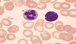
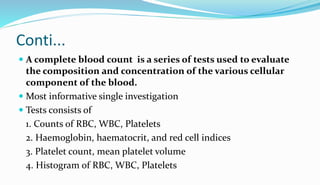
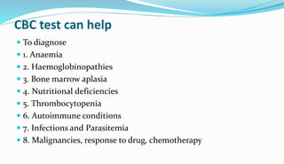
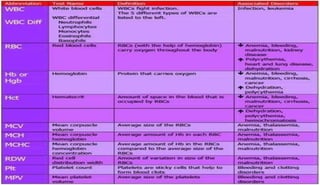
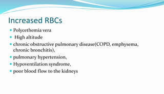
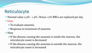
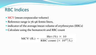
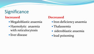
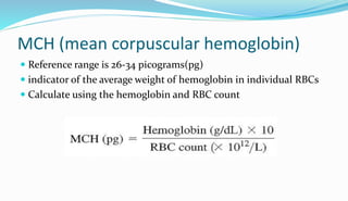
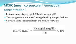
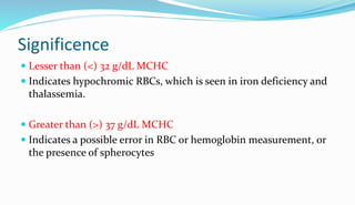
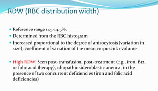
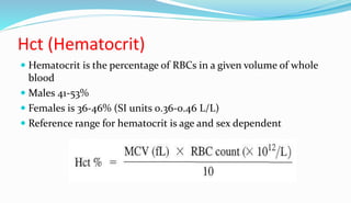
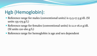
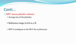
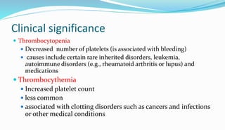
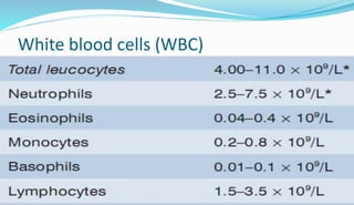
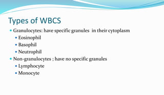
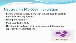
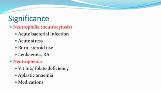
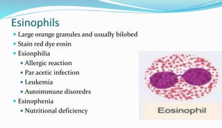
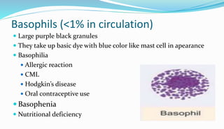
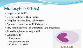
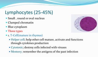
Prepared by
Muhammad Tariq
Medical lab scientist
Khyber medical university
Introduction
 The human body is primarily made up of water and cells
 Stationary cells; skin, muscles, bones and organs(heart, lungs,
kidneys)
 Circulating cells move throughout the body by travelling in the
blood provide oxygen to all of the stationary cells
 Help in fight infection, stop bleeding
 Information about these cells can provide important clues about
the overall health of the body.
Cont...
 CBC, is a lab test that provides information about these circulating
cells
 Sample of blood is collected commonly in EDTA tube and sent to
the lab
 This tube contains EDTA (ethylene diamine tetra acetic acid),
which acts as a potent anticoagulant by binding to calcium in the
blood
 A lab instrument then automatically/manually counts the number
of each type of circulating cell

Conti...
 A complete blood count is a series of tests used to evaluate
the composition and concentration of the various cellular
component of the blood.
 Most informative single investigation
 Tests consists of
1. Counts of RBC, WBC, Platelets
2. Haemoglobin, haematocrit, and red cell indices
3. Platelet count, mean platelet volume
4. Histogram of RBC, WBC, Platelets
CBC test can help
 To diagnose
 1. Anaemia
 2. Haemoglobinopathies
 3. Bone marrow aplasia
 4. Nutritional deficiencies
 5. Thrombocytopenia
 6. Autoimmune conditions
 7. Infections and Parasitemia
 8. Malignancies, response to drug, chemotherapy
Red blood cells
 RBC produced in marrow and requires
 Iron, copper, cobalt
 Vitamins; especially B12, folic acid
 Regulated by erythropoietin, thyroid hormone, androgens
 Life span - 120 days
Conti...
 Biconcave, disk like structure.
 Have smooth contours
 Appears to be of the same size as the nucleus of the small
lymphocyte.
 Have no nucleus
 1/3 cell is filled with the HAEMOGLOBIN
Normal values
 Newborn 4.1-6.1 million/mm3
 Children 3.6-5.5 million/mm3
 Adult (M) 4.6-6.0 million/mm3
 Adult (F) 4.2-5.0 million/mm3
Decreased RBCs
•Blood loss
•Trauma
•Surgery
•GL bleeding
•Impaired production
•Pure red cell aplasia
•Pernicious anaemia
•Megaloblastic anaemia
•Iron deficiency anaemia
•Thalassemia etc
•Increased destruction
•Hereditary spherocytosis
•Sickle cell anaemia
•G6PD
•Autoimmune
•Haemolytic disease of
newborn
•Mismatch transfusion
•DIC etc
Increased RBCs
 Polycethemia vera
 High altitude
 chronic obstructive pulmonary disease(COPD, emphysema,
chronic bronchitis),
 pulmonary hypertension,
 Hypoventilation syndrome,
 poor blood flow to the kidneys
Reticulocyte
 Normal value 0.5% - 1.5%. Hence <2% RBCs are replaced per day
 Uses
 To evaluate anaemia
 Response to treatment of anaemia
 Note
 If the disease causing the anaemia is inside the marrow, the
reticulocyte count is decreased
 If the disease causing the anaemia is outside the marrow, the
reticulocyte count is increased
RBC Indices
 MCV (mean corpuscular volume)
 Reference range is 76-96 femto liters,
 indicator of the average/mean volume of erythrocytes (RBCs)
 Calculate using the hematocrit and RBC count
Significance
Increased Decreased
 Megaloblastic anaemia
 Haemolytic anaemia
with reticulocytosis
 liver disease
 Iron deficiency anaemia
 Thalassemia
 sideroblastic anaemia
 lead poisoning
MCH (mean corpuscular hemoglobin)
 Reference range is 26-34 picograms(pg)
 indicator of the average weight of hemoglobin in individual RBCs
 Calculate using the hemoglobin and RBC count
Significance
 Increased in
 Macrocytic anaemia
 Decreased in
 Microcytic
 Hypochromic anaemia
MCHC (mean corpuscular hemoglobin
concentration)
 Reference range is 32-37 g/dL (SI units 320-370 g/L)
 The average concentration of hemoglobin in grams per deciliter
 Calculate using the hemoglobin and hematocrit values
Significence
 Lesser than (<) 32 g/dL MCHC
 Indicates hypochromic RBCs, which is seen in iron deficiency and
thalassemia.
 Greater than (>) 37 g/dL MCHC
 Indicates a possible error in RBC or hemoglobin measurement, or
the presence of spherocytes
RDW (RBC distribution width)
 Reference range 11.5-14.5%.
 Determined from the RBC histogram
 Increased proportional to the degree of anisocytosis (variation in
size); coefficient of variation of the mean corpuscular volume
 High RDW: Seen post-transfusion, post-treatment (e.g., iron, B12,
or folic acid therapy), idiopathic sideroblastic anemia, in the
presence of two concurrent deficiencies (iron and folic acid
deficiencies)
Hct (Hematocrit)
 Hematocrit is the percentage of RBCs in a given volume of whole
blood
 Males 41-53%
 Females is 36-46% (SI units 0.36-0.46 L/L)
 Reference range for hematocrit is age and sex dependent
Hgb (Hemoglobin):
 Reference range for males (conventional units) is 13.5-17.5 g/dL (SI
units 135-175 g/L)
 Reference range for females (conventional units) is 12.0-16.0 g/dL
(SI units 120-160 g/L)
 Reference range for hemoglobin is age and sex dependent
Platelets
 PLT (Platelets)
 Reference range (SI units) is 150-450 X 109/L (conventional units
150,000-450,000/^L)
 Cytoplasmic fragments of large megakaryocyte
 2-3 mm in diameter with granules
 Promotes clot formation
 8-20 per oil immersion field
Conti...
 MPV (mean platelet volume)
 Average size of the platelets
 Reference range is 6.8-10.2 fL
 MPV is analogous to the MCV for erythrocytes
Clinical significance
 Thrombocytopenia
 Decreased number of platelets (is associated with bleeding)
 causes include certain rare inherited disorders, leukemia,
autoimmune disorders (e.g., rheumatoid arthritis or lupus) and
medications
 Thrombocythemia
 Increased platelet count
 less common
 associated with clotting disorders such as cancers and infections
or other medical conditions
White blood cells (WBC)
Types of WBCS
 Granulocytes: have specific granules in their cytoplasm
 Eosinophil
 Basophil
 Neutrophil
 Non-granulocytes ; have no specific granules
 Lymphocyte
 Monocyte
Neutrophils (45-65% in cirulation)
 Polymorphonuclear cells along with esinophils and basophils(
multi lobulated 2-5 lobules)
 Small purple garnules
 Most abundant of WBC
 Highly motile and play role in acute phase of inflammation
especially bacterial infections
Significance
 Neutrophilia (neutrocytosis)
 Acute bacterial infection
 Acute stress
 Burn, steroid use
 Leukaemia, RA
 Neutrophenia
 Vit b12/ folate deficiency
 Aplastic anaemia
 Medications
Esinophils
 Large orange granules and usually bilobed
 Stain red dye eosin
 Esionphilia
 Allergic reaction
 Par acetic infection
 Leukemia
 Autoimmune disoredrs
 Esinophenia
 Nutritional deficiency
Basophils (<1% in circulation)
 Large purple black granules
 They take up basic dye with blue color like mast cell in apearance
 Basophilia
 Allergic reaction
 CML
 Hodgkin’s disease
 Oral contraceptive use
 Basophenia
 Nutritional deficiency
Monocytes (3-10%)
 Largest of all WBCs
 Grey cytoplasm with vacuoles
 Irregular nucleus, linear chromatin
 Approach three time of RBC diameter
 Play role in chronic inflammation, viral infections
 Stored in spleen and very motile
 What they do
 Phagocytosis
 Antigen presentation
 Cytokines production
Lymphocytes (25-45%)
 Small , round or oval nucleus
 Clumped chromatin
 Blue cytoplasm
 Three types
 1. T-Cell(mature in thymus)
 Helper cell; help other cell mature, activate and functions
through cytokines production
 Cytotoxic; destroy cells infected with viruses
 Memory; remember the antigens of the past infection
 2.B-cells(formed in bone marrow)
 Plasma-produce large amount of antibodies
 Memory-remember past infection
 3.Natural killer cells(NK)
 Similar role like cytotoxic T-cell, kill virus infected cells and tumour cells
signifincence
 Lymphocytosis;
 Viral infections
 Leukemias
 Adrenal insufficiency
 Lymphocytopenia
 HIV-destroy T-cells
 Aplastic anaemia
The complete blood count (cbc)

The complete blood count (cbc)

  • 1.
    Prepared by Muhammad Tariq Medicallab scientist Khyber medical university
  • 3.
    Introduction  The humanbody is primarily made up of water and cells  Stationary cells; skin, muscles, bones and organs(heart, lungs, kidneys)  Circulating cells move throughout the body by travelling in the blood provide oxygen to all of the stationary cells  Help in fight infection, stop bleeding  Information about these cells can provide important clues about the overall health of the body.
  • 4.
    Cont...  CBC, isa lab test that provides information about these circulating cells  Sample of blood is collected commonly in EDTA tube and sent to the lab  This tube contains EDTA (ethylene diamine tetra acetic acid), which acts as a potent anticoagulant by binding to calcium in the blood  A lab instrument then automatically/manually counts the number of each type of circulating cell
  • 5.
  • 6.
    Conti...  A completeblood count is a series of tests used to evaluate the composition and concentration of the various cellular component of the blood.  Most informative single investigation  Tests consists of 1. Counts of RBC, WBC, Platelets 2. Haemoglobin, haematocrit, and red cell indices 3. Platelet count, mean platelet volume 4. Histogram of RBC, WBC, Platelets
  • 7.
    CBC test canhelp  To diagnose  1. Anaemia  2. Haemoglobinopathies  3. Bone marrow aplasia  4. Nutritional deficiencies  5. Thrombocytopenia  6. Autoimmune conditions  7. Infections and Parasitemia  8. Malignancies, response to drug, chemotherapy
  • 9.
    Red blood cells RBC produced in marrow and requires  Iron, copper, cobalt  Vitamins; especially B12, folic acid  Regulated by erythropoietin, thyroid hormone, androgens  Life span - 120 days
  • 10.
    Conti...  Biconcave, disklike structure.  Have smooth contours  Appears to be of the same size as the nucleus of the small lymphocyte.  Have no nucleus  1/3 cell is filled with the HAEMOGLOBIN
  • 11.
    Normal values  Newborn4.1-6.1 million/mm3  Children 3.6-5.5 million/mm3  Adult (M) 4.6-6.0 million/mm3  Adult (F) 4.2-5.0 million/mm3
  • 12.
    Decreased RBCs •Blood loss •Trauma •Surgery •GLbleeding •Impaired production •Pure red cell aplasia •Pernicious anaemia •Megaloblastic anaemia •Iron deficiency anaemia •Thalassemia etc •Increased destruction •Hereditary spherocytosis •Sickle cell anaemia •G6PD •Autoimmune •Haemolytic disease of newborn •Mismatch transfusion •DIC etc
  • 13.
    Increased RBCs  Polycethemiavera  High altitude  chronic obstructive pulmonary disease(COPD, emphysema, chronic bronchitis),  pulmonary hypertension,  Hypoventilation syndrome,  poor blood flow to the kidneys
  • 14.
    Reticulocyte  Normal value0.5% - 1.5%. Hence <2% RBCs are replaced per day  Uses  To evaluate anaemia  Response to treatment of anaemia  Note  If the disease causing the anaemia is inside the marrow, the reticulocyte count is decreased  If the disease causing the anaemia is outside the marrow, the reticulocyte count is increased
  • 15.
    RBC Indices  MCV(mean corpuscular volume)  Reference range is 76-96 femto liters,  indicator of the average/mean volume of erythrocytes (RBCs)  Calculate using the hematocrit and RBC count
  • 16.
    Significance Increased Decreased  Megaloblasticanaemia  Haemolytic anaemia with reticulocytosis  liver disease  Iron deficiency anaemia  Thalassemia  sideroblastic anaemia  lead poisoning
  • 17.
    MCH (mean corpuscularhemoglobin)  Reference range is 26-34 picograms(pg)  indicator of the average weight of hemoglobin in individual RBCs  Calculate using the hemoglobin and RBC count
  • 18.
    Significance  Increased in Macrocytic anaemia  Decreased in  Microcytic  Hypochromic anaemia
  • 19.
    MCHC (mean corpuscularhemoglobin concentration)  Reference range is 32-37 g/dL (SI units 320-370 g/L)  The average concentration of hemoglobin in grams per deciliter  Calculate using the hemoglobin and hematocrit values
  • 20.
    Significence  Lesser than(<) 32 g/dL MCHC  Indicates hypochromic RBCs, which is seen in iron deficiency and thalassemia.  Greater than (>) 37 g/dL MCHC  Indicates a possible error in RBC or hemoglobin measurement, or the presence of spherocytes
  • 21.
    RDW (RBC distributionwidth)  Reference range 11.5-14.5%.  Determined from the RBC histogram  Increased proportional to the degree of anisocytosis (variation in size); coefficient of variation of the mean corpuscular volume  High RDW: Seen post-transfusion, post-treatment (e.g., iron, B12, or folic acid therapy), idiopathic sideroblastic anemia, in the presence of two concurrent deficiencies (iron and folic acid deficiencies)
  • 22.
    Hct (Hematocrit)  Hematocritis the percentage of RBCs in a given volume of whole blood  Males 41-53%  Females is 36-46% (SI units 0.36-0.46 L/L)  Reference range for hematocrit is age and sex dependent
  • 23.
    Hgb (Hemoglobin):  Referencerange for males (conventional units) is 13.5-17.5 g/dL (SI units 135-175 g/L)  Reference range for females (conventional units) is 12.0-16.0 g/dL (SI units 120-160 g/L)  Reference range for hemoglobin is age and sex dependent
  • 24.
    Platelets  PLT (Platelets) Reference range (SI units) is 150-450 X 109/L (conventional units 150,000-450,000/^L)  Cytoplasmic fragments of large megakaryocyte  2-3 mm in diameter with granules  Promotes clot formation  8-20 per oil immersion field
  • 25.
    Conti...  MPV (meanplatelet volume)  Average size of the platelets  Reference range is 6.8-10.2 fL  MPV is analogous to the MCV for erythrocytes
  • 26.
    Clinical significance  Thrombocytopenia Decreased number of platelets (is associated with bleeding)  causes include certain rare inherited disorders, leukemia, autoimmune disorders (e.g., rheumatoid arthritis or lupus) and medications  Thrombocythemia  Increased platelet count  less common  associated with clotting disorders such as cancers and infections or other medical conditions
  • 27.
  • 28.
    Types of WBCS Granulocytes: have specific granules in their cytoplasm  Eosinophil  Basophil  Neutrophil  Non-granulocytes ; have no specific granules  Lymphocyte  Monocyte
  • 29.
    Neutrophils (45-65% incirulation)  Polymorphonuclear cells along with esinophils and basophils( multi lobulated 2-5 lobules)  Small purple garnules  Most abundant of WBC  Highly motile and play role in acute phase of inflammation especially bacterial infections
  • 30.
    Significance  Neutrophilia (neutrocytosis) Acute bacterial infection  Acute stress  Burn, steroid use  Leukaemia, RA  Neutrophenia  Vit b12/ folate deficiency  Aplastic anaemia  Medications
  • 31.
    Esinophils  Large orangegranules and usually bilobed  Stain red dye eosin  Esionphilia  Allergic reaction  Par acetic infection  Leukemia  Autoimmune disoredrs  Esinophenia  Nutritional deficiency
  • 32.
    Basophils (<1% incirculation)  Large purple black granules  They take up basic dye with blue color like mast cell in apearance  Basophilia  Allergic reaction  CML  Hodgkin’s disease  Oral contraceptive use  Basophenia  Nutritional deficiency
  • 33.
    Monocytes (3-10%)  Largestof all WBCs  Grey cytoplasm with vacuoles  Irregular nucleus, linear chromatin  Approach three time of RBC diameter  Play role in chronic inflammation, viral infections  Stored in spleen and very motile  What they do  Phagocytosis  Antigen presentation  Cytokines production
  • 34.
    Lymphocytes (25-45%)  Small, round or oval nucleus  Clumped chromatin  Blue cytoplasm  Three types  1. T-Cell(mature in thymus)  Helper cell; help other cell mature, activate and functions through cytokines production  Cytotoxic; destroy cells infected with viruses  Memory; remember the antigens of the past infection
  • 35.
     2.B-cells(formed inbone marrow)  Plasma-produce large amount of antibodies  Memory-remember past infection  3.Natural killer cells(NK)  Similar role like cytotoxic T-cell, kill virus infected cells and tumour cells
  • 36.
    signifincence  Lymphocytosis;  Viralinfections  Leukemias  Adrenal insufficiency  Lymphocytopenia  HIV-destroy T-cells  Aplastic anaemia