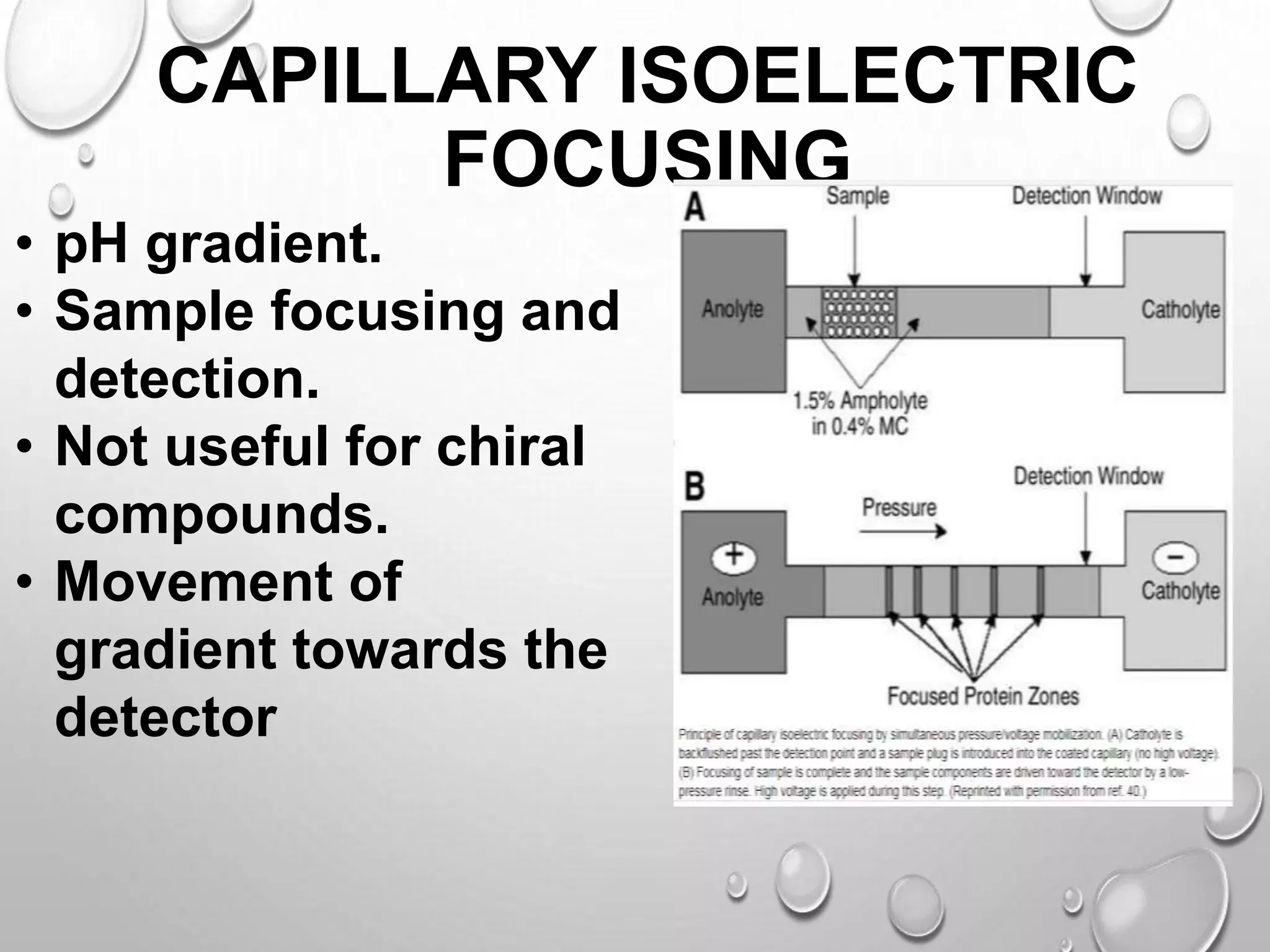
TOPIC
ISOELECTRI
C
FOCUSING
INTRODUCTI
ON
• Proteins are separated in
a pH gradient according
to their isoelectric points.
• Basic principle involved is
electrophoresis.
• Proteins are subjected to
electric field in a pH
gradient.
• Requires a solid surface
normally polyacrilamide.
ISOELECTRIC POINT
(PI)
• The pH at which net charge on protein
becomes zero.
1. Below PI – positive charge.
2. Above PI – negative charge.
• Proteins move toward the electrode with
the opposite charge.
• During motion , proteins will either pick or
loose protons.
• Different from conventional electrophoresis
migrate to steady state.
PRINCIPLE
• All proteins have an isoelectric point pH .
• A procedure to determine the isoelectric point
of proteins , thus a mixture of proteins can be
electrophorised through a solution having a
state pH gradient in form the anode to the
cathode and each protein will migrate to the
position in the pH gradient according to its
isoelectric point . This is called ISOELECTRIC
FOCUSING.
• Protein migrate into the point where its net
CAPILLARY ISOELECTRIC
FOCUSING
• pH gradient.
• Sample focusing and
detection.
• Not useful for chiral
compounds.
• Movement of
gradient towards the
detector
AMPHOLYTES
• Establishment of stable pH gradient is
important.
• Achieved by means of commercially
available synthetic carrier amphoteric
electrolytes.
• 600-900 Da .
• Closely spaced PI and high conductivity.
• The curve is determined by pH interval
covered by the ampholytes and the
distance between electrodes.
WORKING
PROCEDURE
Following chemicals
are required-
1. Acrylamide
solution
2. Water
3. Ampholyte solution
pH 3.5-10
4.Ampholyte solution
pH 4-6
5. Urea
Procedure:
• Spin gently to mix urea.
• Add 10% APS and TEMED at
the end.
• Remove bubbles.
• Fill the cassette completely with
solution.
• Allow to polymerize at room
temperature.
• Centrifuge the sample to
remove any
• aggregate.
• Apply the supernatant to the
wells with a disposable tip or
Hamilton syringe.
• Run the get at 150v for 30 min
and then at
• 200v for 2 hr.
A typical isoelectric focussing gel track 1 contains a mixture of standard
proteins of known isoelectric points. Tracks 2–5 show increasing loadings of
venom from the Japanese water moccasin snake.
SET UP GEL
• Remove the comb carefully after gel has
polymerized.
• Attach gel to the electrophoresis tank
according the instructions of
manufacturer.
• Add catholyte (sodium hydroxide) to the
upper buffer chamber and anolyte
(phosphoric acid) to the lower buffer
chamber.
SAMPLE PREPARATION AND
LOADING
Mix protein sample with equal volume of 2x
loading buffer.
• Loading buffer includes the following reagents-
1. Urea.
2. Ampholyte solution ph 3.5-10.
3. Ampholyte solution ph 4-6.
4. Triton X-100.
5. 2- mercaptoethanol.
6. 1% bromophenol blue.
2D GEL
ELECTROPHORESIS
• Technique of IEF and SDS PAGE combined.
• Protein separated in two dimensions.
1. On the base of PI.
2. On the basis of molecular weight in
normal SDS PAGE.
• Procedure can be adapted by combining IEF
and page.
• Series of spots formed in gel.
TRADITIONAL EQUIPMENT FOR
ISOELECTRIC FOCUSING
ADVANTAG
ES• Proteins that by as little as 0.001 pH units can be
separated.
• As spreading of bands is minimized due to
application of the applied field and the pH gradient ,
high resolution can be achieved.
• Isoelectric focusing (IEF) is a powerful analytical
tool for the separation of proteins.
• Performing IEF is easier because the placement of
sample application is not important.
DISADVANTAG
ES
• Carrier ampholytes are generally used in
high concentration , a high voltage (upto
2000v) is necessary. As a result the
electrophoretic matrix must be cooled which
sometimes makes it difficult.
• Limited stability of solutions.
• Lot-to-lot inconsistency.
• Inadequate purity for application as a
standard.
APPLICATIONS
• For separating proteins and peptides.
• Used in limit test when the density of band is
compared with the density of band of STD
preparation.
• For research in taxonomy , cytology and
immunology etc.
• IEF gel is used as identity test when migration
pattern on gel is compared with STD preparation.
• Isoelectric focusing (IEF) offers an effective
alternative to conventional electrophoresis for
Isoelectric focusing

Isoelectric focusing

  • 1.
  • 2.
    INTRODUCTI ON • Proteins areseparated in a pH gradient according to their isoelectric points. • Basic principle involved is electrophoresis. • Proteins are subjected to electric field in a pH gradient. • Requires a solid surface normally polyacrilamide.
  • 4.
    ISOELECTRIC POINT (PI) • ThepH at which net charge on protein becomes zero. 1. Below PI – positive charge. 2. Above PI – negative charge. • Proteins move toward the electrode with the opposite charge. • During motion , proteins will either pick or loose protons. • Different from conventional electrophoresis migrate to steady state.
  • 5.
    PRINCIPLE • All proteinshave an isoelectric point pH . • A procedure to determine the isoelectric point of proteins , thus a mixture of proteins can be electrophorised through a solution having a state pH gradient in form the anode to the cathode and each protein will migrate to the position in the pH gradient according to its isoelectric point . This is called ISOELECTRIC FOCUSING. • Protein migrate into the point where its net
  • 7.
    CAPILLARY ISOELECTRIC FOCUSING • pHgradient. • Sample focusing and detection. • Not useful for chiral compounds. • Movement of gradient towards the detector
  • 8.
    AMPHOLYTES • Establishment ofstable pH gradient is important. • Achieved by means of commercially available synthetic carrier amphoteric electrolytes. • 600-900 Da . • Closely spaced PI and high conductivity. • The curve is determined by pH interval covered by the ampholytes and the distance between electrodes.
  • 9.
    WORKING PROCEDURE Following chemicals are required- 1.Acrylamide solution 2. Water 3. Ampholyte solution pH 3.5-10 4.Ampholyte solution pH 4-6 5. Urea Procedure: • Spin gently to mix urea. • Add 10% APS and TEMED at the end. • Remove bubbles. • Fill the cassette completely with solution. • Allow to polymerize at room temperature. • Centrifuge the sample to remove any • aggregate. • Apply the supernatant to the wells with a disposable tip or Hamilton syringe. • Run the get at 150v for 30 min and then at • 200v for 2 hr.
  • 10.
    A typical isoelectricfocussing gel track 1 contains a mixture of standard proteins of known isoelectric points. Tracks 2–5 show increasing loadings of venom from the Japanese water moccasin snake.
  • 11.
    SET UP GEL •Remove the comb carefully after gel has polymerized. • Attach gel to the electrophoresis tank according the instructions of manufacturer. • Add catholyte (sodium hydroxide) to the upper buffer chamber and anolyte (phosphoric acid) to the lower buffer chamber.
  • 12.
    SAMPLE PREPARATION AND LOADING Mixprotein sample with equal volume of 2x loading buffer. • Loading buffer includes the following reagents- 1. Urea. 2. Ampholyte solution ph 3.5-10. 3. Ampholyte solution ph 4-6. 4. Triton X-100. 5. 2- mercaptoethanol. 6. 1% bromophenol blue.
  • 13.
    2D GEL ELECTROPHORESIS • Techniqueof IEF and SDS PAGE combined. • Protein separated in two dimensions. 1. On the base of PI. 2. On the basis of molecular weight in normal SDS PAGE. • Procedure can be adapted by combining IEF and page. • Series of spots formed in gel.
  • 15.
  • 16.
    ADVANTAG ES• Proteins thatby as little as 0.001 pH units can be separated. • As spreading of bands is minimized due to application of the applied field and the pH gradient , high resolution can be achieved. • Isoelectric focusing (IEF) is a powerful analytical tool for the separation of proteins. • Performing IEF is easier because the placement of sample application is not important.
  • 17.
    DISADVANTAG ES • Carrier ampholytesare generally used in high concentration , a high voltage (upto 2000v) is necessary. As a result the electrophoretic matrix must be cooled which sometimes makes it difficult. • Limited stability of solutions. • Lot-to-lot inconsistency. • Inadequate purity for application as a standard.
  • 18.
    APPLICATIONS • For separatingproteins and peptides. • Used in limit test when the density of band is compared with the density of band of STD preparation. • For research in taxonomy , cytology and immunology etc. • IEF gel is used as identity test when migration pattern on gel is compared with STD preparation. • Isoelectric focusing (IEF) offers an effective alternative to conventional electrophoresis for