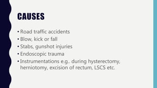
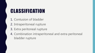
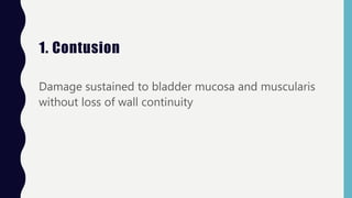
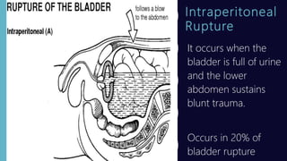
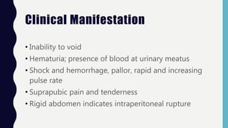
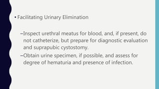
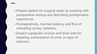
BLADDER INJURY
K A N C H A N K U S A T H A
• Bladder injury simply means trauma to the bladder
arises usually with some mechanical force that may be
blunt or penetrating one.
• Traumatic injury to the bladder is uncommon
CAUSES
• Road traffic accidents
• Blow, kick or fall
• Stabs, gunshot injuries
• Endoscopic trauma
• Instrumentations e.g.. during hysterectomy,
herniotomy, excision of rectum, LSCS etc.
CLASSIFICATION
1. Contusion of bladder
2. Intraperitoneal rupture
3. Extra peritoneal rupture
4. Combination intraperitoneal and extra peritoneal
bladder rupture
1. Contusion
Damage sustained to bladder mucosa and muscularis
without loss of wall continuity
Intraperitoneal
Rupture
It occurs when the
bladder is full of urine
and the lower
abdomen sustains
blunt trauma.
Occurs in 20% of
bladder rupture
Extraperitoneal
Rupture
It occurs when the lower
bladder is perforated by a
bony fragment during
pelvic fracture or with a
sharp instrument during
surgery.
Occurs in 80% of bladder
rupture cases
Clinical Manifestation
• Inability to void
• Hematuria; presence of blood at urinary meatus
• Shock and hemorrhage, pallor, rapid and increasing
pulse rate
• Suprapubic pain and tenderness
• Rigid abdomen indicates intraperitoneal rupture
Diagnostic Procedure
• Physical examination; abdominal and genitourinary
• Plain x-ray shows ground glass appearance
• Retrograde cystography
• CT scan abdomen
Medical Management
• Treatment instituted for shock and hemorrhage.
• Surgical intervention carried out for Intraperitoneal
and Large extra peritoneal rupture.
• Small extra peritoneal bladder ruptures will heal
spontaneously with indwelling suprapubic or urethral
catheter drainage.
• Medications: Analgesics, Antibiotics, Antispasmodics
Nursing Management
• Stabilizing Circulatory Volume
–Monitor vital signs and CVP frequently as indicated
by condition.
–Establish I.V. access, and replace blood and fluids as
ordered.
• Facilitating Urinary Elimination
–Inspect urethral meatus for blood, and, if present, do
not catheterize, but prepare for diagnostic evaluation
and suprapubic cystostomy.
–Obtain urine specimen, if possible, and assess for
degree of hematuria and presence of infection.
CONT.…..
–Prepare patient for surgical repair by assisting with
preoperative workup and describing postoperative
experiences.
–Postoperatively, maintain patency and flow of
indwelling urinary catheters.
–Inspect suprapubic incision and drain area for
bleeding, extravasation of urine, or signs of
infection.
• Controlling Pain
–Administer analgesics as ordered
–Assess patient's response to pain control medications.
–Position for comfort (usually semi-Fowler's position) if
not contraindicated by other injuries, and prevent
pulling of catheter tubing.
• Relieving Fear
–Provide information to the conscious patient
throughout the stabilization and evaluation phase;
prepare for surgery if impending.
–Keep patient's family or significant others informed of
condition and progress.
–Provide information on long-term outcome of
treatment.
• Patient Education and Health Maintenance
–Teach patient to care for indwelling catheters that
will remain in place during healing or after surgery.
–Empty frequently.
–Clean catheter and insertion area with soap and
water.
–Inspect urine for blood, cloudiness, or
concentration.
CONT.…
–Drink plenty of fluids to keep urine flowing.
–Teach patient to report signs and symptoms of UTI.
–Instruct patient (after surgical repair of bladder
rupture) that bladder capacity may be temporarily
decreased causing frequency and nocturia; this
resolves over time.

Bladder injury

  • 1.
    BLADDER INJURY K AN C H A N K U S A T H A
  • 2.
    • Bladder injurysimply means trauma to the bladder arises usually with some mechanical force that may be blunt or penetrating one. • Traumatic injury to the bladder is uncommon
  • 3.
    CAUSES • Road trafficaccidents • Blow, kick or fall • Stabs, gunshot injuries • Endoscopic trauma • Instrumentations e.g.. during hysterectomy, herniotomy, excision of rectum, LSCS etc.
  • 4.
    CLASSIFICATION 1. Contusion ofbladder 2. Intraperitoneal rupture 3. Extra peritoneal rupture 4. Combination intraperitoneal and extra peritoneal bladder rupture
  • 5.
    1. Contusion Damage sustainedto bladder mucosa and muscularis without loss of wall continuity
  • 6.
    Intraperitoneal Rupture It occurs whenthe bladder is full of urine and the lower abdomen sustains blunt trauma. Occurs in 20% of bladder rupture
  • 7.
    Extraperitoneal Rupture It occurs whenthe lower bladder is perforated by a bony fragment during pelvic fracture or with a sharp instrument during surgery. Occurs in 80% of bladder rupture cases
  • 8.
    Clinical Manifestation • Inabilityto void • Hematuria; presence of blood at urinary meatus • Shock and hemorrhage, pallor, rapid and increasing pulse rate • Suprapubic pain and tenderness • Rigid abdomen indicates intraperitoneal rupture
  • 9.
    Diagnostic Procedure • Physicalexamination; abdominal and genitourinary • Plain x-ray shows ground glass appearance • Retrograde cystography • CT scan abdomen
  • 10.
    Medical Management • Treatmentinstituted for shock and hemorrhage. • Surgical intervention carried out for Intraperitoneal and Large extra peritoneal rupture. • Small extra peritoneal bladder ruptures will heal spontaneously with indwelling suprapubic or urethral catheter drainage. • Medications: Analgesics, Antibiotics, Antispasmodics
  • 11.
    Nursing Management • StabilizingCirculatory Volume –Monitor vital signs and CVP frequently as indicated by condition. –Establish I.V. access, and replace blood and fluids as ordered.
  • 12.
    • Facilitating UrinaryElimination –Inspect urethral meatus for blood, and, if present, do not catheterize, but prepare for diagnostic evaluation and suprapubic cystostomy. –Obtain urine specimen, if possible, and assess for degree of hematuria and presence of infection.
  • 13.
    CONT.….. –Prepare patient forsurgical repair by assisting with preoperative workup and describing postoperative experiences. –Postoperatively, maintain patency and flow of indwelling urinary catheters. –Inspect suprapubic incision and drain area for bleeding, extravasation of urine, or signs of infection.
  • 14.
    • Controlling Pain –Administeranalgesics as ordered –Assess patient's response to pain control medications. –Position for comfort (usually semi-Fowler's position) if not contraindicated by other injuries, and prevent pulling of catheter tubing.
  • 15.
    • Relieving Fear –Provideinformation to the conscious patient throughout the stabilization and evaluation phase; prepare for surgery if impending. –Keep patient's family or significant others informed of condition and progress. –Provide information on long-term outcome of treatment.
  • 16.
    • Patient Educationand Health Maintenance –Teach patient to care for indwelling catheters that will remain in place during healing or after surgery. –Empty frequently. –Clean catheter and insertion area with soap and water. –Inspect urine for blood, cloudiness, or concentration.
  • 17.
    CONT.… –Drink plenty offluids to keep urine flowing. –Teach patient to report signs and symptoms of UTI. –Instruct patient (after surgical repair of bladder rupture) that bladder capacity may be temporarily decreased causing frequency and nocturia; this resolves over time.