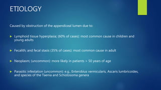
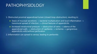
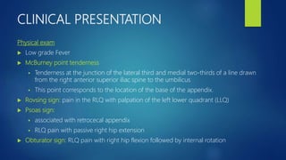
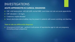
ACUTE APPENDICITIS
DR. ASIF ALI
DEFINITIONS
Appendicitis:
 acute inflammation of the vermiform appendix
Uncomplicated appendicitis:
 appendicitis with no evidence of an appendiceal fecalith, an appendiceal tumor, or
complications, such as perforation, gangrene, abscess, or mass
Complicated appendicitis:
 appendicitis associated with perforation, gangrene, abscess, an inflammatory mass, an
appendiceal fecalith, or an appendiceal tumor
ETIOLOGY
Caused by obstruction of the appendiceal lumen due to:
 Lymphoid tissue hyperplasia; (60% of cases): most common cause in children and
young adults
 Fecalith; and fecal stasis (35% of cases): most common cause in adult
 Neoplasm; (uncommon): more likely in patients > 50 years of age
 Parasitic infestation (uncommon): e.g., Enterobius vermicularis, Ascaris lumbricoides,
and species of the Taenia and Schistosoma genera
PATHOPHYSIOLOGY
1. Obstructed proximal appendiceal lumen (closed-loop obstruction), resulting in:
 Stasis of mucosal secretions → bacterial multiplication and local inflammation →
transmural spread of infection → clinical features of appendicitis
 Increased intraluminal pressure → obstruction of veins → edema of the
appendiceal walls → obstruction of capillaries → ischemia → gangrenous
appendicitis with/without perforation
2. Inflammation can spread to serosa, leading to peritonitis
CLINICAL PRESENTATION
Symptoms
 Abdominal pain: initial periumbilical pain with migration to the right lower
quadrant (RLQ)
 Anorexia
 Nausea
 Vomiting
 Diarrhea
 Constipation
 Indigestion
CLINICAL PRESENTATION
Physical exam
 Low grade Fever
 McBurney point tenderness
 Tenderness at the junction of the lateral third and medial two-thirds of a line drawn
from the right anterior superior iliac spine to the umbilicus
 This point corresponds to the location of the base of the appendix.
 Rovsing sign: pain in the RLQ with palpation of the left lower quadrant (LLQ)
 Psoas sign:
 associated with retrocecal appendix
 RLQ pain with passive right hip extension
 Obturator sign: RLQ pain with right hip flexion followed by internal rotation
OTHER CLINICAL SIGNS
 Hyperesthesia within Sherren triangle: formed by the anterior superior iliac spine,
umbilicus, and symphysis pubis
 Lanz point tenderness: at the junction of the right third and left two-thirds of a line
connecting both the anterior superior iliac spines
 Pain in the Pouch of Douglas: pain elicited by palpating the recto uterine pouch on
rectal examination
 Baldwin sign: pain in the flank when flexing the right hip (suggests an inflamed
retrocecal appendix)
INVESTIGATIONS
 CBC: mild leukocytosis with left shift; normal WBC count does not rule out acute appendicitis
 CRP: elevated (> 10 mg/L)
 Creatinine: maybe elevated
 Serum electrolyte abnormalities may be present in patients with severe vomiting; and diarrhea
Tests to rule out differential diagnoses
 Urine/serum β-hCG test; : perform in all women of reproductive age to rule out pregnancy
(including ectopic pregnancy)
ACUTE APPENDICITIS IS CLINICAL DIAGNOSIS
Radiological Investigation
 CT abdomen with IV contrast: preferred initial imaging modality in adults
(except for pregnant women)
 MRI abdomen without IV contrast: pregnant patients with inconclusive
ultrasound findings
 Abdominal ultrasound:
 Preferred initial imaging modality in children or pregnant patients
 As an alternative to CT scan if CT findings are inconclusive
 Abdominal ultrasound is more reliable for confirming acute
appendicitis than ruling it out.
DIFFERENTIAL DIAGNOSIS
 Ectopic pregnancy
 Renal colic
 Psoas abscess
 Epiploid appendagitis
 Constipation
 Irritable Bowel syndrome
DIAGNOSTIC CRITERIA – ALVARDO SCORING
TREATMENT
Supportive care
 Bowel rest - Nil by mouth (NPO)
 Intravenous fluids: Ringer Lactate and Normal saline
 Electrolyte repletion as needed
 IV analgesics: Ketorolac 30mg (Toradol) OR Tramadol 100mg (Tramol)
 IV antiemetics as needed: Metoclopramide Or Dimenhydrinate
 Antipyretic therapy: IV Paracetamol 1g/100ml x TDS/BD
 IV antibiotic therapy: Ceftriaxone 1g x BD, + Metronidazole 500mg/100ml x TDS
SURGICAL TREATEMNT
Non-perforated appendicitis
 Appendectomy (laparoscopic or open)
 should be performed within 12 hours of diagnosis
 laparoscopic approach is more common and popular
Perforated appendicitis with hemodynamic instability, sepsis, free perforation, or
peritonitis
 emergency appendectomy: irrigation and drainage of peritoneal cavity, colon resection if
needed
Stable perforated appendicitis
 initial nonoperative management: IV antibiotics, percutaneous drainage of abscess if present
 Rescue appendectomy for patients who do not respond to antibiotics
COMPLICATIONS
 Appendiceal abscess
 Perforation
 Sepsis
 Peritonitis
 Hemodynamic instability
 Death
THANK YOU

Acute appendicitis

  • 1.
  • 2.
    DEFINITIONS Appendicitis:  acute inflammationof the vermiform appendix Uncomplicated appendicitis:  appendicitis with no evidence of an appendiceal fecalith, an appendiceal tumor, or complications, such as perforation, gangrene, abscess, or mass Complicated appendicitis:  appendicitis associated with perforation, gangrene, abscess, an inflammatory mass, an appendiceal fecalith, or an appendiceal tumor
  • 3.
    ETIOLOGY Caused by obstructionof the appendiceal lumen due to:  Lymphoid tissue hyperplasia; (60% of cases): most common cause in children and young adults  Fecalith; and fecal stasis (35% of cases): most common cause in adult  Neoplasm; (uncommon): more likely in patients > 50 years of age  Parasitic infestation (uncommon): e.g., Enterobius vermicularis, Ascaris lumbricoides, and species of the Taenia and Schistosoma genera
  • 4.
    PATHOPHYSIOLOGY 1. Obstructed proximalappendiceal lumen (closed-loop obstruction), resulting in:  Stasis of mucosal secretions → bacterial multiplication and local inflammation → transmural spread of infection → clinical features of appendicitis  Increased intraluminal pressure → obstruction of veins → edema of the appendiceal walls → obstruction of capillaries → ischemia → gangrenous appendicitis with/without perforation 2. Inflammation can spread to serosa, leading to peritonitis
  • 5.
    CLINICAL PRESENTATION Symptoms  Abdominalpain: initial periumbilical pain with migration to the right lower quadrant (RLQ)  Anorexia  Nausea  Vomiting  Diarrhea  Constipation  Indigestion
  • 6.
    CLINICAL PRESENTATION Physical exam Low grade Fever  McBurney point tenderness  Tenderness at the junction of the lateral third and medial two-thirds of a line drawn from the right anterior superior iliac spine to the umbilicus  This point corresponds to the location of the base of the appendix.  Rovsing sign: pain in the RLQ with palpation of the left lower quadrant (LLQ)  Psoas sign:  associated with retrocecal appendix  RLQ pain with passive right hip extension  Obturator sign: RLQ pain with right hip flexion followed by internal rotation
  • 7.
    OTHER CLINICAL SIGNS Hyperesthesia within Sherren triangle: formed by the anterior superior iliac spine, umbilicus, and symphysis pubis  Lanz point tenderness: at the junction of the right third and left two-thirds of a line connecting both the anterior superior iliac spines  Pain in the Pouch of Douglas: pain elicited by palpating the recto uterine pouch on rectal examination  Baldwin sign: pain in the flank when flexing the right hip (suggests an inflamed retrocecal appendix)
  • 8.
    INVESTIGATIONS  CBC: mildleukocytosis with left shift; normal WBC count does not rule out acute appendicitis  CRP: elevated (> 10 mg/L)  Creatinine: maybe elevated  Serum electrolyte abnormalities may be present in patients with severe vomiting; and diarrhea Tests to rule out differential diagnoses  Urine/serum β-hCG test; : perform in all women of reproductive age to rule out pregnancy (including ectopic pregnancy) ACUTE APPENDICITIS IS CLINICAL DIAGNOSIS
  • 9.
    Radiological Investigation  CTabdomen with IV contrast: preferred initial imaging modality in adults (except for pregnant women)  MRI abdomen without IV contrast: pregnant patients with inconclusive ultrasound findings  Abdominal ultrasound:  Preferred initial imaging modality in children or pregnant patients  As an alternative to CT scan if CT findings are inconclusive  Abdominal ultrasound is more reliable for confirming acute appendicitis than ruling it out.
  • 10.
    DIFFERENTIAL DIAGNOSIS  Ectopicpregnancy  Renal colic  Psoas abscess  Epiploid appendagitis  Constipation  Irritable Bowel syndrome
  • 11.
    DIAGNOSTIC CRITERIA –ALVARDO SCORING
  • 12.
    TREATMENT Supportive care  Bowelrest - Nil by mouth (NPO)  Intravenous fluids: Ringer Lactate and Normal saline  Electrolyte repletion as needed  IV analgesics: Ketorolac 30mg (Toradol) OR Tramadol 100mg (Tramol)  IV antiemetics as needed: Metoclopramide Or Dimenhydrinate  Antipyretic therapy: IV Paracetamol 1g/100ml x TDS/BD  IV antibiotic therapy: Ceftriaxone 1g x BD, + Metronidazole 500mg/100ml x TDS
  • 13.
    SURGICAL TREATEMNT Non-perforated appendicitis Appendectomy (laparoscopic or open)  should be performed within 12 hours of diagnosis  laparoscopic approach is more common and popular Perforated appendicitis with hemodynamic instability, sepsis, free perforation, or peritonitis  emergency appendectomy: irrigation and drainage of peritoneal cavity, colon resection if needed Stable perforated appendicitis  initial nonoperative management: IV antibiotics, percutaneous drainage of abscess if present  Rescue appendectomy for patients who do not respond to antibiotics
  • 14.
    COMPLICATIONS  Appendiceal abscess Perforation  Sepsis  Peritonitis  Hemodynamic instability  Death
  • 15.