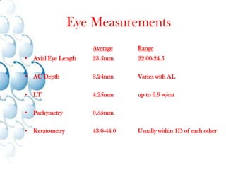
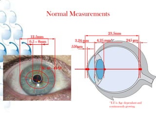
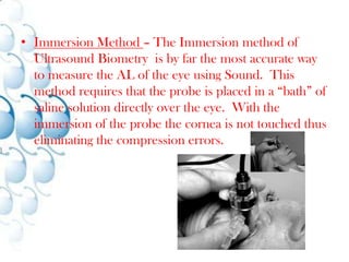
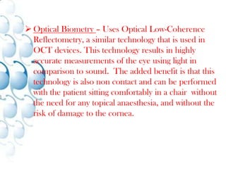
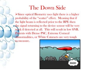
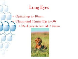
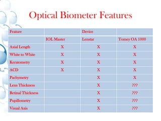
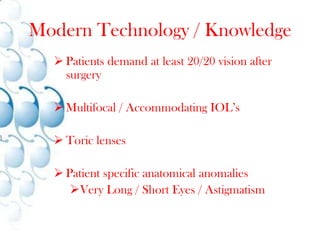
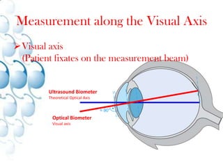
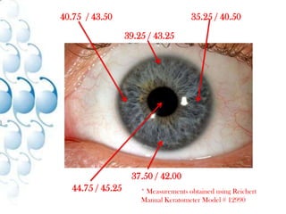
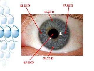
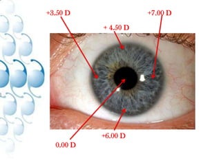
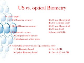
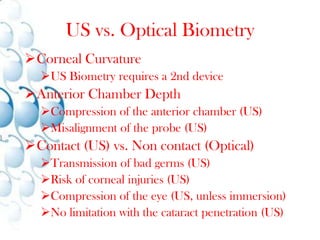
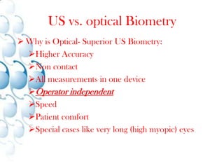
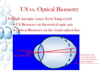
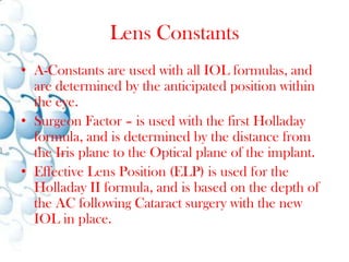
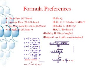
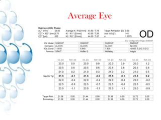
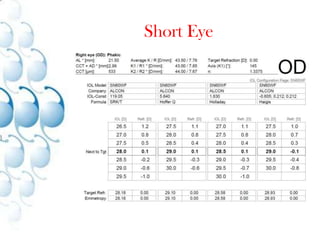
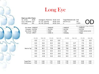
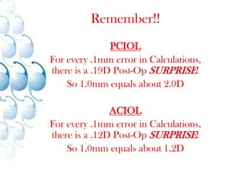
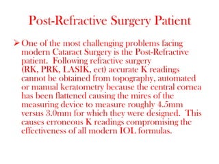
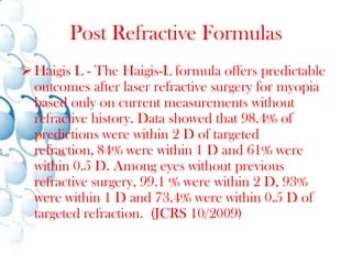
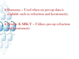
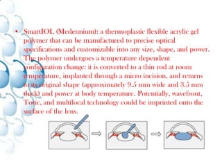
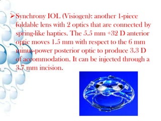
Optical Biometry Measurements for Future IOL’sAaron Graham, BA, CCOA
Financial Disclosure
Ground Rules
The GoodExclusive 	Эксклюзивные 	Exclusif 	ExclusivaSuperior	верхний 		Supérieur 	SuperiorElite		Элитные 		élite 		elisiónSpecialist	Специалист 		Spécialiste 	Especialista
The Good
The Good
The Good
The Good
The Good
The Good
The Good
The Good
The Good
    Good        καλός        Buono    Versus       έναντι       Contro    Bad	        κακός       Cattivo
The BadIndifferent	Безразлично 	Indifférent 	IndiferenteInferior	Нижняя 	Inférieur 	InferiorGeneric	родовой 	Générique 	GenéricoApathetic	апатичный 	Apathique 	Apático
The Bad
The Bad
The Bad
The Bad
The Bad
The Bad
The Bad
The Bad
The Bad
Finer PointsEstimates of the resolution of the human eye are some where around 576 mega pixels (24000 x 24000 pixels) for a 120 degree field of view.
However, it must be noted that the human eye itself has only a small spot of sharp vision in the middle of the retina, the fovea centralis, the rest of the field of view being blurry.
The angle of the sharp vision being just few degrees in the middle of the view, the sharp area thus barely achieves even a single mega pixel resolution.
The experience of wide sharp human vision is in fact based on turning the eyes towards the current point of interest in the field of view, the brain thus perceiving an observation of a wide sharp field of view. First surgical procedure 6th century B.C.
"couching", in which a curved needle was used to push the lens into the rear of the eye and out of the field of vision. The eye would later be soaked with warm clarified butter and then bandaged.
Couching continued to be used throughout the Middle Ages and is still used in some parts of Africa and in Yemen.
1748 First lens extraction (Modern Era)
1884 First local anesthetics (Cocaine Drops)
1940 First IOL (Harold Ridley)
1960 PhacoemulsificationCataract Surgery Saga
StatsAccording to surveys of members of the American Society of Cataract and Refractive Surgery, approximately 2.85 million cataract procedures were performed in the United States during 2004 and 2.79 million in 2005.Eye MeasurementsAverageRangeAxial Eye Length	23.5mm		22.00-24.5AC Depth		3.24mm		Varies with ALLT			4.25mm		up to 6.9 w/catPachymetry		0.55mm		Keratometry		43.0-44.0	Usually within 1D of each other
Normal Measurements23.5mm12.5mm245 μm4.25 mm*3.24 mm0.5 – 8mm550μmK:	43 - 44 D*LT is Age dependant andcontinuously growing
A-Scan MethodsContact Method – Contact Biometry is the most common technique used for measuring eye length, but is by far the most inaccurate.  This method requires placing the A-Scan probe directly on the Cornea.  This will result on Corneal compression and the amount of compression will vary depending on the experience and technique of the user.
Studies have shown that this method can result in erroneous eye length measurements between 0.14mm and 0.36mm too short. (.25D - .75D)
Immersion Method – The Immersion method of Ultrasound Biometry  is by far the most accurate way to measure the AL of the eye using Sound.  This method requires that the probe is placed in a “bath” of saline solution directly over the eye.  With the immersion of the probe the cornea is not touched thus eliminating the compression errors.
Optical Biometry – Uses Optical Low-Coherence Reflectometry, a similar technology that is used in OCT devices. This technology results in highly accurate measurements of the eye using light in comparison to sound.  The added benefit is that this technology is also non contact and can be performed with the patient sitting comfortably in a chair  without the need for any topical anaesthesia, and without the risk of damage to the cornea.
The Down SideSince optical Biometry uses light there is a higher probability of the “scatter” effect.  Meaning that if the light beam is reflected prior to the RPE then the signal returning to the device sensor will be very weak if detected at all.  This will result in low SNR.  Patients with Dense PSC, Extreme Corneal Abnormalities, or White Cataracts are very tough to measure.Long EyesOptical up to 40mmUltrasound 45mm (Up to 60)	< 5% of patients have AL > 26mm
Lenstar LS 900 (Haag-Streit)
IOL Master (Carl Zeiss)
OA 1000 (Tomey) to be launched 2000?Manufacturers
Optical Biometer Features
Patients demand at least 20/20 vision after surgery
Multifocal / Accommodating IOL’s
Toric lenses
Patient specific anatomical anomalies
Very Long / Short Eyes / AstigmatismModern Technology / Knowledge
Measurement along the Visual AxisVisual axis(Patient fixates on the measurement beam)Ultrasound BiometerTheoretical Optical Axis≈ 90°Optical BiometerVisual axis
What about the “Dust Cover”?aka CorneaTo ensure proper K value along central visual axis Keratometry should be measured at the same time as Axial Length.
Poor Alignment* Image courtesy of Karen Bachman and Cara Fletcher from the Cincinnati Eye Institute
40.75  / 43.50 35.25 / 40.50 39.25 / 43.25 37.50 / 42.00 44.75 / 45.25 * Measurements obtained using Reichert Manual Keratometer Model # 12990
42.12 D37.88 D41.25 D39.75 D45.00 D
IOL DataAlcon SN60WF
SRK/T using 119.05
www.doctor-hill.com
AL = 23.50
CCT + AD = 3.7923.0 D27.5 D24.0 D25.5 D19.5 D
+3.50 D+7.00 D+ 4.50 D+6.00 D0.00 D
US vs. optical BiometryAxial length
US Biometry accuracy 	±0.04 mm (theoretical)±0.1 to 0.12 mm (real)
Optical Biometry accuracy 	±0.03 mm (theoretical)	±0.03 mm (real)
US depends on user (0.1mm ≡ 0.28 D)
Compression of the eye
Misalignment of the probe
Achievable accuracy in post-op. refractive error
US Biometry based	St. Dev.: 0.8D
Optical Biometry based	St. Dev.: 0.25 to 0.5DUS vs. Optical BiometryCorneal Curvature
US Biometry requires a 2nd device
Anterior Chamber Depth
Compression of the anterior chamber (US)
Misalignment of the probe (US)
Contact (US) vs. Non contact (Optical)
Transmission of bad germs (US)
Risk of corneal injuries (US)
Compression of the eye (US, unless immersion)
No limitation with the cataract penetration (US)US vs. optical BiometryWhy is Optical- Superior US Biometry:
Higher Accuracy
Non contact
All measurements in one device
Operator independent
Speed
Patient comfort

Optical Biometry Measurements For Future Iol’S