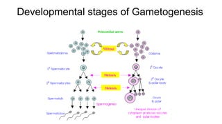
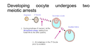
GAMETOGENESIS
Introduction
• Process by which diploid or haploid cells undergo cell division
and differentiation to form mature haploid gametes.
• The process of production of haploid sex cells.
• Gametes have one-half the genetic material (haploid
chromosome numbers) from the germ cell of each parent.
• The process of gametes formation.
• It occurs in the gonads (ovary or testis).
Four Phases
• Gametogenesis is divided into four phases
1. Extra-gonadal origin of primordial germ cells
2. Proliferation of germ cells by mitosis
3. Meiosis
4. Structural and functional maturation of ova and spermatozoa
Primordial Germ Cells
• Are the earliest precursors of all germ cells
• Are formed in the early stages of embryonic development
• Proliferate and migrate into the gonad (testis or ovary)
• Differentiate into male or female germ cells (determined by sex
chromosomes)
• Gonial cells are the designation given to germ cells before they
enter meiosis.
• Germ cells residing in the primordial gonad are often referred to
as gonocytes or gonial cells. As primordial germ cells (PGCs)
enter the primordial gonad, these cells acquire the capacity as
gonial cells to develop into either eggs or sperms.
• Replicate mitotically.
• Genetically identical to the original zygote.
• When they begin meiosis, they are called spermatocytes and
oocytes.
Developmental stages of Gametogenesis
Spermatogenesis
• The process of formation of the male germ cells (sperm
formation).
• It occurs in the seminiferous tubules of the testis.
• It is thread-like in shape.
• Occurs from puberty to old age, producing immense numbers of
spermatozoa (mature male gamete) at an average rate of 1.5
million spermatozoa per minute.
• The sperm will mature in the epididymis, nourished by Sertoli
cells for up to 10 weeks.
• Humans aged 13-90 can make 1 billion sperm a day.
• The Sertoli cells are supporting cells that have several functions.
• They form the blood-testes barrier: nutrients and circulating
substances do not directly reach the germ cells
• The Sertoli cells determine which substances reach the germ
cells.
• The spermatogonia are outside the blood-testis barrier.
• They form invaginations surrounding the spermatocytes,
spermatids, and developing spermatozoa and are nutritive to
them.
• They also produce antigen-binding proteins, which are necessary
for spermiogenesis (morphological development of spermatids
(immature male gamete) to spermatozoa).
• Spermatogenesis is the process by which male primary sperm cells
undergo meiosis, and produce a number of cells termed
spermatogonia, from which the primary spermatocytes are derived.
• Each primary spermatocyte divides into two secondary
spermatocytes and each secondary spermatocyte into two
spermatids or young spermatozoa.
• These develop into mature spermatozoa, also known as sperm
cells.
• The primary spermatocyte gives rise to two cells, the secondary
spermatocytes, and the two secondary spermatocytes by their
subdivision produce four spermatozoa.
Developmental stages of the male Germ
Cells
Spermatogonium
• It is an intermediary male gametogonium (a kind of germ cell).
• Diploid in number: 44 autosomes and 2 sex cells
• It is very small and in the process of development.
• Undergoing G1-S
Primary Spermatocytes
• Diploid in number: 44 autosomes and 2 sex cells
• Increases in size
• Undergoing G2.
Secondary Spermatocytes
• Haploid in number: 22 autosomes (double-stranded) and 1 sex cell
each.
• Can see the cleavage furrow
• Undergoing Meiosis I (Reduction Division)
• Chromosomes are reduced into half number.
 Spermatid
• Haploid in number: 22 (single-stranded) autosomes and 1 sex cell.
• Undergoing Meiosis II (Equational Division)
Spermatozoa
• It is a motile sperm cell or moving form of the haploid cell that is the
male gamete.
• It joins an ovum to form a zygote.
• Matured Sperm Cells are ready to fertilize the egg.
Spermiogenesis
• Morphological development of spermatids to spermatozoa. It
involves:
• Nuclear changes
• The nucleus becomes condensed and heterochromatic -
histones are replaced by protamines, which allow a high
degree of DNA compaction and make DNA inaccessible for
transcription
• Cytoplasmic changes – These are directed to the formation of
a motile sperm cell capable of penetrating the ovum and involve
the following changes:
• The Golgi apparatus at one pole of the cell forms an acrosome
containing proteolytic enzymes
• The centriole at the opposite pole organizes the formation of
microtubules to form a flagellum
• Alignment of mitochondria in a spiral around the base of the
flagellum – this forms the mid-piece of the spermatozoon
• The excess residual cytoplasm accumulates at one side of the cell
and becomes detached to form a residual body
Oogenesis
• The process of formation of the female germ cells (egg
formation).
• It happens in the Ovary.
• It is Spherical in shape.
• Oogenesis begins in early foetal life.
• All oocytes formed in females are produced during foetal life. Many
of them degenerate with time and at birth, the ovaries contain about
2 million oocytes.
• All the oocytes go into a meiotic arrest when they reach the first
meiotic division during foetal life.
• The primary oocytes remain in the prophase of the first meiotic
division until the time of puberty when they are gradually released to
complete meiosis at regular intervals known as the ovarian cycle.
• On average only one oocyte matures during each cycle, which
occurs at approximately monthly intervals so that the total amount of
oocytes to be ovulated is about 500 oocytes in a lifetime.
Developing oocyte undergoes two
meiotic arrests
Graafian Follicle
• A fluid-filled structure in the mammalian ovary within which an ovum develops
prior to ovulation.
• It provides for the maturation and release of a fertilizable oocyte. It also forms
the corpus luteum, which promotes and maintains the implantation of the
embryo. For these processes to occur, the follicle has to grow, ovulate, and
luteinizing.
Zona pellucida: Thick transparent membrane surrounding a
mammalian ovum before implantation.
• Is secreted by the oocyte
• Consists of glycoprotein and glycosaminoglycans.
• Contains sperm receptors necessary for fertilization
Cumulus oophorus: an appearance in the ovary in which multiple
granulosa cells enlarge around a developing oocyte. These support
cells ("cumulus cells") serve multiple functions in the maturation of
the oocyte.
Theca cells: A group of endocrine cells in the ovary made up of
connective tissue surrounding the follicle. They have many diverse
functions, including folliculogenesis.
Oogonium/Oogonia
• Diploid in number: 44 autosomes and 2 sex cells
• It is very small and in the process of development.
• Undergoing G1-S.
Primary Oocytes
• It is a cell whose function is to divide by the process of meiosis.
• It is also diploid in number: 44 autosomes and 2 sex cells.
• Increase in size.
• Undergoing G2.
Secondary Oocytes
• Undergoing Meiosis I (Reduction Division)
• Chromosomes are reduced into half number.
• Begins during embryonic development but stops in the
diplotene stage of prophase I until puberty.
• Primary oocytes continue to develop in each menstrual cycle;
however, synapsis occurs and tetrads form, enabling
chromosomal crossover to occur.
• Results of meiosis I, the primary oocyte has now developed into
the secondary oocyte and the first polar body.
Ovum
• After meiosis I, the haploid secondary oocyte initiates meiosis II
(Equational Division).
• This process also stops at the metaphase II stage until
fertilization.
• When meiosis II has completed, an ootid and another polar body
have now been created.
Polar Bodies
• Both polar bodies disintegrate at the end of Meiosis II, leaving only
the ootid, which then eventually undergoes maturation into a
mature ovum.
• The function of the forming polar bodies is to discard the extra
haploid sets of chromosomes that have resulted because of
meiosis.

GAMETOGENESIS.pptx

  • 1.
  • 2.
    Introduction • Process bywhich diploid or haploid cells undergo cell division and differentiation to form mature haploid gametes. • The process of production of haploid sex cells. • Gametes have one-half the genetic material (haploid chromosome numbers) from the germ cell of each parent. • The process of gametes formation. • It occurs in the gonads (ovary or testis).
  • 3.
    Four Phases • Gametogenesisis divided into four phases 1. Extra-gonadal origin of primordial germ cells 2. Proliferation of germ cells by mitosis 3. Meiosis 4. Structural and functional maturation of ova and spermatozoa
  • 4.
    Primordial Germ Cells •Are the earliest precursors of all germ cells • Are formed in the early stages of embryonic development • Proliferate and migrate into the gonad (testis or ovary) • Differentiate into male or female germ cells (determined by sex chromosomes)
  • 5.
    • Gonial cellsare the designation given to germ cells before they enter meiosis. • Germ cells residing in the primordial gonad are often referred to as gonocytes or gonial cells. As primordial germ cells (PGCs) enter the primordial gonad, these cells acquire the capacity as gonial cells to develop into either eggs or sperms. • Replicate mitotically. • Genetically identical to the original zygote. • When they begin meiosis, they are called spermatocytes and oocytes.
  • 6.
  • 7.
    Spermatogenesis • The processof formation of the male germ cells (sperm formation). • It occurs in the seminiferous tubules of the testis. • It is thread-like in shape. • Occurs from puberty to old age, producing immense numbers of spermatozoa (mature male gamete) at an average rate of 1.5 million spermatozoa per minute. • The sperm will mature in the epididymis, nourished by Sertoli cells for up to 10 weeks. • Humans aged 13-90 can make 1 billion sperm a day.
  • 9.
    • The Sertolicells are supporting cells that have several functions. • They form the blood-testes barrier: nutrients and circulating substances do not directly reach the germ cells • The Sertoli cells determine which substances reach the germ cells. • The spermatogonia are outside the blood-testis barrier. • They form invaginations surrounding the spermatocytes, spermatids, and developing spermatozoa and are nutritive to them. • They also produce antigen-binding proteins, which are necessary for spermiogenesis (morphological development of spermatids (immature male gamete) to spermatozoa).
  • 10.
    • Spermatogenesis isthe process by which male primary sperm cells undergo meiosis, and produce a number of cells termed spermatogonia, from which the primary spermatocytes are derived. • Each primary spermatocyte divides into two secondary spermatocytes and each secondary spermatocyte into two spermatids or young spermatozoa. • These develop into mature spermatozoa, also known as sperm cells. • The primary spermatocyte gives rise to two cells, the secondary spermatocytes, and the two secondary spermatocytes by their subdivision produce four spermatozoa.
  • 11.
    Developmental stages ofthe male Germ Cells
  • 12.
    Spermatogonium • It isan intermediary male gametogonium (a kind of germ cell). • Diploid in number: 44 autosomes and 2 sex cells • It is very small and in the process of development. • Undergoing G1-S Primary Spermatocytes • Diploid in number: 44 autosomes and 2 sex cells • Increases in size • Undergoing G2.
  • 13.
    Secondary Spermatocytes • Haploidin number: 22 autosomes (double-stranded) and 1 sex cell each. • Can see the cleavage furrow • Undergoing Meiosis I (Reduction Division) • Chromosomes are reduced into half number.  Spermatid • Haploid in number: 22 (single-stranded) autosomes and 1 sex cell. • Undergoing Meiosis II (Equational Division) Spermatozoa • It is a motile sperm cell or moving form of the haploid cell that is the male gamete. • It joins an ovum to form a zygote. • Matured Sperm Cells are ready to fertilize the egg.
  • 14.
    Spermiogenesis • Morphological developmentof spermatids to spermatozoa. It involves: • Nuclear changes • The nucleus becomes condensed and heterochromatic - histones are replaced by protamines, which allow a high degree of DNA compaction and make DNA inaccessible for transcription
  • 15.
    • Cytoplasmic changes– These are directed to the formation of a motile sperm cell capable of penetrating the ovum and involve the following changes: • The Golgi apparatus at one pole of the cell forms an acrosome containing proteolytic enzymes • The centriole at the opposite pole organizes the formation of microtubules to form a flagellum • Alignment of mitochondria in a spiral around the base of the flagellum – this forms the mid-piece of the spermatozoon • The excess residual cytoplasm accumulates at one side of the cell and becomes detached to form a residual body
  • 16.
    Oogenesis • The processof formation of the female germ cells (egg formation). • It happens in the Ovary. • It is Spherical in shape.
  • 17.
    • Oogenesis beginsin early foetal life. • All oocytes formed in females are produced during foetal life. Many of them degenerate with time and at birth, the ovaries contain about 2 million oocytes. • All the oocytes go into a meiotic arrest when they reach the first meiotic division during foetal life. • The primary oocytes remain in the prophase of the first meiotic division until the time of puberty when they are gradually released to complete meiosis at regular intervals known as the ovarian cycle. • On average only one oocyte matures during each cycle, which occurs at approximately monthly intervals so that the total amount of oocytes to be ovulated is about 500 oocytes in a lifetime.
  • 18.
    Developing oocyte undergoestwo meiotic arrests
  • 19.
    Graafian Follicle • Afluid-filled structure in the mammalian ovary within which an ovum develops prior to ovulation. • It provides for the maturation and release of a fertilizable oocyte. It also forms the corpus luteum, which promotes and maintains the implantation of the embryo. For these processes to occur, the follicle has to grow, ovulate, and luteinizing.
  • 20.
    Zona pellucida: Thicktransparent membrane surrounding a mammalian ovum before implantation. • Is secreted by the oocyte • Consists of glycoprotein and glycosaminoglycans. • Contains sperm receptors necessary for fertilization Cumulus oophorus: an appearance in the ovary in which multiple granulosa cells enlarge around a developing oocyte. These support cells ("cumulus cells") serve multiple functions in the maturation of the oocyte. Theca cells: A group of endocrine cells in the ovary made up of connective tissue surrounding the follicle. They have many diverse functions, including folliculogenesis.
  • 21.
    Oogonium/Oogonia • Diploid innumber: 44 autosomes and 2 sex cells • It is very small and in the process of development. • Undergoing G1-S. Primary Oocytes • It is a cell whose function is to divide by the process of meiosis. • It is also diploid in number: 44 autosomes and 2 sex cells. • Increase in size. • Undergoing G2.
  • 22.
    Secondary Oocytes • UndergoingMeiosis I (Reduction Division) • Chromosomes are reduced into half number. • Begins during embryonic development but stops in the diplotene stage of prophase I until puberty. • Primary oocytes continue to develop in each menstrual cycle; however, synapsis occurs and tetrads form, enabling chromosomal crossover to occur. • Results of meiosis I, the primary oocyte has now developed into the secondary oocyte and the first polar body.
  • 23.
    Ovum • After meiosisI, the haploid secondary oocyte initiates meiosis II (Equational Division). • This process also stops at the metaphase II stage until fertilization. • When meiosis II has completed, an ootid and another polar body have now been created. Polar Bodies • Both polar bodies disintegrate at the end of Meiosis II, leaving only the ootid, which then eventually undergoes maturation into a mature ovum. • The function of the forming polar bodies is to discard the extra haploid sets of chromosomes that have resulted because of meiosis.