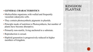
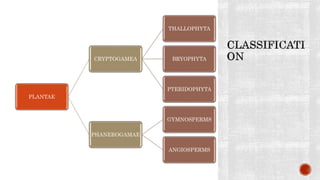
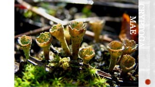
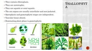
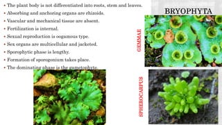
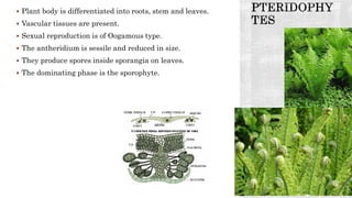
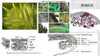
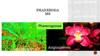
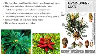
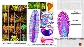
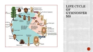
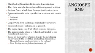
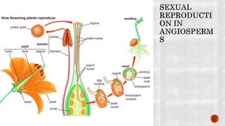
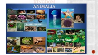
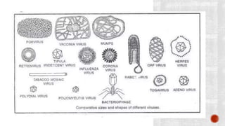
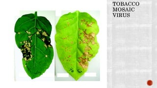
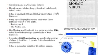
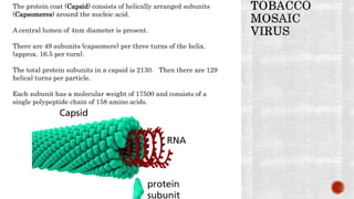
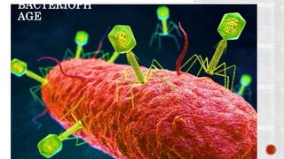
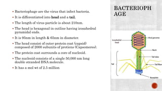
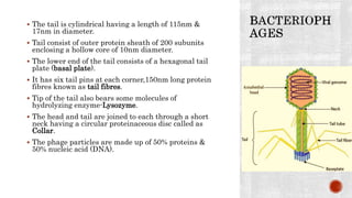
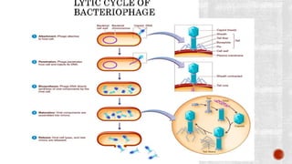
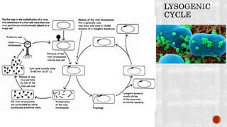
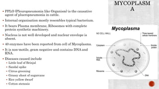
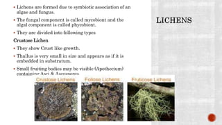
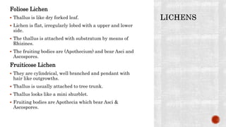
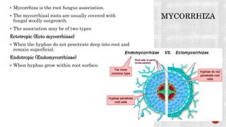
KINGDOM PLANTAE, KINGDOM ANIMALIA, VIRUS &
MYCOPLASMA.
 GENERAL CHARACTERISTICS
 Multicellular organisms with walled and frequently
vacuolate eukaryotic cells.
 They contain photosynthetic pigments in plastids.
 Principle mode of nutrition is Photosynthesis, but number of
plants have become absorptive.
 Primarily non-motile, living anchored to a substrate.
 Reproduction is sexual.
 Haploid generation is progressively reduced in higher
members of kingdom.
PLANTAE
CRYPTOGAMEA
THALLOPHYTA
BRYOPHYTA
PTERIDOPHYTA
PHANEROGAMAE
GYMNOSPERMS
ANGIOSPERMS
CRYPTOGA
MAE
 They contain chloroplasts.
 They are autotrophic.
 They are aquatic or semi-aquatic.
 The sex organs are usually unicellular and non-jacketed.
 Sporophytic and gametophytic stages are independent.
 Vascular tissue absent.
 Dominating plant phase is gametophytic.
 The plant body is not differentiated into roots, stem and leaves.
 Absorbing and anchoring organs are rhizoids.
 Vascular and mechanical tissue are absent.
 Fertilization is internal.
 Sexual reproduction is oogamous type.
 Sex organs are multicellular and jacketed.
 Sporophytic phase is lengthy.
 Formation of sporogonium takes place.
 The dominating phase is the gametophyte.
SPHEROCARPUSGEMMAE
 Plant body is differentiated into roots, stem and leaves.
 Vascular tissues are present.
 Sexual reproduction is of Oogamous type.
 The antheridium is sessile and reduced in size.
 They produce spores inside sporangia on leaves.
 The dominating phase is the sporophyte.
 The plant body is differentiated into roots, leaves and stem.
 They have vascular and mechanical tissue in them.
 Roots have symbiotic association with mycorrhiza.
 Fertilization is siphonogamous i.e. by pollen tube.
 Due development of cambium, they show secondary growth.
 Seeds are borne on structure called cones.
 The seeds are exposed and naked.
DIFFERENT TYPE OF CONES
MALE CONE (MICROSPOROPHYLL)
FEMALE CONE (MEGASPOROPHYLL)
 Plant body differentiated into roots, leaves & stem.
 They have vascular & mechanical tissue present in them.
 Produce flower which bear the reproductive structures.
 Stamens form the male reproductive structure and consist
of
 Anther &
 Filament.
 Carpel/Pistil form the female reproductive structure.
 Process of double fertilization is present.
 The ovary ripens into fruit which contains the seeds.
 The gametophytic phase is reduced and limited to the
formation of gametes.
 Based on the number of cotyledons in the developing
embryo inside the seed they are further classified as
 Monocots (having single cotyledon in the embryo)
 Dicot (having two cotyledons in the embryo)
KINGDOM
ANIMALIA
 They are multicellular organisms.
 They bear eukaryotic cells and lack cell walls.
 They are devoid of plastids and photosynthetic
pigments.
 Nutrition is primarily ingestive with digestion in an
internal cavity, but some forms are absorptive and
number of groups lack internal digestive cavity.
 Level of organization and tissue differentiation in
higher forms far exceeding that of other kingdoms,
with evolution of sensory neuromotor systems.
 They have developed cells with the ability to contract
(muscle cells) or transmit impulses (nerve cells).
 Reproduction is mainly sexual.
 Haploid stage other than gametes almost lacking
above lower phyla.
 It represents the better relationships in regard to
both levels of organization and nutritive modes.
 The red and brown algae and the fungi are better
placed.
 This system is better in coherence and definable
characters of kingdoms as units of classification.
 Unicellular eukaryotes like Euglena had earlier been
included in both plants and animals. By grouping
them in Protista, this anomaly has been removed.
 It succeeds in indicating the gradual evolution of
early organisms into plants and animals.
 Fungi bear its own biochemical, physiological and
structural organisation. The separation of kingdom
fungi from plants was long overdue.
 The distinction of the unicellular virus, the
multicellular and multinucleate conditions becomes
the line of division and difficulty.
 The Protista is a group of diverse organisms of
divergent directions of evolution.
 Viruses & Bacteriophages are much smaller than bacteria.
 In one sense viruses are not living organisms but large
nucleoprotein particles which enter specific kind of plant or
animal or bacteria and multiply to form new virus particles.
 Bacteriophages are also viruses which parasitize bacteria
(phage-eater).
 When viruses are outside the host they are metabolically
inert.
 They reproduce using the metabolic machinery of the host
cell, and infect chemically.
 They are made up of one of the nucleic acids DNA or RNA
(one of them) and proteins.
 Genetic material is enclosed within a protective coat of
protein.
 They show the property of living organisms, i.e they have the
property of replication.
 Since they lack a cell or cell organelles they are placed just
below the level of cellular orangisation.
 Scientific name is Protovirus tabacci.
 The virus particle is a long cylindrical, rod shaped,
helical virus.
 It has a length of 300 nm (3000Å) and 17.5nm (175Å)
diameter.
 X-ray crystallographic studies show that these
particles consist of two parts,
 Protein coat &
 The Nucleic acid.
 The Nucleic acid (nucleoid) is a single stranded RNA,
helically coiled forming a central core of 8nm
diameter.
 It consist of 6300 nucleotides an a molecular weight
of about 2.2 million. It consist 5.6% of total virus
composition
 It has a molecular weight of 40 million approx.
The protein coat (Capsid) consists of helically arranged subunits
(Capsomeres) around the nucleic acid.
A central lumen of 4nm diameter is present.
There are 49 subunits (capsomere) per three turns of the helix.
(approx. 16.5 per turn).
The total protein subunits in a capsid is 2130. Then there are 129
helical turns per particle.
Each subunit has a molecular weight of 17500 and consists of a
single polypeptide chain of 158 amino acids.
BACTERIOPH
AGE
 Bacteriophage are the virus that infect bacteria.
 It is differentiated into head and a tail.
 The length of virus particle is about 210nm.
 The head is hexagonal in outline having icosahedral
pyramidal ends.
 It is 95nm in length & 65nm in diameter.
 The head consist of outer protein coat (capsid)
composed of 2000 subunits of protiens (Capsomeres).
 The protein coat surrounds a core of nucleoid.
 The nucleoid consists of a single 50,000 nm long
double stranded DNA molecule.
 It has a mol wt of 2.5 million
 The tail is cylindrical having a length of 115nm &
17nm in diameter.
 Tail consist of outer protein sheath of 200 subunits
enclosing a hollow core of 10nm diameter.
 The lower end of the tail consists of a hexagonal tail
plate (basal plate).
 It has six tail pins at each corner,150nm long protein
fibres known as tail fibres.
 Tip of the tail also bears some molecules of
hydrolyzing enzyme-Lysozyme.
 The head and tail are joined to each through a short
neck having a circular proteinaceous disc called as
Collar.
 The phage particles are made up of 50% proteins &
50% nucleic acid (DNA).
 PPLO (Pleuropneumonia like Organism) is the causative
agent of plueropneumonia in cattle.
 Internal organisation mostly resembles typical bacterium.
 It bears Plasma membrane, Ribosomes with complete
protein synthetic machinery.
 Nucleus is not well developed and nuclear envelope is
absent.
 40 enzymes have been reported from cell of Mycoplasma.
 It is non-motile, gram negative and contains DNA and
RNA.
 Diseases caused include
 Little leaf of Brinjal
 Sandal spike
 Citrus greening
 Grassy shoot of sugarcane
 Rice yellow dwarf
 Cotton stenosis
 Lichens are formed due to symbiotic association of an
algae and fungus.
 The fungal component is called mycobiont and the
algal component is called phycobiont.
 They are divided into following types
Crustose Lichen
 They show Crust like growth.
 Thallus is very small in size and appears as if it is
embedded in substratum.
 Small fruiting bodies may be visible (Apothecium)
containing Asci & Ascospores.
Foliose Lichen
 Thallus is like dry forked leaf.
 Lichen is flat, irregularly lobed with a upper and lower
side.
 The thallus is attached with substratum by means of
Rhizines.
 The fruiting bodies are (Apothecium) and bear Asci and
Ascospores.
Fruiticose Lichen
 They are cylindrical, well branched and pendant with
hair like outgrowths.
 Thallus is usually attached to tree trunk.
 Thallus looks like a mini shurblet.
 Fruiting bodies are Apothecia which bear Asci &
Ascospores.
 Mycorrhiza is the root fungus association.
 The mycorrhizal roots are usually covered with
fungal woolly outgrowth.
 The association may be of two types
Ectotropic (Ecto mycorrhizae)
 When the hyphae do not penetrate deep into root and
remain superficial.
Endotropic (Endomycorrhizae)
 When hyphae grow within root surface.
Biological classification Part-IV

Biological classification Part-IV

  • 1.
    KINGDOM PLANTAE, KINGDOMANIMALIA, VIRUS & MYCOPLASMA.
  • 3.
     GENERAL CHARACTERISTICS Multicellular organisms with walled and frequently vacuolate eukaryotic cells.  They contain photosynthetic pigments in plastids.  Principle mode of nutrition is Photosynthesis, but number of plants have become absorptive.  Primarily non-motile, living anchored to a substrate.  Reproduction is sexual.  Haploid generation is progressively reduced in higher members of kingdom.
  • 4.
  • 5.
  • 6.
     They containchloroplasts.  They are autotrophic.  They are aquatic or semi-aquatic.  The sex organs are usually unicellular and non-jacketed.  Sporophytic and gametophytic stages are independent.  Vascular tissue absent.  Dominating plant phase is gametophytic.
  • 7.
     The plantbody is not differentiated into roots, stem and leaves.  Absorbing and anchoring organs are rhizoids.  Vascular and mechanical tissue are absent.  Fertilization is internal.  Sexual reproduction is oogamous type.  Sex organs are multicellular and jacketed.  Sporophytic phase is lengthy.  Formation of sporogonium takes place.  The dominating phase is the gametophyte. SPHEROCARPUSGEMMAE
  • 8.
     Plant bodyis differentiated into roots, stem and leaves.  Vascular tissues are present.  Sexual reproduction is of Oogamous type.  The antheridium is sessile and reduced in size.  They produce spores inside sporangia on leaves.  The dominating phase is the sporophyte.
  • 11.
     The plantbody is differentiated into roots, leaves and stem.  They have vascular and mechanical tissue in them.  Roots have symbiotic association with mycorrhiza.  Fertilization is siphonogamous i.e. by pollen tube.  Due development of cambium, they show secondary growth.  Seeds are borne on structure called cones.  The seeds are exposed and naked.
  • 12.
    DIFFERENT TYPE OFCONES MALE CONE (MICROSPOROPHYLL) FEMALE CONE (MEGASPOROPHYLL)
  • 14.
     Plant bodydifferentiated into roots, leaves & stem.  They have vascular & mechanical tissue present in them.  Produce flower which bear the reproductive structures.  Stamens form the male reproductive structure and consist of  Anther &  Filament.  Carpel/Pistil form the female reproductive structure.  Process of double fertilization is present.  The ovary ripens into fruit which contains the seeds.  The gametophytic phase is reduced and limited to the formation of gametes.  Based on the number of cotyledons in the developing embryo inside the seed they are further classified as  Monocots (having single cotyledon in the embryo)  Dicot (having two cotyledons in the embryo)
  • 16.
  • 17.
     They aremulticellular organisms.  They bear eukaryotic cells and lack cell walls.  They are devoid of plastids and photosynthetic pigments.  Nutrition is primarily ingestive with digestion in an internal cavity, but some forms are absorptive and number of groups lack internal digestive cavity.  Level of organization and tissue differentiation in higher forms far exceeding that of other kingdoms, with evolution of sensory neuromotor systems.  They have developed cells with the ability to contract (muscle cells) or transmit impulses (nerve cells).  Reproduction is mainly sexual.  Haploid stage other than gametes almost lacking above lower phyla.
  • 20.
     It representsthe better relationships in regard to both levels of organization and nutritive modes.  The red and brown algae and the fungi are better placed.  This system is better in coherence and definable characters of kingdoms as units of classification.  Unicellular eukaryotes like Euglena had earlier been included in both plants and animals. By grouping them in Protista, this anomaly has been removed.  It succeeds in indicating the gradual evolution of early organisms into plants and animals.  Fungi bear its own biochemical, physiological and structural organisation. The separation of kingdom fungi from plants was long overdue.
  • 21.
     The distinctionof the unicellular virus, the multicellular and multinucleate conditions becomes the line of division and difficulty.  The Protista is a group of diverse organisms of divergent directions of evolution.
  • 23.
     Viruses &Bacteriophages are much smaller than bacteria.  In one sense viruses are not living organisms but large nucleoprotein particles which enter specific kind of plant or animal or bacteria and multiply to form new virus particles.  Bacteriophages are also viruses which parasitize bacteria (phage-eater).  When viruses are outside the host they are metabolically inert.  They reproduce using the metabolic machinery of the host cell, and infect chemically.  They are made up of one of the nucleic acids DNA or RNA (one of them) and proteins.  Genetic material is enclosed within a protective coat of protein.  They show the property of living organisms, i.e they have the property of replication.  Since they lack a cell or cell organelles they are placed just below the level of cellular orangisation.
  • 26.
     Scientific nameis Protovirus tabacci.  The virus particle is a long cylindrical, rod shaped, helical virus.  It has a length of 300 nm (3000Å) and 17.5nm (175Å) diameter.  X-ray crystallographic studies show that these particles consist of two parts,  Protein coat &  The Nucleic acid.  The Nucleic acid (nucleoid) is a single stranded RNA, helically coiled forming a central core of 8nm diameter.  It consist of 6300 nucleotides an a molecular weight of about 2.2 million. It consist 5.6% of total virus composition  It has a molecular weight of 40 million approx.
  • 27.
    The protein coat(Capsid) consists of helically arranged subunits (Capsomeres) around the nucleic acid. A central lumen of 4nm diameter is present. There are 49 subunits (capsomere) per three turns of the helix. (approx. 16.5 per turn). The total protein subunits in a capsid is 2130. Then there are 129 helical turns per particle. Each subunit has a molecular weight of 17500 and consists of a single polypeptide chain of 158 amino acids.
  • 28.
  • 29.
     Bacteriophage arethe virus that infect bacteria.  It is differentiated into head and a tail.  The length of virus particle is about 210nm.  The head is hexagonal in outline having icosahedral pyramidal ends.  It is 95nm in length & 65nm in diameter.  The head consist of outer protein coat (capsid) composed of 2000 subunits of protiens (Capsomeres).  The protein coat surrounds a core of nucleoid.  The nucleoid consists of a single 50,000 nm long double stranded DNA molecule.  It has a mol wt of 2.5 million
  • 30.
     The tailis cylindrical having a length of 115nm & 17nm in diameter.  Tail consist of outer protein sheath of 200 subunits enclosing a hollow core of 10nm diameter.  The lower end of the tail consists of a hexagonal tail plate (basal plate).  It has six tail pins at each corner,150nm long protein fibres known as tail fibres.  Tip of the tail also bears some molecules of hydrolyzing enzyme-Lysozyme.  The head and tail are joined to each through a short neck having a circular proteinaceous disc called as Collar.  The phage particles are made up of 50% proteins & 50% nucleic acid (DNA).
  • 33.
     PPLO (Pleuropneumonialike Organism) is the causative agent of plueropneumonia in cattle.  Internal organisation mostly resembles typical bacterium.  It bears Plasma membrane, Ribosomes with complete protein synthetic machinery.  Nucleus is not well developed and nuclear envelope is absent.  40 enzymes have been reported from cell of Mycoplasma.  It is non-motile, gram negative and contains DNA and RNA.  Diseases caused include  Little leaf of Brinjal  Sandal spike  Citrus greening  Grassy shoot of sugarcane  Rice yellow dwarf  Cotton stenosis
  • 34.
     Lichens areformed due to symbiotic association of an algae and fungus.  The fungal component is called mycobiont and the algal component is called phycobiont.  They are divided into following types Crustose Lichen  They show Crust like growth.  Thallus is very small in size and appears as if it is embedded in substratum.  Small fruiting bodies may be visible (Apothecium) containing Asci & Ascospores.
  • 35.
    Foliose Lichen  Thallusis like dry forked leaf.  Lichen is flat, irregularly lobed with a upper and lower side.  The thallus is attached with substratum by means of Rhizines.  The fruiting bodies are (Apothecium) and bear Asci and Ascospores. Fruiticose Lichen  They are cylindrical, well branched and pendant with hair like outgrowths.  Thallus is usually attached to tree trunk.  Thallus looks like a mini shurblet.  Fruiting bodies are Apothecia which bear Asci & Ascospores.
  • 36.
     Mycorrhiza isthe root fungus association.  The mycorrhizal roots are usually covered with fungal woolly outgrowth.  The association may be of two types Ectotropic (Ecto mycorrhizae)  When the hyphae do not penetrate deep into root and remain superficial. Endotropic (Endomycorrhizae)  When hyphae grow within root surface.