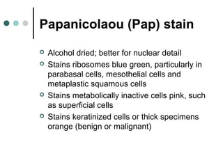
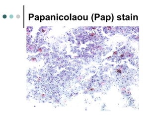
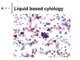
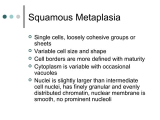
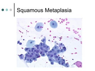
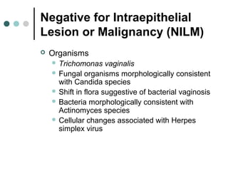
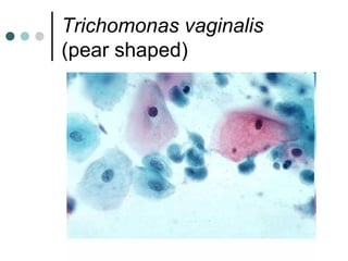
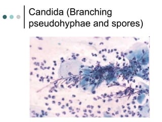
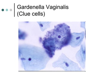
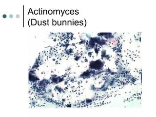
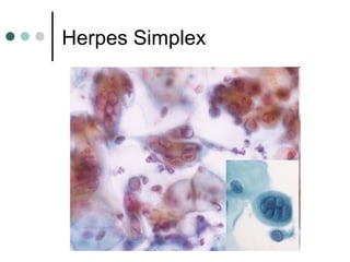
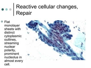
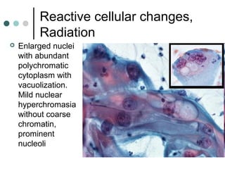
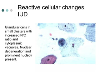
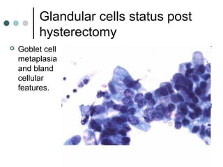
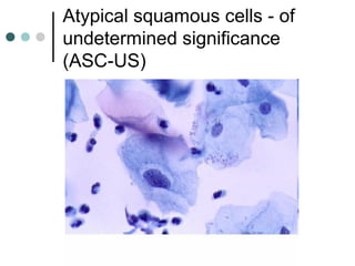
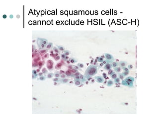
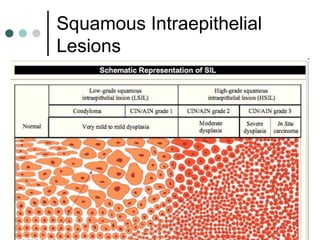
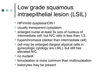
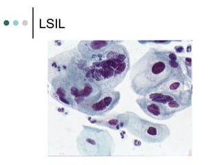
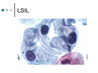
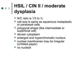
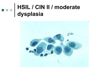
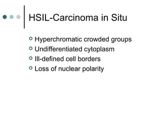
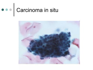
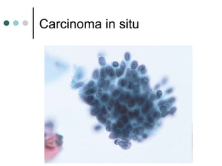
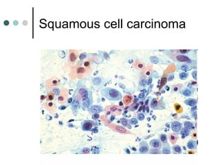
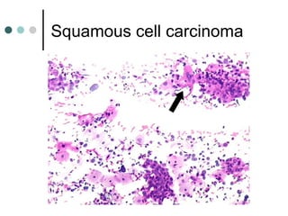
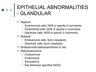
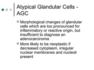
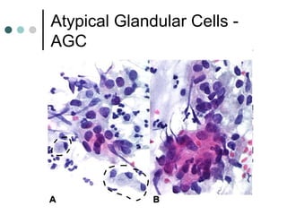
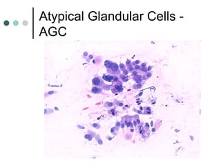
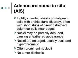
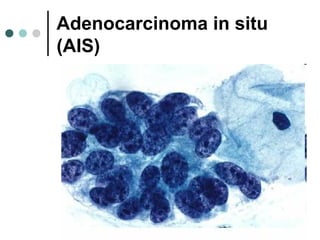
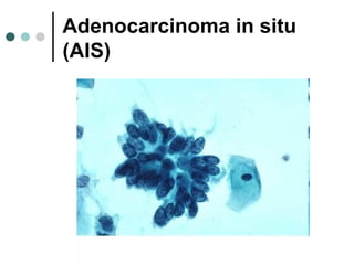
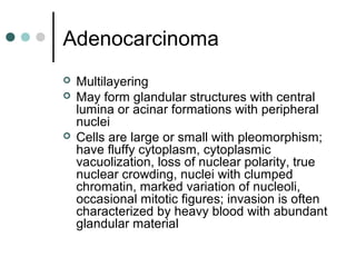
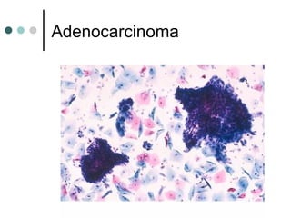
The document provides information on cervical cytology screening and the Bethesda System for reporting results. It describes the Papanicolaou staining technique and liquid-based cytology collection methods. The Bethesda System categories for specimen adequacy, interpretation, and epithelial cell abnormalities are outlined. Normal cervical cell types are defined. Reactive, infectious, and other non-neoplastic findings are described. Atypical squamous cells of undetermined significance, low-grade squamous intraepithelial lesions, and high-grade squamous intraepithelial lesions are defined. Adenocarcinoma in situ and adenocarcinoma are also summarized.
4. Liquid based cytology
Head of spatula, where cells are lodged, is
broken off into small glass vial containing
preservative fluid, or rinsed directly into
preservative fluid
Sample is sent to lab, then spun and treated
to remove mucus, pus or other obscuring
material
Random sample of remaining cells is taken
and deposited onto a slide
Reduces number of inadequate smears and
need for repeat smears
6. Diff-Quik stain
An air dried, Giemsa-type stain
Better for background material or to
assess adequacy of endocervical
smears to detect C. trachomatis
Used for fine needle aspirates, not for
cervical smears
9. Parabasal cells
Cytoplasm is round, dense, basophilic
Nucleus is vesicular, central, round
and relatively large (50μm²)
May see naked nuclei
Higher N/C ratio and smaller size than
intermediate cells
11. Intermediate Cells
Cytoplasm is polygonal, transparent,
basophilic
Nucleus is about the size of a red
blood cell (35 μm²), is vesicular,
round/oval
Nuclear texture and size is reference
for dysplasia
17. Squamous Metaplasia
Single cells, loosely cohesive groups or
sheets
Variable cell size and shape
Cell borders are more defined with maturity
Cytoplasm is variable with occasional
vacuoles
Nuclei is slightly larger than intermediate
cell nuclei, has finely granular and evenly
distributed chromatin, nuclear membrane is
smooth, no prominent nucleoli
20. BETHESDA SYSTEM
Uniform terminology introduced in
1988 and revised in 1991 and 2001
To establish uniform terminology and
standardize diagnostic reports.
In addition, it introduced a
standardized approach for reporting if
an individual specimen is adequate for
evaluation.
21. BETHESDA SYSTEM, 2001
Specimen adequacy
General categorization (optional)
Interpretation/result
Automated review and ancillary
testing (include if appropriate)
Educational notes and suggestions
(optional)
22. Adequacy
Satisfactory for evaluation (describe
presence or absence of
endocervical/transformation zone
component and all other quality
indicators, such as partially obscuring
blood, inflammation, etc.)
Unsatisfactory for evaluation
(indicate reason)
Specimen rejected/not processed
(indicate reason)
23. Assessment of adequacy
Adequate number of squamous cells
(conventional smear should have 8000-12000
cells, liquid-based preparation should have
5000 cells)
The presence or absence of endocervical
cells should be reported; an adequate number
of endocervical cells (at least 10 well-
preserved endocervical or metaplastic cells)
confirms sampling of transition zone
Specimen with more than 75% of cells
obscured by inflammation and bacteria is
unsatisfactory (however, should still report
presence of abnormal cells)
26. Interpretation/result
Negative for Intraepithelial Lesion or
Malignancy (NILM)
Epithelial Cell Abnormality
Other malignant neoplasm (Specify)
27. Negative for Intraepithelial
Lesion or Malignancy (NILM)
Organisms
Trichomonas vaginalis
Fungal organisms morphologically consistent
with Candida species
Shift in flora suggestive of bacterial vaginosis
Bacteria morphologically consistent with
Actinomyces species
Cellular changes associated with Herpes
simplex virus
33. Other non-neoplastic findings (optional to
report, list is not inclusive)
Reactive cellular changes associated with:
• inflammation (includes typical repair)
• irradiation
• Intrauterine contraceptive device (IUD)
Glandular cells status post hysterectomy
Atrophy
34. Reactive cellular changes,
Repair
Flat
monolayer
sheets with
distinct
cytoplasmic
outlines,
streaming
nuclear
polarity,
prominent
nucleolus in
almost every
cell.
35. Reactive cellular changes,
Radiation
Enlarged nuclei
with abundant
polychromatic
cytoplasm with
vacuolization.
Mild nuclear
hyperchromasia
without coarse
chromatin,
prominent
nucleoli
36. Reactive cellular changes,
IUD
Glandular cells in
small clusters with
increased N/C
ratio and
cytoplasmic
vacuoles. Nuclear
degeneration and
prominent nucleoli
present.
37. Glandular cells status post
hysterectomy
Goblet cell
metaplasia
and bland
cellular
features.
38. Atrophy
Sheets of
uniform orderly
parabasal
cells. Some
nuclei may
show grooves,
but chromatin
pattern is fine.
Atrophic cells
may have
nucleoli.
39. Other
Endometrial cells (in a woman greater
than or equal to 40 years of age;
specify if “negative for squamous
intraepithelial lesion”)
40. Endometrial cells
Cells occur in small
clusters. Small,
round nuclei similar
in size to a normal
intermediate cell
nucleus.
Inconspicuous or
absent nucleoli.
Cytoplasm is scant,
basophilic and
sometimes
vacuolated. Cell
borders are ill-
defined.
41. Epithelial Cell Abnormality
SQUAMOUS CELL
Atypical squamous cells
- of undetermined significance (ASC-US)
- cannot exclude HSIL (ASC-H)
Low grade squamous intraepithelial lesion (LSIL)
- encompassing HPV/mild dysplasia/CIN I
High grade squamous intraepithelial lesion (HSIL)
- encompassing: moderate and severe
dysplasia/CIN2/CIN3/CIS
- with features suspicious for invasion (if
invasion suspected)
Squamous cell carcinoma
42. Atypical squamous cells - of
undetermined significance
(ASC-US)
May be neoplastic (HPV related, LSIL,
HSIL) or reactive
Nuclear changes more marked than
reactive, less than LSIL; nucleus is 2.5-3x
size of intermediate cell nucleus
In perimenopausal women (40-55 years),
cells with bland nuclear enlargement (2-3x
size of intermediate cell nuclei), smooth
nuclear membranes and fine chromatin are
likely to be negative for SIL/malignancy
46. Low grade squamous
intraepithelial lesion (LSIL)
HPV/mild dysplasia/CIN I
usually transparent cytoplasm
enlarged nuclei at least 3x size of nucleus of
intermediate cell, but N/C ratio is less than 1/3
hyperchromasia (darker than intermediate cell)
cell may be enlarged (largest atypical cells in
gynecologic cytology are LSIL), but still has
elevated N/C
no nucleoli
binucleation is more common than multinucleation
koilocytes may be present
49. HSIL / CIN II / moderate
dysplasia
N/C ratio is 1/3 to ½
cell size is same as squamous metaplastic
or parabasal cells
polygonal shape (like intermediate or
superficial cell)
denser cytoplasm
enlarged and hyperchromatic nucleus
nuclear membranes may be irregular
(crinkled paper)
no nucleoli
52. HSIL / CIN III / severe
dysplasia
Usually single, small, round/oval cells
scant cytoplasm
nuclei same size as dysplastic cells but
increased N/C ratio (greater than 1/2) since
only minimal rim of cytoplasm
nuclei vary markedly in contour and have
irregular coarsely clumped chromatin
no macronucleoli
no tumor diathesis
61. EPITHELIAL ABNORMALITIES
- GLANDULAR
Atypical
Endocervical cells, NOS or specify in comments
Endometrial cells, NOS or specify in comments
Glandular cells, NOS or specify in comments
Atypical
Endocervical cells, favor neoplastic
Glandular cells, favor neoplastic
Endocervical adenocarcinoma in situ
Adenocarcinoma
Endocervical
Endometrial
Extrauterine
Not otherwise specified (NOS)
62. Atypical Glandular Cells -
AGC
Morphological changes of glandular
cells which are too pronounced for
inflammatory or reactive origin, but
insufficient to diagnose an
adenocarcinoma
More likely to be neoplastic if
decreased cytoplasm, irregular
nuclear membranes and nucleoli
present
65. Adenocarcinoma in situ
(AIS)
Tightly crowded sheets of malignant
cells with architectural disarray, often
with short strips of pseudostratified
columnar cells near edges
Nuclei may be partially denuded,
causing a feathered appearance
Nuclei are enlarged, usually oval, and
hyperchromatic
Often prominent nucleoli
No tumor diathesis
68. Adenocarcinoma
Multilayering
May form glandular structures with central
lumina or acinar formations with peripheral
nuclei
Cells are large or small with pleomorphism;
have fluffy cytoplasm, cytoplasmic
vacuolization, loss of nuclear polarity, true
nuclear crowding, nuclei with clumped
chromatin, marked variation of nucleoli,
occasional mitotic figures; invasion is often
characterized by heavy blood with abundant
glandular material