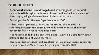
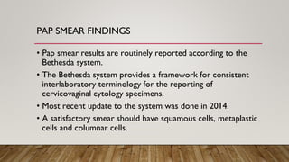
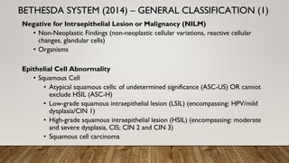
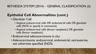
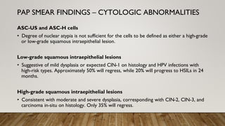
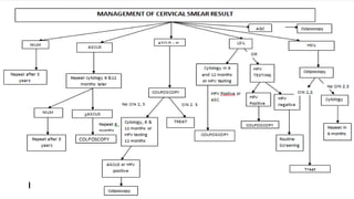
2. OUTLINE
• Introduction: Pap smear
• Overview: Cervical cancer
• Pap smear procedure
• Pap smear findings
• Management
• Conclusion
• References
3. • A cervical smear is a cytology-based screening test for cervical
cancer in which vaginal cells are collected and stained as a means of
detecting cytologic abnormalities of the uterine cervix.
• Developed by Dr George Papanicolaou in 1943.
• It has been implemented in countries around the world as a
screening strategy, and subsequent reductions in rates of cervical
cancer by 50% or more have been seen.
• It is recommended to be performed once every 3-5 years for women
between the ages of 25 to 65 years.
• The reported sensitivity and specificity of Pap smear varies: sensitivity
ranges from 30-87%, and specificity ranges from 86-100%.
INTRODUCTION
4. INTRODUCTION
• For more than 95% of women, cervical cytology is normal
and normal squamous cells are seen.
• Abnormal cervical cytology shows squamous cells at different
stages of maturity(dyskaryosis).
• When abnormal cells are detected on the pap smear,
diagnostic testing in the form of colposcopy is often indicated.
5. • 2nd most common cancer among women in developing
countries.
• Most common gynaecological cancer and a leading cause of
cancer deaths among women in Nigeria.
• Established aetiology is a persistent Human Papillomavirus
infection (most implicated serotypes are high-risk 16 and 18),
mainly acquired from sexual contact.
• Risk factors include: early coitarche, multiple sexual partners,
promiscuous male partners, history of STIs,
immunosuppression (HIV), multiparity, COCP use, smoking.
CERVICAL CANCER
6. PAP SMEAR PROCEDURE
Preparation
• Cervical screening should be done when the patient is not menstruating.
• Avoid vaginal intercourse, douching, use of tampons, use of medicinal
vaginal or contraceptive cream for 24-48 hours prior to the test.
• Ideally, pre-existing cervicitis should be treated prior to cervical screening.
• Screening should proceed in the presence of bleeding or cervicitis, as
these symptoms may be related to cervical dysplasia or neoplasia.
7. PAP SMEAR PROCEDURE
Positioning
• Patient should be supine, in dorsal
lithotomy position to correctly
perform the procedure.
• The coccyx of the patient should be
at the edge of the examination table
to provide adequate visualization of
the cervix once the speculum is
inserted.
8. • Cervix is visualized using a speculum.
• Samples of the cervix are taken from the transformation zone; where the
ectocervix and endocervix meet and where dysplasia is most likely to be
identified.
• Samples are taken using an Ayres spatula.
• Smear spread on a slide and the slide fixed in 90% ethanol.
• Smear is sent to a cytopathologist, who provides a cytologic analysis.
PAP SMEAR PROCEDURE
9.
10. PAP SMEAR FINDINGS
• Pap smear results are routinely reported according to the
Bethesda system.
• The Bethesda system provides a framework for consistent
interlaboratory terminology for the reporting of
cervicovaginal cytology specimens.
• Most recent update to the system was done in 2014.
• A satisfactory smear should have squamous cells, metaplastic
cells and columnar cells.
11. THE BETHESDA SYSTEM
• Specimen Type
• Specimen Adequacy
• General categorization*
• Interpretation/Results
• Ancillary Testing
• Automated Review
• Educational Notes & Suggestions
12. Negative for Intraepithelial Lesion or Malignancy (NILM)
• Non-Neoplastic Findings (non-neoplastic cellular variations, reactive cellular
changes, glandular cells)
• Organisms
Epithelial Cell Abnormality
• Squamous Cell
• Atypical squamous cells: of undetermined significance (ASC-US) OR cannot
exclude HSIL (ASC-H)
• Low-grade squamous intraepithelial lesion (LSIL) (encompassing: HPV/mild
dysplasia/CIN 1)
• High-grade squamous intraepithelial lesion (HSIL) (encompassing: moderate
and severe dysplasia, CIS; CIN 2 and CIN 3)
• Squamous cell carcinoma
BETHESDA SYSTEM (2014) – GENERAL CLASSIFICATION (1)
13. Epithelial Cell Abnormalities (cont.)
• Glandular Cell
• Atypical endocervical cells OR endometrial cells OR glandular
cells (NOS or specify in comments)
• Atypical endocervical cells (favour neoplastic) OR glandular
cells (favour neoplastic)
• Endocervical adenocarcinoma in situ
• Adenocarcinoma: endocervical, endometrial, extrauterine,
not otherwise specified (NOS)
BETHESDA SYSTEM (2014) – GENERAL CLASSIFICATION (2)
14. ASC-US and ASC-H cells
• Degree of nuclear atypia is not sufficient for the cells to be defined as either a high-grade
or low-grade squamous intraepithelial lesion.
Low-grade squamous intraepithelial lesions
• Suggestive of mild dysplasia or expected CIN-1 on histology and HPV infections with
high-risk types. Approximately 50% will regress, while 20% will progress to HSILs in 24
months.
High-grade squamous intraepithelial lesions
• Consistent with moderate and severe dysplasia, corresponding with CIN-2, CIN-3, and
carcinoma in-situ on histology. Only 35% will regress.
PAP SMEAR FINDINGS – CYTOLOGIC ABNORMALITIES
15. • Cervical Intraepithelial Neoplasia (CIN) is a precancerous lesion of the cervix
• Identified in cytology by:
• Koilocytes (atypical cells with a perinuclear cavitation or halo in the
cytoplasm)
• Dysplastic cells with increased nucleo-cytoplasmic ratio.
• Low-grade CIN/CIN-1: one-third of epithelial thickness has dysplastic
changes
• CIN-2: half to two-thirds of epithelial thickness has dysplastic changes
• CIN-3: full-thickness involvement of epithelium
• Carcinoma in-situ: dysplasia seen throughout the epithelium, resembles
cervical cancer, but no basement membrane invasion.
PAP SMEAR FINDINGS – CIN
16. NILM
• Repeat in 3-5 years as recommended
ASC-US
• Repeat cytology in 1 year; if negative, repeat cytology in 3 years
• HPV testing is preferred; if negative, repeat cotesting in 3 years; if
positive, perform colposcopy
ASC-H cells, LSIL, HSIL
• Perform colposcopy
Colposcopy involves visualization the cervix and obtaining biopsies of
lesions identified by acetic acid stain (sharp, distinct, well defined, dense
aceto-white lesions).
MANAGEMENT (1)
17.
18.
19. Precancerous lesions can be treated via:
• Ablative Methods
• Cryotherapy
• Laser
• Cold coagulation
• Excisional Methods
• Large Loop Excision of the Transformation Zone (LLETZ)/Loop Electrosurgical Excision
Procedure (LEEP)
• Laser cone biopsy
• Cold knife cone biopsy
• Hysterectomy (in cases of co-existing uterine conditions such as fibroids)
Annual follow-up with Pap smears following treatment of precancerous lesions.
MANAGEMENT (2)
20.
21. CONCLUSION
Cervical/Pap smear is a useful screening tool to control the most common gynaecological
cancer among women in Nigeria.
22. REFERENCES
1. Cervical Screening: Overview, Human Papillomavirus, Papanicolaou Test. 2021 Aug 22
[cited 2022 Mar 10]; Available from: https://emedicine.medscape.com/article/1618870-
overview
1. Gynaecology by ten teachers, 20E
1. HPV and premalignant disease of the cervix by Prof Anorlu