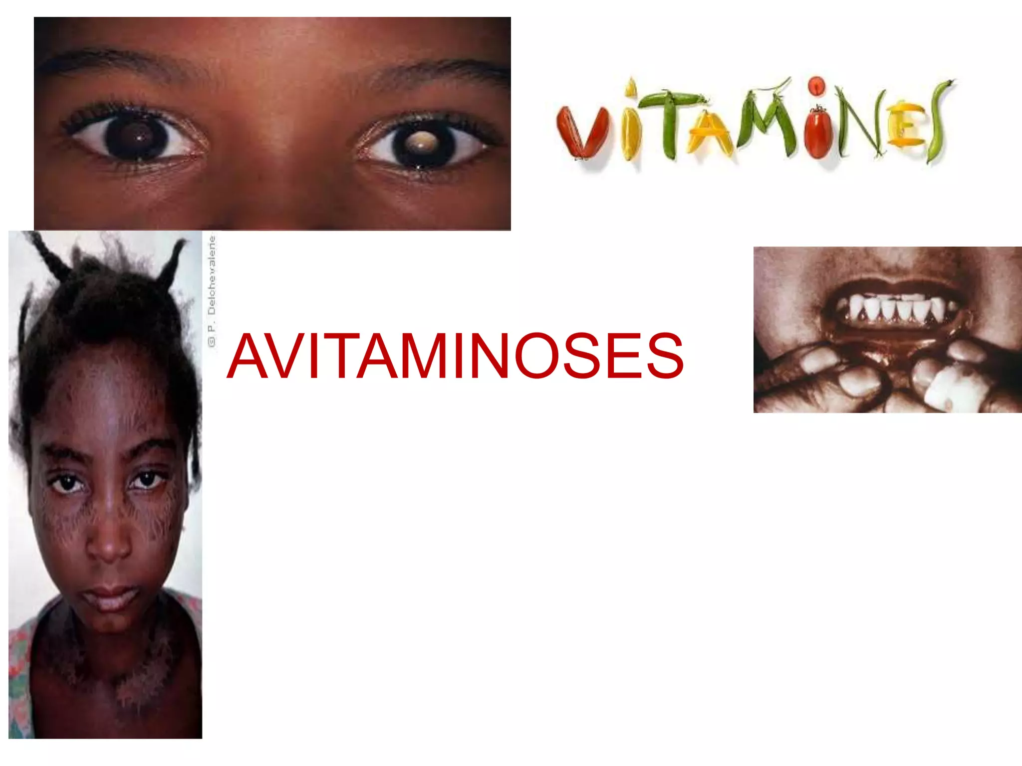
Avitaminoses are a assorted groups of diseases, and as unrelated to each other as the chemical constituents of the various vitamins, they too share enough group of diseases..
2. Contents
• Introduction
• Fat Soluble Vitamins
– Vit-A
– Vit- D
– Vit-E
• Water Soluble Vitamins
– Vit –K
– Vit-C
– Vit-B
• Conclusion
• References
3. Vitamins
Vitamin is defined as the organic substance i.e. not made by the body, and is soluble
either in FAT or WATER and is ordinarily needed in only one minute quantities to act as a co-factor
in varieties of metabolic reasons.
Those vitamins that occurs naturally in physiologically inactive states are called PROVITAMINS.
The various forms of available vitamins are:
Fat soluble:
– Vit-A
– Vit-D
– Vit-E
Water Soluble:
– Vit-K
– Vit-C
4. Avitaminoses
Avitaminoses are a assorted groups of diseases, and as unrelated to each other as the chemical
constituents of the various vitamins, they too share enough group of diseases..
Avitaminoses are due to the ABSENCE of minute amounts of biologically important materials
rather then to the presence of minute amounts of biologically active materials(infectious
agents).
They cause disease not in apositive way but in a negative way. “THE DEFFICIENCY IS THE
DISEASE.”
6. Vitamin A
The primary manifestation of Vit-A defficiency is impaired , low light vision,
i.e. Night Blindness.
Xerophthalmia, Keratomalacia
Current research indicates Vit-A has its additional role in Vitamin and lysosomal
stability. Also its role in regulation of epithelial differentiation.
Intracellular receptors transport Vit- A molecules to the nucleus where they interact
with DNA to direct cellular differentiation.
Hence its deficiency leads to failure of cell to differentiate which leads to the loss of
specificity in basal layer and tends to form stratified squamous epithelium with
keratin production, independent of the type of cell formed by basal cells. This
condition called KETANINIZING METAPLASIA of epithelial cells. This results
affects TRACHEA, CONJUCTIVA, URETER, SALIVARY & other GLANDS.
7. Dental Manifestation:
The odontogenic epithelium fails to undergo normal histodifferentiation and
morphodifferentiation and results in increased rate of cellular proliferation. Hence,
epithelial invasion of pulpal tissue is characterstic manifestation of Vit-A defficiency.
Eruption rate is retarded.
Rate of formation of alveolar bone is retarded.
Hyperplastic gingival epithelium prolonged cases shows keratinization.
Periodontal diseases and microabscess formation.
Major and minor salivary glands undergo typical keratinizing metaplasia.
NOTE: most of the cases are reversible with feeding of Vit-A
8. Vitamin D
Vitamin D (1,25-dihydroxycholecalciferol) is one of the compounds that
are grouped together as the hydroxylated cholecalciferols. Vitamin- D is
commonly referred as anthracitic vitamin.
Defficiency of Vit-D leads to
1. Vit-D deficient Rickets
2. Osteomalacia
3. Vit-D resistant Rickets
4. Renal Rickets
5. Hypophosphatasia
6. Pseudo-Hypophosphatasia
9. Vit- D Deficient Rickets
Rickets is defective mineralization of bones before epiphyseal closure
in immature mammals due to deficiency or impaired metabolism of vitamin
D , phosphorus or calcium, potentially leading to fractures and deformity. Rickets is among
the most frequent childhood disease in many developing countries. The predominant
cause is a vitamin D deficiency, but lack of adequate calcium in the diet may also lead to
rickets (cases of severe diarrhea and vomiting may be the cause of the deficiency).
Although it can occur in adults, the majority of cases occur in children suffering from
severe malnutrition, usually resulting from famine or starvation during the early stages of
childhood.
Failure of endochondral calcification i.e. hypomineralised bone
matrix is rickets.
Oral manifestation:
developmental abnormalities of enamel and dentin.
Delayed eruption
Malalignment of teeth
High caries index
Eruption rate of decidious and permanent teeth altered.
10. Osteomalacia/ Adult Rickets
It is the adult equivalent of Juvenile 9Vit-D deficient) rickets.
Usually flat bones and diaphyses of long bones affected.
Seen usually on post-menopausal females with history of low calcium intake and UV
exposure.
Clinical Features
Softening and distortion of skeleton
Increased tendancy towards fracture.
Pelvic deformities
Oral manifestation
Severe periodontitis
11. Vit- D resistant Rickets
familial hypophosphatemia, Refractory Rickets, Phosphate Diabetes
It is a isolated renal tubular defects, associated with an inability to reabsorb certain
metabolites such as water, calcium, phosphate and potassium.
Clinical features:
Skull deformities
Sitting deformities of legs
Short strature
Muscular weakness and atonybowing of legs
Continuing osteomalacia
Persence of pseudo fractures
Oral manifestation:
Wide spread formation of globular, hypocalcified dentin, with clefts and tubular defects
occuring in pulpal horns resultingbof enolgation of pulpal horns making it ease of acess
to micro organisms.
Multiple gingival fistulas
Abnormal cementum
Absence of lamina dura or pooly defined
Abnormal pattern of alveolar bone
12. Renal Rickets
Renal Ostoedystrophy
It is acondition charactirised by inability of diesased kidney to synthesize 1-alpha-hydroxylase and
convert 25-hydroxycholecalciferol to the active form of Vit-D.
Cinical Features:
Painful, crippling bone disease
HYPOPHOSPHATESIA
It is a heredetary disease, transmittedas a recessive autosomal characterstic.
Characterised by deficiency of enzyme alkaline phosphatase in serrum or tissue and excretion of
phosphotenanolamine in the urine.
Clinical features:
Severe rickets, hypercalcemia, bone abnormalities, and failure to thrive.
Growth retardation
Rachitic like deformities
Deformed extremities
Chostrochondral junction enlargement
Failure of clavirium to calcify
Spontaneous fracture, osseous radioliciencies
13. Oral Manifestation:
Prefature exfolliation
gingivitis
PseudoHypophosphatasia
It is a disease resembling classical hypophosphatasia but withn normal serum alkaline
phosphatase level.
Clinical features:
Osteopathy of long bones
Premature loss of decidious teeth
Hypotonia
Hypercalcemia
phosphoethanolaminiuria
14. Vitamin E
The main function of Vit-E is to prevent peroxidation of polyunsaturated fatty acids.
Deficiency of Vit-E leads to:
Multisystem disoredes
Decreased male fertility
Impaired fetal-maternal vascular interrelationship
Nutritional muscular dystrophy
Encephalomalacia
Increased vascular disruption and hemolysis
16. VITAMIN-K
Vit-K is necessary for post-transistional carboxylation of glutamic acid necessary for
calcium binding to gamma carboxylated protiens such as prothrombin, factors VII, IX, X,
Protien-C, Protien-S and proteins found in the bone.
Vit-K deficiency is rare in humans however newborns are prone to it.
Well the main manifestation of Vit-K deficiency is gingival bleeding
17. VITAMIN C
The deficiency of Vit-B leads to Scurvy
Clinical Features:
Gingival and periodontal tissues are affected.
The interdental and marginal gingiva is swollen, smooth, shiny surface
In full developed scurvy gingiva become boogy, ulcerates,and bleeds. The color
changes to violaces red.
In infants the enlarged tissue may cover the teeth
Loss of bone and loosening of teeth ultimately leading to exfolliation
18. VITAMIN B COMPLEX
Unlike other vitamin deficiencies the Vit-B deficiencies sign primarily
occurs in soft tissue: the tongue, mucus membranes, gingiva, and lips.
The vitamin B groups contains 11 well characterised vitamins.
1. Thiamin
2. Riboflavin
3. Niacin
4. Pyridoxine
5. Pantothenic acid
6. Biotin
7. Follic acid
8. Vit-B 12
9. Inositol
10. Paraaminobenzoic acid
11. choline
19. THIAMIN
It is a colorless basic organic compound composed of sulfated pyrimidine ring. It is readily
absorbed from intestine. It is phosphorylated mainly by liver and to a lesser extent by kidney.
It is required for carbohydrate and branched chain amino acid metabolism.
It also plays role in peripheral nerve conduction.
Thiamin deficiency
Its deficiency leads to BeriBeri.
It is of two types
1. Dry
2. Wet
It is generally insidious in onset, chronic in course, and may lead to sudden death.
In either form patient complain of pain and parasthesia.
Wet beriberi manifests with cardiovascular symptomsdue to impaired myocardial energy
metabolism, dysautonomia, cardiomegaly, highoutput cardiac failure, peripheral edema
and peripheral neuritis.
Dry edema same symptoms seen except for edema.
20. RIBOFLAVIN
It is fully dialyzable, intensely yellow water soluble pigment which is decomposed by light. It
is readily absorbed from intestine tract and is phosphorylated from walls of intestine as well
as in other tissues of body.
Riboflavin Defficiency:
It is usually seen in infants who have less intake of milk.
The diagnostic features of ariboflavinosis are limited to mouth and perioral regions.
In mild deficiency:
Glossitis with soreness of the tip/lateral margins of tongue
The filliform papillae becomes atropic whilr the fungiform papillae remain normal or
become engorged and mushroom shaped giving tongue surface a reddened, coarsly
granular appearance.
Paleness of the lips especially at angle of mouth is earliest sign of the deficiency.
It also affect nasolabial folds and alae nasi which exhibit a scaly, greasy dermatitis. It
may also occur in hand, valva, anus, perineum.
Ocular changes consisting of corneal vascularization, photophobia, and a superficial and
interstitial keratitis.
21. 3. NIACIN
The deficiency of niacin leads to pellagra in human beings and black
tongue in dogs.
(Pellegra- means rough skin)
Clinical features:
The mucus membrane affecting tongue, oral cavity, vagina are earliest signs
Others lesions common in pellegra are typical dermal lesions of bilaterally
assymetry, sharply outlined, roughened, keratotic areas.
Mental symptoms and weight loss also occurs.
Loss of apetite
Vauge gastrointestinal symptoms
Generall weakness, lassitude, mental confusion, forgetfullness, other ill-defined
symptoms
Burning sensation in tongue which becomes swollen and presses against teeth
leaving indentations. The tip and lateral margins becomes red.
22. The acute stage of pellegra, the entire oral mucosa becoms red and painful.
The mouth feels as if like scalded. Salivation is profuse. The epithelium of the entire tongue
desquamates.
Tenderness, pain, redness and ulceration begins at the interdental gingiva papillae and
spread rapidly.
Superimposed necrotizing ulcerative gingivostomatitis or Vincet’s infection involving gingiva
tongue and oral mucosa is common sequale.
Pantothenic Acid
No evidence of Pantothenic Acid defficiency is noted.
23. PYRIDOXINE
VIT-B6
Pyridoxine is a complex of three related substances i.e. pyridoxine, pyridoxal, pyridoxamine.
It plays important role in protein metabolism.
Clinical features of prridoxine defficiency:
Mental depression
Albuminiuria
Leukopenia
Oral lesion shows striking resembelance
to pellagerous stomatitis.
24. CHOLINE
It is the important constituent of lecithin, certain sphingomyleins and acetylcholine.
CHOLINE deficiency however does not occurs.
Biotin
It is unlikely that biotin deficiency ever develops spontaneously in man. No symptoms are
described in biotin deficient animals.
Inositol
Well, inositol also has no definitive symptoms in its depletion
25. FOLLIC ACID
The follic acid is necessary for purine synthesis. The synthesis of DNA is impossible in
absence of follic acid.
Clinical features of follic acid deficiency:
Glossitis
Diarrhea
Macrocytic anemia
26. Conclussion
• Avitaminoses are due to the ABSENCE of minute amounts of biologically important materials
rather then to the presence of minute amounts of biologically active materials(infectious
agents).
• They cause disease not in a positive way but in a negative way. “THE DEFFICIENCY IS
THE DISEASE.”
• Most of the disease are reversible with proper dietary intake and requirements.
• The minimum dietary allowance should always be fulfilled.