Basics of Protein Translation
•
5 likes•470 views

The present ppt is covers all aspects of protein translation in bacteria as well as in eukaryotes. It also includes a brief introduction to ribosomes and tRNA which are among the key components of the translation machinery.

Recommended
More Related Content
What's hot
What's hot (20)
Similar to Basics of Protein Translation
Similar to Basics of Protein Translation (20)
More from Riddhi Datta
More from Riddhi Datta (14)
Recently uploaded
Recently uploaded (20)
Basics of Protein Translation
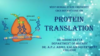
- 1. PROTEIN Translation West Bengal State University cbcs Botany Core viii Dr. Riddhi Datta Department of Botany Dr. A.P.J. Abdul Kalam Government College
- 2. Translation • The information for the proteins found in a cell is encoded in genes of the genome of the cell. • A protein-coding gene is expressed by transcription of the gene to produce an mRNA, followed by translation of the mRNA. • Translation involves the conversion of the base sequence of the mRNA into the amino acid sequence of a polypeptide. • The base sequence information that specifies the amino acid sequence of a polypeptide is called the genetic code. Dr. Riddhi Datta
- 3. Translation • Polypeptide synthesis takes place on ribosomes, where the genetic message encoded in mRNA is translated. • The mRNA is translated in the 5’-to-3’ direction. • The polypeptide is made in the N- terminal–to–C-terminal direction. • Amino acids are brought to the ribosome bound to tRNA molecules. Dr. Riddhi Datta
- 4. • Polypeptide synthesis takes place on ribosomes. • Several thousands of ribosomes are present in each cell. • Ribosomes bind to mRNA and facilitate the binding of the tRNA to the mRNA (codon-anticodon pairing) so that a polypeptide chain can be synthesized. • In both prokaryotes and eukaryotes, ribosomes consist of two unequally sized subunits—the large and small ribosomal subunits. Dr. Riddhi Datta RIBOSOME • Each subunit contains one or more rRNA molecules and a large number of ribosomal proteins.
- 5. Bacterial ribosome: • Size of 70S (S = sedimentation coefficient/ Sevedberg unit) • Consists of two subunits of sizes 50S (large subunit) and 30S (small subunit) • Contain three rRNA molecules— – 23S rRNA and 5S rRNA in the large subunit – 16S rRNA in the small subunit Dr. Riddhi Datta RIBOSOME
- 6. Eukaryotic (mammalian) ribosome: • Size of 80S • Consists of two subunits of sizes 60S (large subunit) and 40S (small subunit) • Contain four rRNA molecules— – 28S rRNA, 5.8S rRNA, and 5S rRNA in the large subunit – 18S rRNA in the small subunit Dr. Riddhi Datta RIBOSOME
- 7. • During translation, the mRNA passes through the small subunit of the ribosome. • It has 3 binding sites for tRNAs: – A (aminoacyl) site is where an incoming aminoacyl–tRNA binds and consists of both small and large subunits – P (peptidyl) site is where the tRNA carrying the growing polypeptide chain is located and consists of both small and large subunits – E (exit) site is where a tRNA binds on its path from the P site to leaving the ribosome and consists only of large subunit Dr. Riddhi Datta RIBOSOME
- 8. • The regions of DNA that contain genes for rRNA are called ribosomal DNA (rDNA) or rRNA transcription units. • E. coli has 7 rRNA transcription units. • Each rRNA transcription unit contains single promoter and one copy each of the 16S, 23S, and 5S rRNA coding sequences, arranged in the order 16S–23S–5S. Dr. Riddhi Datta RIBOSOMal RNA genes IN BACTERIA
- 9. • Transcription by RNA polymerase produces a precursor rRNA (pre- rRNA) molecule which also contain some non-rRNA spacer sequences. • The pre-rRNA is processed to remove the spacers, releasing the three rRNAs. • The transcript-processing events take place in a large ribonucleoprotein complex and specific associations of the rRNAs with ribosomal proteins generate the functional ribosomal subunits. Dr. Riddhi Datta RIBOSOMal RNA genes IN BACTERIA
- 10. • The genes for 18S, 5.8S, and 28S rRNAs are found adjacent to one another in the order 18S–5.8S–28S, with each set tandemly repeated 100 to 1,000 times to form clusters of rDNA repeat units. • Due to active transcription of the repeat units, a nucleolus forms around each cluster and the multiple nucleoli then fuse to form one nucleolus. • Each eukaryotic rDNA repeat unit is transcribed by RNA polymerase I to produce a pre-rRNA molecule which has some non-rRNA spacer sequences. Dr. Riddhi Datta RIBOSOMal RNA genes IN EUKARYOTES
- 11. • The 5S rRNA is produced by transcription of the 5S rRNA genes (typically located elsewhere in the genome) by RNA polymerase III. • The pre-rRNA is processed to remove the spacers, releasing the three rRNAs. • The pre-rRNA processing takes place in complexes formed between the pre-rRNA, 5S rRNA, and ribosomal proteins which ultimately forms the functional 60S and 40S ribosomal subunits and are then transported to the cytosol. Dr. Riddhi Datta RIBOSOMal RNA genes IN EUKARYOTES
- 12. Transfer RNA • During translation of mRNA, each transfer RNA (tRNA) brings a specific amino acid to the ribosome which is to be added to a growing polypeptide chain. • The correct amino acid sequence of a polypeptide is achieved as a result of: – the binding of each amino acid to a specific tRNA – the binding between the codon of the mRNA and the complementary anticodon in the tRNA. Dr. Riddhi Datta
- 13. Structure of Transfer RNA • tRNAs are 75 to 90 nucleotides long having a cloverleaf structure. • The differences in tRNA sequences explain the ability of a particular tRNA molecule to bind a specific amino acid. • The cloverleaf results from complementary base pairing between different sections of the molecule, producing 4 base-paired ―stems‖ separated by 4 loops: I, II, III, and IV. • Loop II contains the three-nucleotide anticodon sequence, which pairs with a three-nucleotide codon sequence in mRNA during translation. Dr. Riddhi Datta
- 14. Structure of Transfer RNA • All tRNAs exhibit an upside-down L-shaped 3D structure in which the 3’ end of the tRNA is at the end of the L that is opposite from the anticodon loop. • All tRNA molecules have the sequence 5’-CCA-3’ at their 3’ ends where the amino acid attaches. • All tRNA molecules have a number of enzymatically modified bases, which include – I: inosine, – T: ribothymidine, – ψ: pseudouridine, – D: dihydrouridine, – Gme: methylguanosine, – GMe2: dimethylguanosine, – Ime: methylinosine. Dr. Riddhi Datta
- 15. Transfer RNA GENES • tRNA genes in bacteria: – found in one or a few copies in the genome – transcribed by the only RNA polymerase found in bacteria • tRNA genes in eukaryotes: – repeated many times in the genome – transcribed by RNA polymerase III • Transcription of tRNA genes in both bacteria and eukaryotes produces precursor tRNA (pre-tRNA) molecules, which are then post-transcriptionally modified, a 5’- CCA-3’ sequence is added to the 3’ end and modification of bases throughout the molecule then take place. • Some tRNA genes in eukaryotes may contain introns between the first and second nucleotides 3’ to the anticodon. Dr. Riddhi Datta
- 16. Aminoacylation of trna • The peptide bond formation between the amino group and carboxyl group of two amino acids is thermodynamically unfavorable. This barrier is overcome by the activation of carboxyl group of the precursor amino acid. • The correct amino acid is attached to the tRNA by an enzyme called aminoacyl–tRNA synthetase. • The process is called aminoacylation, or charging or activation, and produces an aminoacyl–tRNA (or charged tRNA). • Aminoacylation uses energy from ATP hydrolysis. • There are 20 different aminoacyl–tRNA synthetases, one for each of the 20 different amino acids. • Each enzyme recognizes particular structural features of the tRNA or tRNAs it aminoacylates. Dr. Riddhi Datta
- 17. Aminoacylation of trna 1 2 3 4 5 Dr. Riddhi Datta
- 18. Aminoacylation of trna 1. First, the amino acid and ATP bind to the specific aminoacyl–tRNA synthetase enzyme. 2. The enzyme then catalyzes a reaction in which the ATP is hydrolyzed to AMP, which covalently binds to the amino acid (carboxyl group) as AMP to form aminoacyl–AMP. L-aa (L-amino acid) + ATP + ENZ (Enzyme) aa-AMP-ENZ (aminoacyl-AMP-enzyme complex) + PPi 3. Next, the tRNA molecule binds to the enzyme. 4. The enzyme then transfers the amino acid from the aminoacyl–AMP to the tRNA, displaces the AMP and releases the aminoacyl–tRNA molecule. This is called tRNA charging. tRNA-3’OH + aa-AMP-ENZ tRNA-aa (aminoacyl-tRNA) + AMP + ENZ 5. The enzyme then returns to its original state. Chemically, the amino acid attaches at the 3’ end of the tRNA by a covalent linkage between the carboxyl group of the amino acid and the 3’-OH or 2’-OH group of the ribose of the adenine nucleotide found at the 3’ end of every tRNA. https://vimeo.com/138715154 Dr. Riddhi Datta
- 19. PROTEIN SYNTHESIS • Requirements of protein synthesis: – A pool of 20 amino acids – 20 aminoacyl-tRNA synthetases – ATP and GTP – Different tRNA molecules – Ribosomes – Initiation factors – Transfer factors – Elongation factors – Release factors – Mg2+ ions – Several co-factors • The three basic stages of protein synthesis are— – Initiation – Elongation – Termination • Apart from these, aminoacylation of tRNA is required to initiate translation. Dr. Riddhi Datta
- 20. Initiation of Translation • Initiation encompasses all of the steps preceding the formation of the peptide bond between the first two amino acids in the polypeptide chain. • Initiation involves: – an mRNA molecule – a ribosome – a specific initiator tRNA – protein initiation factors (IF) – GTP – Mg2+ ions Dr. Riddhi Datta
- 21. Initiation of Translation IN BACTERIA • Initiation of translation begins with the formation of the initiation complex. • The complex consists of: – 30S ribosomal subunit – three initiation factors, IF1, IF2 and IF3 – mRNA molecule to be translated – GTP – Mg2+ ions • First, the 30S subunit interacts with IF1 and IF3 and then binds with the mRNA molecule near the translation start site. • IF3 aids in the binding of the 30S subunit to mRNA and prevents binding of the 50S ribosomal subunit to the 30S subunit. Dr. Riddhi Datta
- 22. Initiation of Translation IN BACTERIA • The AUG initiating codon signals the start of translation where the 30S subunit actually binds. • Binding also requires a sequence upstream of the AUG, called the ribosome binding site (RBS) or Shine-Dalgarno sequence. • It is a purine rich sequence (AGGAG or similar) and is complementary to a pyrimidine rich region (UCCUC) at the 3’ end of the 16S rRNA in ribosome. • Most RBS are 8-12 nucleotides upstream from AUG. • There is a formation of complementary base pairs between the mRNA and 16S rRNA of the 30S subunit which locates the RBS of the mRNA for the initiation of protein synthesis. Dr. Riddhi Datta
- 23. Initiation of Translation IN BACTERIA • The next step is the binding of a special initiator tRNA to the AUG start codon to which the 30S subunit is bound. • In both prokaryotes and eukaryotes, the AUG initiator codon specifies methionine. • In many cases, the methionine is removed later. • In bacteria, the initiator tRNA is tRNA.fMet, which has the anticodon 5’-CAU-3’ to bind to the AUG start codon. • This tRNA carries a modified form of methionine, formylmethionine (fMet). • First, methionyl–tRNA synthetase catalyzes the addition of methionine to the tRNA. Then the enzyme transformylase adds the formyl group to the amino group of methionine. • The resulting molecule is designated fMet–tRNA.fMet. • When an AUG codon in an mRNA molecule is encountered at a position other than the start of the amino acid-coding sequence, a different tRNA, called tRNA.Met, is used to insert methionine at that point in the polypeptide chain.Dr. Riddhi Datta
- 24. Initiation of Translation IN BACTERIA • The initiator tRNA, fMet–tRNA.fMet, is brought to the 30S subunit–mRNA complex by IF2, which also carries a molecule of GTP. • The 70S ribosome has 3 binding sites for aminoacyl-tRNA: – Exit (E) site – Peptidyl (P) site – Aminoacyl (A) site • The initiator tRNA binds to the subunit in the P site guided by IF2. • IF1 blocks the A site. • IF3 prevents association of the 50S subunit. • This forms the 30S initiation complex. Dr. Riddhi Datta
- 25. Initiation of Translation IN BACTERIA • Next, the 50S ribosomal subunit binds, leading to GTP hydrolysis and the release of the three initiation factors. • This final complex is called the 70S initiation complex. Dr. Riddhi Datta https://www.youtube.com/watch?v=3ooX hZqboqE
- 26. Initiation of Translation IN Eukaryotes • Very similar to that in bacteria • However the process is more complex • It involves many more initiation factors, called eukaryotic initiation factors (eIF) • Differences: – The initiator methionine is unmodified, although a special initiator tRNA brings it to the ribosome. – Shine–Dalgarno sequences are not found in eukaryotic mRNAs. Instead, the initiation complex forms at the 5’ end of the mRNA. Dr. Riddhi Datta
- 27. Initiation of Translation IN Eukaryotes • A cap-binding protein (CBP) binds to the 7- methyl guanosine cap at the 5’ terminus of the mRNA. • Then, other initiation factors (eIFs) bind to the CBP-mRNA complex. • Then the small (40S) subunit of the ribosome binds. • Eukaryotes contain a special initiator tRNA, tRNAiMet (―i‖ for initiator), but the amino group of the methionyl-tRNAiMet is not formylated. • The initiator methionyl-tRNAiMet interacts with a soluble eIF and enters the P site directly during the initiation process, just as in E. coli. Dr. Riddhi Datta
- 28. Initiation of Translation IN Eukaryotes • The entire initiation complex moves 5’ → 3’ along the mRNA molecule, searching for an AUG codon. • The AUG codon is embedded in a short sequence—called the Kozak sequence, after Marilyn Kozak—which indicates that it is the initiator codon. • The optimal initiation sequence is 5’-GCC(A or G)CCAUGG-3’. • This process is called the scanning model for initiation. • When an AUG triplet is found, the initiation factors dissociate from the complex. • Now the large (60S) subunit binds to the methionyl-tRNA/mRNA/40S subunit complex, forming the complete (80S) ribosome. • The 80S ribosome/mRNA/tRNA complex is ready to begin the second phase of translation, chain elongation. Dr. Riddhi Datta
- 29. Elongation of Translation • The elongation involves addition of amino acids to the growing polypeptide chain one by one. • This phase has three steps: – Aminoacyl–tRNA (charged tRNA) binds to the ribosome in the A site. – A peptide bond forms. – The ribosome moves (translocates) along the mRNA one codon. • Elongation requires accessory protein factors, called elongation factors (EF), and GTP. • Elongation is similar in eukaryotes. Dr. Riddhi Datta
- 30. Elongation of Translation STEP 1: Binding of Aminoacyl–tRNA • The anticodon of fMet–tRNA is hydrogen bonded to the AUG initiation codon in the P site of the ribosome. • The next codon in the mRNA is in the A site (Ex: UCC codon specifies serine) where the appropriate aminoacyl–tRNA binds (here, Ser– tRNA.Ser). • This aminoacyl–tRNA is brought to the ribosome bound to EF-Tu–GTP complex. • When the aminoacyl-tRNA binds to the codon in the A site, GTP hydrolysis releases EF-Tu–GDP and EF-Tu is recycled. Dr. Riddhi Datta
- 31. Elongation of Translation STEP 1: Recycling of EF-Tu: • First, a second elongation factor, EF-Ts, binds to EF-Tu and displaces the GDP. • Next, GTP binds to the EF-Tu–EF-Ts complex to produce an EF-Tu–GTP complex simultaneously releasing EF-Ts. • Another aminoacyl-tRNA binds to the EF-Tu–GTP, and the entire process is repeated. • The process is highly similar in eukaryotes, with eEF-1A playing the role of EF-Tu, and eEF-1B playing the role of EF-Ts. Dr. Riddhi Datta
- 32. Elongation of Translation Dr. Riddhi Datta STEP 1:
- 33. Elongation of Translation STEP 2: Peptide Bond Formation: • The ribosome maintains the two aminoacyl– tRNAs in the P and A sites in the correct positions for a peptide bond formation. • First, the bond between the amino acid and the tRNA in the P site (here, fMet and its tRNA) is cleaved. • Second, the peptide bond is formed between the now-freed fMet and the Ser attached to the tRNA in the A site in a reaction catalyzed by peptidyl transferase. • The 23S rRNA molecule of the large ribosomal subunit functions as the peptidyl transferase activity. Dr. Riddhi Datta STEP 2:
- 34. Elongation of Translation STEP 2: Peptide Bond Formation: • Once the peptide bond has formed, a tRNA without an attached amino acid (an uncharged tRNA) is left in the P site. • The tRNA in the A site, now called peptidyl–tRNA, has the first two amino acids of the polypeptide chain attached to it (here, fMet–Ser). Dr. Riddhi Datta
- 35. Elongation of Translation STEP 3: Translocation: • In the last step in the elongation cycle, the ribosome moves one codon along the mRNA toward the 3’ end. • Translocation requires the activity of another elongation factor, EF-G. • The steps involve: – EF-G–GTP complex binds to the ribosome – GTP is hydrolyzed – translocation of ribosome occurs displacing the uncharged tRNA away from the P site • Translocation is similar in eukaryotes; the elongation factor in this case is eEF-2, which functions like bacterial EF-G. Dr. Riddhi Datta
- 36. Elongation of Translation STEP 3: Translocation: • During the translocation step, the peptidyl–tRNA remains attached to its codon on the mRNA. • As the ribosome moves, the uncharged tRNA moves from the P site to the E site. • The peptidyl–tRNA is now located in the P site. • When translocation is complete, the uncharged tRNA is then released from the ribosome and EF-G is released and recycled. • The A site is also vacant where an aminoacyl–tRNA with correct anticodon binds to the newly exposed codon. Dr. Riddhi Datta
- 37. Dr. Riddhi Datta Elongation of Translation STEP 3:
- 38. Elongation of TranslationSTEP 3: Translocation: • The entire elongation process is then repeated until a stop codon is encountered. • In both bacteria and eukaryotes, once the ribosome moves away from the initiation site on the mRNA, another initiation event occurs on another ribosome. Dr. Riddhi Datta • Typically, several ribosomes are translating each mRNA simultaneously together forming a complex called polysome or polyribosome. https://www.youtube.com/watch?v=bNFKt6RVPf4
- 39. TERMINATION of Translation • The termination of translation is signaled by one of three stop codons: UAG, UAA, and UGA. • Stop codons are same in prokaryotes and eukaryotes. • The stop codons do not code for any amino acid, so have no tRNAs for them. • The ribosome recognizes a stop codon with the help of proteins called release factors (RF). • In E. coli, there are three RFs: RF1, RF2 and RF3. • RF1 recognizes UAA and UAG and RF2 recognizes UAA and UGA. Dr. Riddhi Datta
- 40. TERMINATION of Translation • The binding of RF1 or RF2 to a stop codon triggers peptidyl transferase to cleave the polypeptide from the tRNA in the P site. • The polypeptide then leaves the ribosome. Dr. Riddhi Datta
- 41. TERMINATION of Translation • Next, RF3–GDP binds to the ribosome, stimulating the release of the RF from the stop codon and the ribosome. • GTP now replaces the GDP on RF3. • RF3 hydrolyses the GTP which allows RF3 to be released from the ribosome. Dr. Riddhi Datta
- 42. TERMINATION of Translation • In E. coli, ribosome recycling factor (RRF) binds to the A site. • Then EF-G binds, causing translocation of the ribosome and thereby moving RRF to the P site and the uncharged tRNA to the E site. • The RRF releases the uncharged tRNA, and EF-G releases RRF, causing the two ribosomal subunits to dissociate from the mRNA. Dr. Riddhi Datta
- 43. TERMINATION of Translation • In eukaryotes, the termination process is similar to that in bacteria. • In this case, a single release factor— eukaryotic release factor 1 (eRF1)— recognizes all three stop codons, and eRF3 stimulates the termination events. • Ribosome recycling occurs in eukaryotes, but there is no equivalent of RRF. Dr. Riddhi Datta https://www.youtube.com/watch?v=mjAuhjJMCws