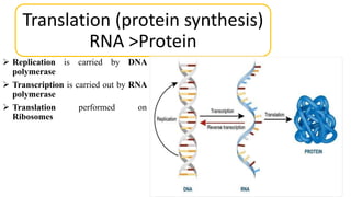
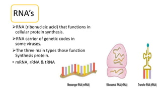
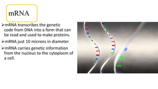
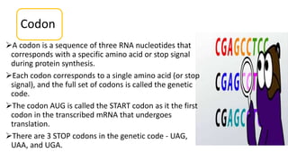
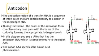
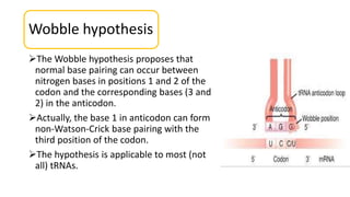
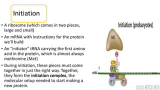
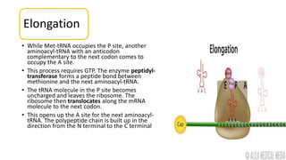
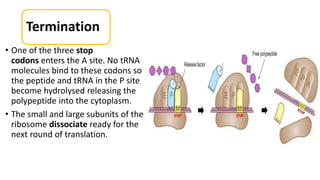
Mumbai Call Girls Service 9910780858 Real Russian Girls Looking Models
Translation (protein synthesis) presentation
1. Tittle
Translation
BS 4th semester presentation
By
Sikandar Ali
Department Of Microbiology UOB,
Quetta Pakistan
WhatsApp number: +923368179410
Gmail: sikanderalisumalani11@gmail.com
2. Translation (protein synthesis)
RNA >Protein
Replication is carried by DNA
polymerase
Transcription is carried out by RNA
polymerase
Translation performed on
Ribosomes
3. Translation
Translation refers to the process of creating protein from mRNA
template.
The sequence of nucleotides on the RNA is translated into the amino
acid sequence.
The protein and this reaction is carried out by ribosomes.
Multiple enzymes in an energy intensive process the uses ATP as well
as GTP
4. RNA’s
RNA (ribonucleic acid) that functions in
cellular protein synthesis.
RNA carrier of genetic codes in
some viruses.
The three main types those function
Synthesis protein.
• mRNA, rRNA & tRNA
5. mRNA
mRNA transcribes the genetic
code from DNA into a form that can
be read and used to make proteins.
mRNA just 10 microns in diameter
mRNA carries genetic information
from the nucleus to the cytoplasm of
a cell.
6. TYPES OF mRNA
The two main types of mRNA:
Monocistronic mRNA
A monocistronic mRNA molecule contains the exon sequences coding
for a single protein.
Most eukaryotic mRNAs are monocistronic
Polycistronic mRNA
A polycistronic mRNA molecule contains the exon coding sequences for
multiple proteins.
Most mRNA of bacteria and bacteriophages (viruses that live in bacterial
hosts) are polycistronic..
7. rRNA
ribonucleic acids (RNA) are a group of large
molecules that have many different roles.
As the name, ribosomal RNA (rRNA) is part of
the ribosome.
rRNA is located in the cytoplasm of a cell,
where ribosomes are found.
8. Ribosome
Ribosomes are approximately 60% rRNA
and 40% protein by weight.
A ribosome contains two subunits, (LSU)
and (SSU)
Prokaryotes have 70S or 50S subunits.
Eukaryotes have 80S or 60S subunits to.
A ribosomes has an mRNA binding site and
three tRNA binding site.
The tRNA binding site are APE
• A:aminocyle tRNA
• P:peptidyl tRNA
• E:exit
9. tRNA
Like rRNA, tRNA is located in the cellular
cytoplasm and is involved in protein
synthesis.
Transfer RNA brings or transfers amino
acids to the ribosome that corresponds to
each three-nucleotide codon of rRNA.
The amino acids then can be joined
together and processed to make
polypeptides and proteins.
10. Codon
A codon is a sequence of three RNA nucleotides that
corresponds with a specific amino acid or stop signal
during protein synthesis.
Each codon corresponds to a single amino acid (or stop
signal), and the full set of codons is called the genetic
code.
The codon AUG is called the START codon as it the first
codon in the transcribed mRNA that undergoes
translation.
There are 3 STOP codons in the genetic code - UAG,
UAA, and UGA.
11.
12.
13. Anticodon
The anticodon region of a transfer RNA is a sequence
of three bases that are complementary to a codon in
the messenger RNA.
During translation , the bases of the anticodon form
complementary base pairs with the bases of the
codon by forming the appropriate hydrogen bonds
In this diagram you see a tRNA that has the
anticodon UUU which is complementary to the codon
AAA.
The codon AAA specifies the amino acid
phenylalanine.
14. Wobble hypothesis
The Wobble hypothesis proposes that
normal base pairing can occur between
nitrogen bases in positions 1 and 2 of the
codon and the corresponding bases (3 and
2) in the anticodon.
Actually, the base 1 in anticodon can form
non-Watson-Crick base pairing with the
third position of the codon.
The hypothesis is applicable to most (not
all) tRNAs.
15.
16. Three phases of translation
• Initiation translation
• Elongation
• Termination
17. Initiation
• A ribosome (which comes in two pieces,
large and small)
• An mRNA with instructions for the protein
we'll build
• An "initiator" tRNA carrying the first amino
acid in the protein, which is almost always
methionine (Met)
• During initiation, these pieces must come
together in just the right way. Together,
they form the initiation complex, the
molecular setup needed to start making a
new protein.
18. Elongation
• While Met-tRNA occupies the P site, another
aminoacyl-tRNA with an anticodon
complementary to the next codon comes to
occupy the A site.
• This process requires GTP. The enzyme peptidyl-
transferase forms a peptide bond between
methionine and the next aminoacyl-tRNA.
• The tRNA molecule in the P site becomes
uncharged and leaves the ribosome. The
ribosome then translocates along the mRNA
molecule to the next codon.
• This opens up the A site for the next aminoacyl-
tRNA. The polypeptide chain is built up in the
direction from the N terminal to the C terminal
19. Termination
• One of the three stop
codons enters the A site. No tRNA
molecules bind to these codons so
the peptide and tRNA in the P site
become hydrolysed releasing the
polypeptide into the cytoplasm.
• The small and large subunits of the
ribosome dissociate ready for the
next round of translation.
Editor's Notes
Energy transfer. GTP is involved in energy transfer within the cell.
So what we should know is 1 ATP (activation) + 4 GTP in each translation cycle, which will be 6 high energy bonds.
Guanosine 5'-Triphosphate
RNA molecule was described by R.W. Holley in 1965. Robert William Holley, (born Jan. 28, 1922). Nobel Prize in Physiology or Medicine in 1968
The existence of mRNA was first suggested by Jacques Monod and François Jacob, and subsequently discovered by Jacob, Sydney Brenner and Matthew Meselson at the California Institute of Technology in 1961.
Bicistronic mRNA
A bicistronic mRNA molecule contains the exon coding sequences for two proteins.
the large ribosomal subunit (LSU) , small ribosomal subunit (SSU)
10 million ribosomes and numerous ribosomes can be connected to the equivalent mRNA strand, this structure is known as a POLYSOME.
the 64 codons, 61 codons code for the 20 amino acids.
Marshall Nirenberg and Heinrich J. Matthaei were the first to reveal the nature of a codon in 1961.
Hypothesis proposed by Francis Crick in 1966 to explain the observed degeneracy in the third position of a codon.