Download as ODP, PPTX


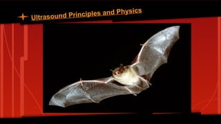
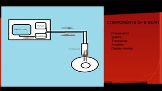
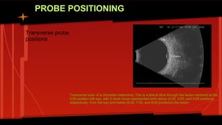
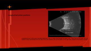
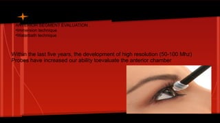
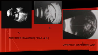
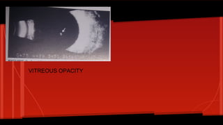
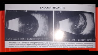
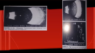
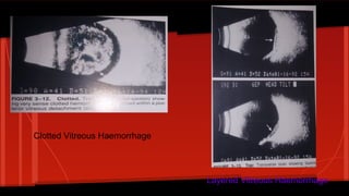
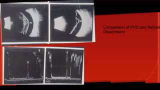
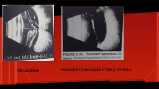
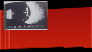
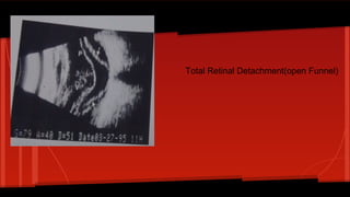
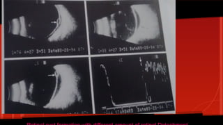
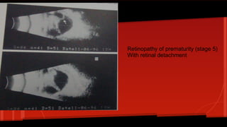
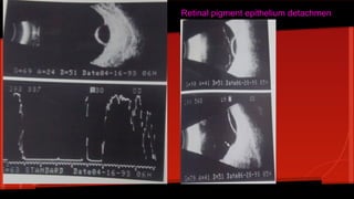
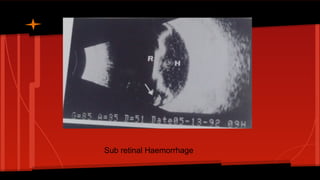
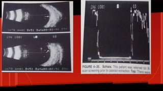
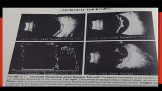
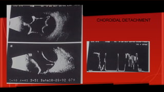

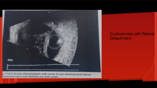
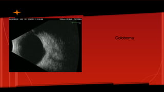
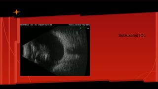


B-scan ultrasonography produces a two-dimensional cross-sectional view of the eye and orbit. It is used to examine conditions where the media is hazy like corneal opacities or dense cataracts. It can also be used to evaluate lesions like tumors even when the media is clear. The B-scan involves a transducer that transmits sound pulses and receives echoes to create images. It can position probes longitudinally or transversely and evaluate both the anterior and posterior segments of the eye. Example images shown include vitreous opacities, retinal detachments, choroidal detachments, and more.


