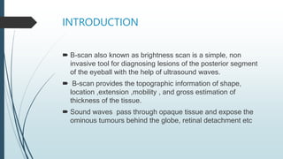
B-scan ultrasonography uses ultrasound waves to non-invasively diagnose posterior segment eye lesions. It provides topographic information on the shape, location, extension, mobility and thickness of tissues. B-scan imaging was developed in the 1950s and 1960s and allows visualization of structures behind opaque tissues. It uses a transducer to transmit ultrasound pulses that are partially reflected by tissues, with the reflections detected to produce images. Different orientations of the transducer probe, such as longitudinal, transverse and axial, allow imaging of different areas of the eye and orbit. B-scan is useful for evaluating a variety of conditions when the ocular media is opaque, including tumors, retinal detachments, intraocular foreign bodies and more.


![INSTRUMENTATION
Ultrasound waves exhibits frequencies above 20kHz which is
not audible to humans . Ultrasound is an acoustic wave in
which compression and rarefactions occur because changes
in density within solid and fluid substances. Ophthalmic
ultrasound ranges frequencies ranges from 8 to 10 mHz.
Higher frequency > shorter wavelength> lesser depth of
penetration >better resolution [ophthalmic use]
4 basic components of B-scan [ a pulser, a transducer,a
receiver and a display system]](https://image.slidesharecdn.com/doc-20230615-wa0003-230629135245-38ba812d/85/DOC-20230615-WA0003-pptx-5-320.jpg)


![PROCEDURE
Explain the procedure to the patient .
Positioning the patient : B-scan is performed with patient
reclining or supine position , certain ocular conditions require
sitting position [eg air bubble in the anterior chamber, to
demonstrate shifting fluid in exudative retinal detachment]
Usually the scan is performed with eyelids closed , using a
coupling jelly over the probe . In case the eyelids need to be
open while performing the scan then topical anesthesia is a
must.
Probe orientation :](https://image.slidesharecdn.com/doc-20230615-wa0003-230629135245-38ba812d/85/DOC-20230615-WA0003-pptx-8-320.jpg)
![CONTI…..
The transducer probe always has a marker [ dot, line ,or logo ]
3 basic probe is present axial ,transverse , and longitudinal
Longitudinal scan: the probe is place such that the marker is
perpendicular to limbus. Hence the beam scans across a
single meridian at any given clock hour . The oriented
meridian is the one exactly opposite to the meridian in which
the probe is placed eg if the probe is placed at 6 o clock
meridian the picture is displayed at 12 0 clock meridian.
Transverse scan : the probe is placed such that the marker is
parallel to limbus . This orientation is useful for lateral extent
of a leision. Depending on the orientation of the probe marker
, transverse scans can be horizontal, vertical or oblique](https://image.slidesharecdn.com/doc-20230615-wa0003-230629135245-38ba812d/85/DOC-20230615-WA0003-pptx-9-320.jpg)


