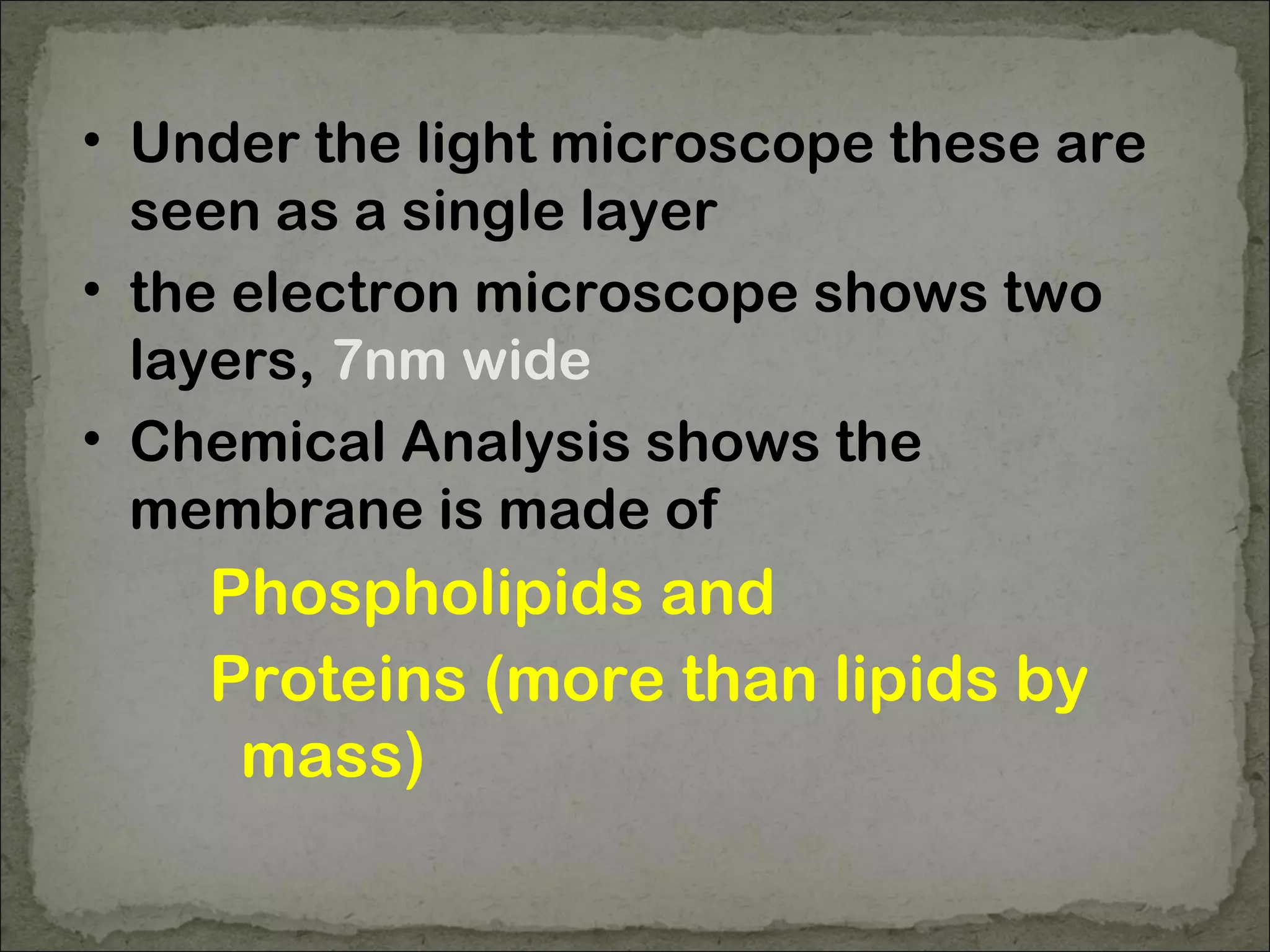
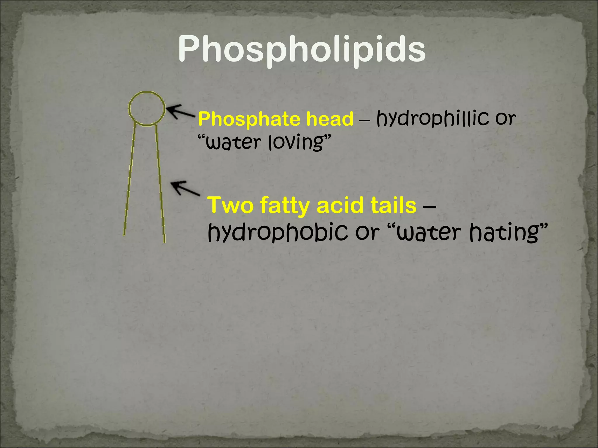
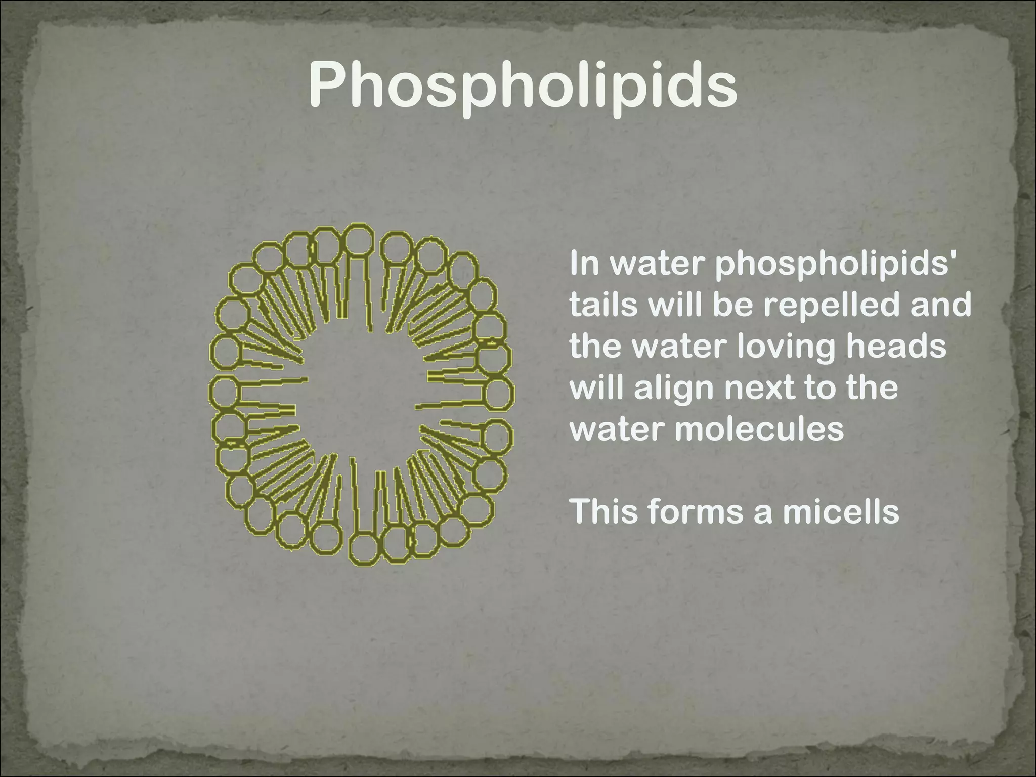
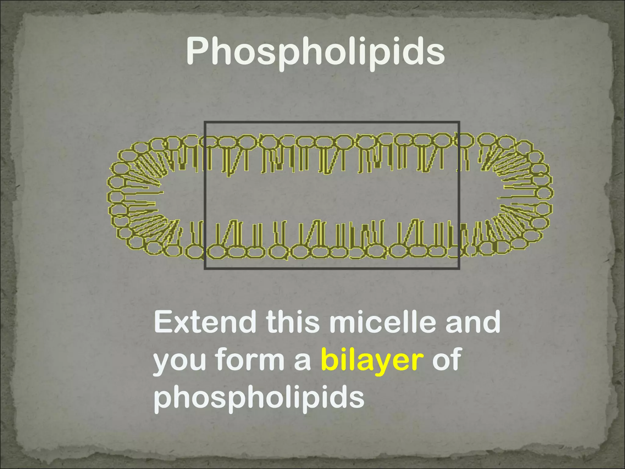
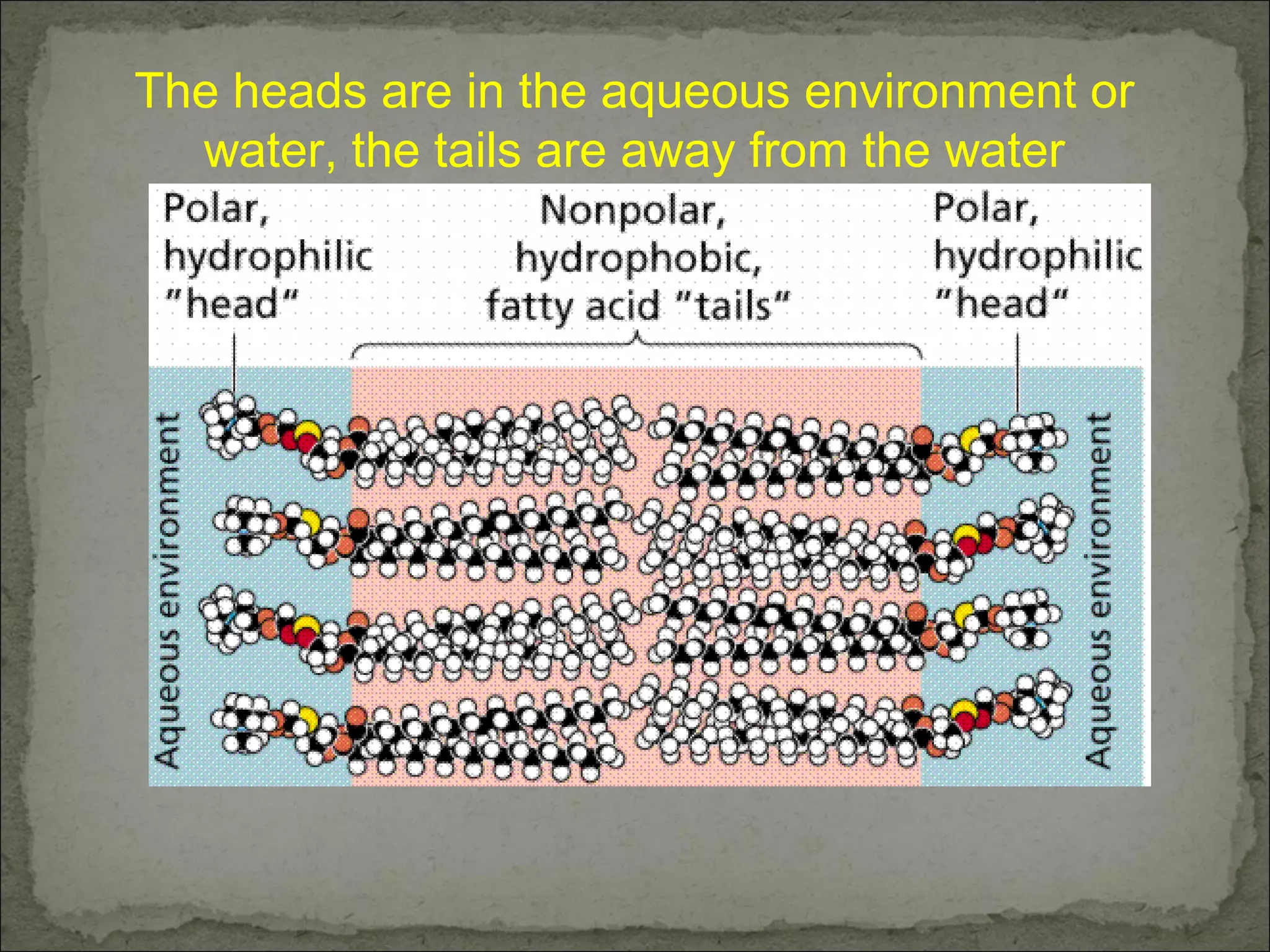
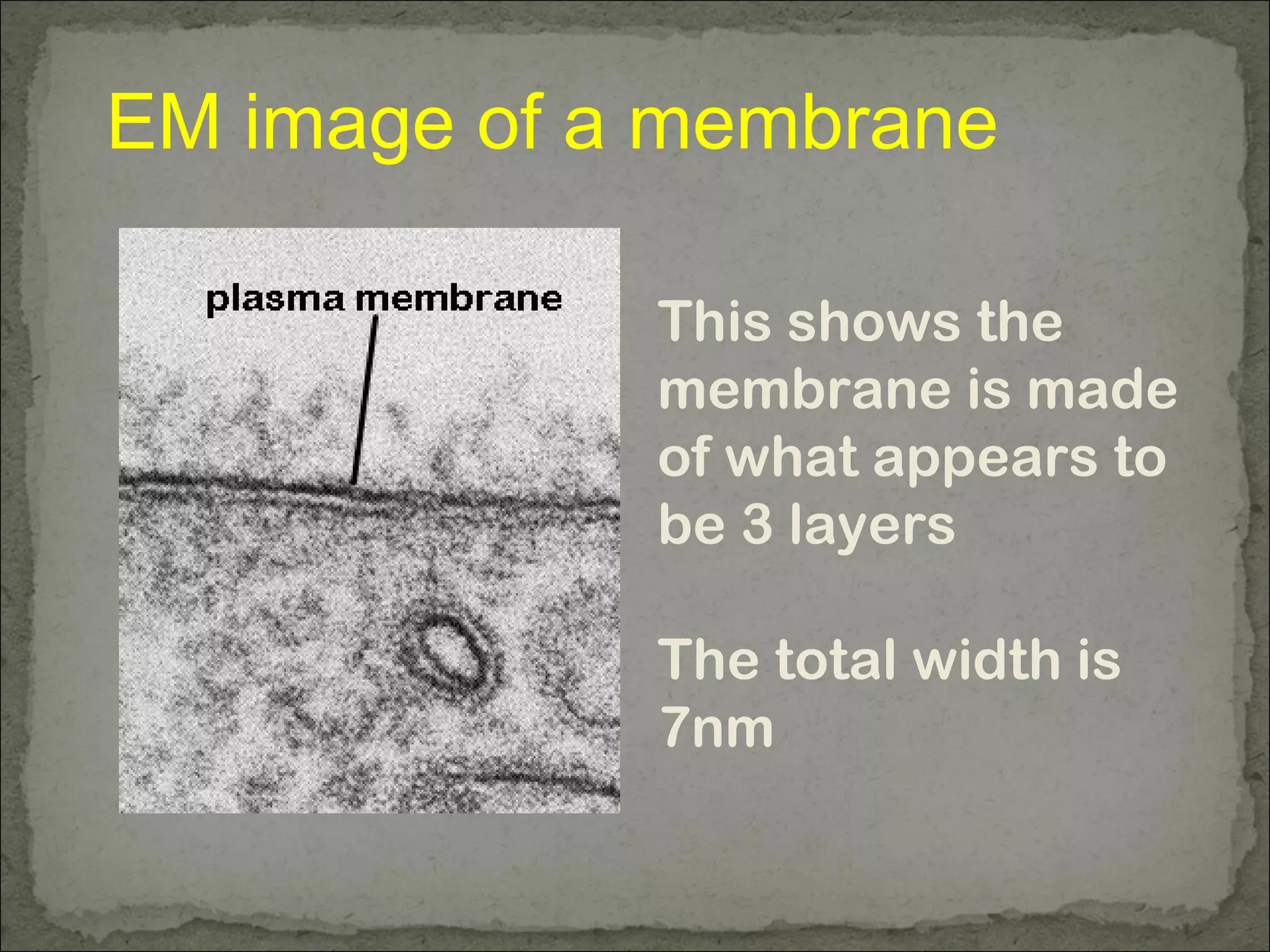
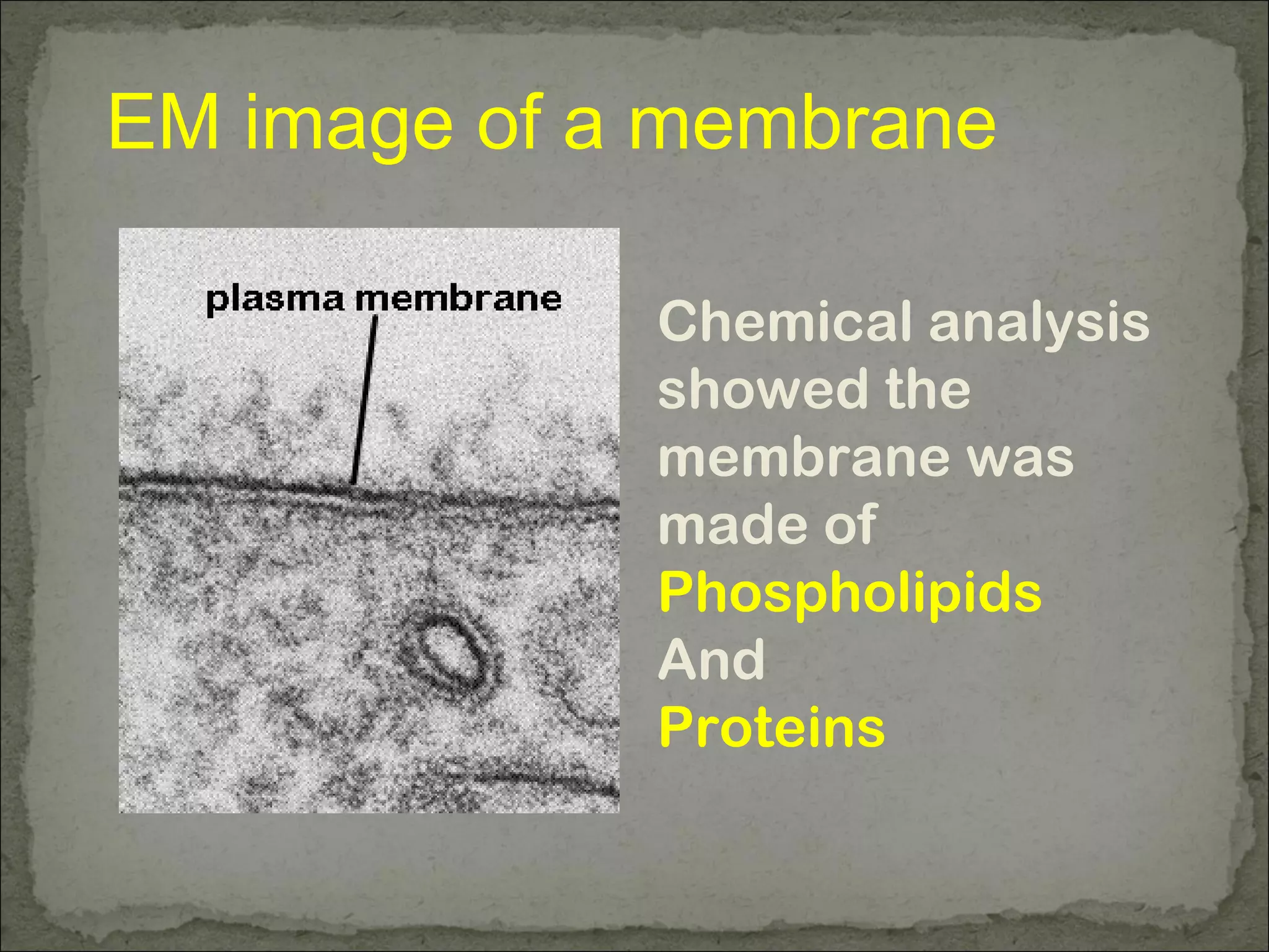
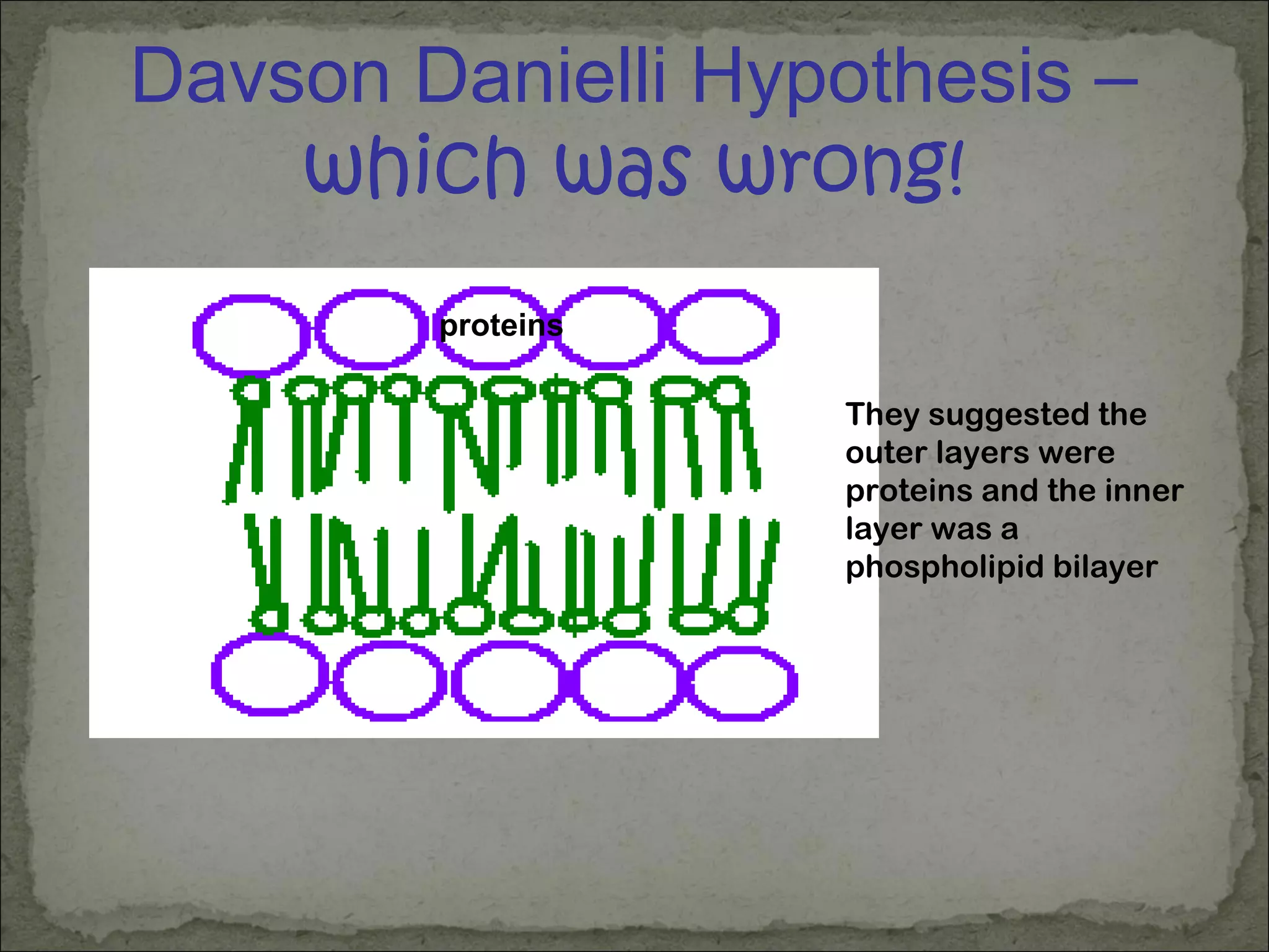
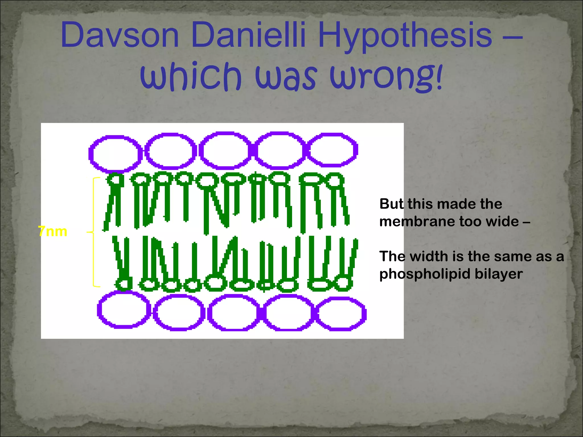
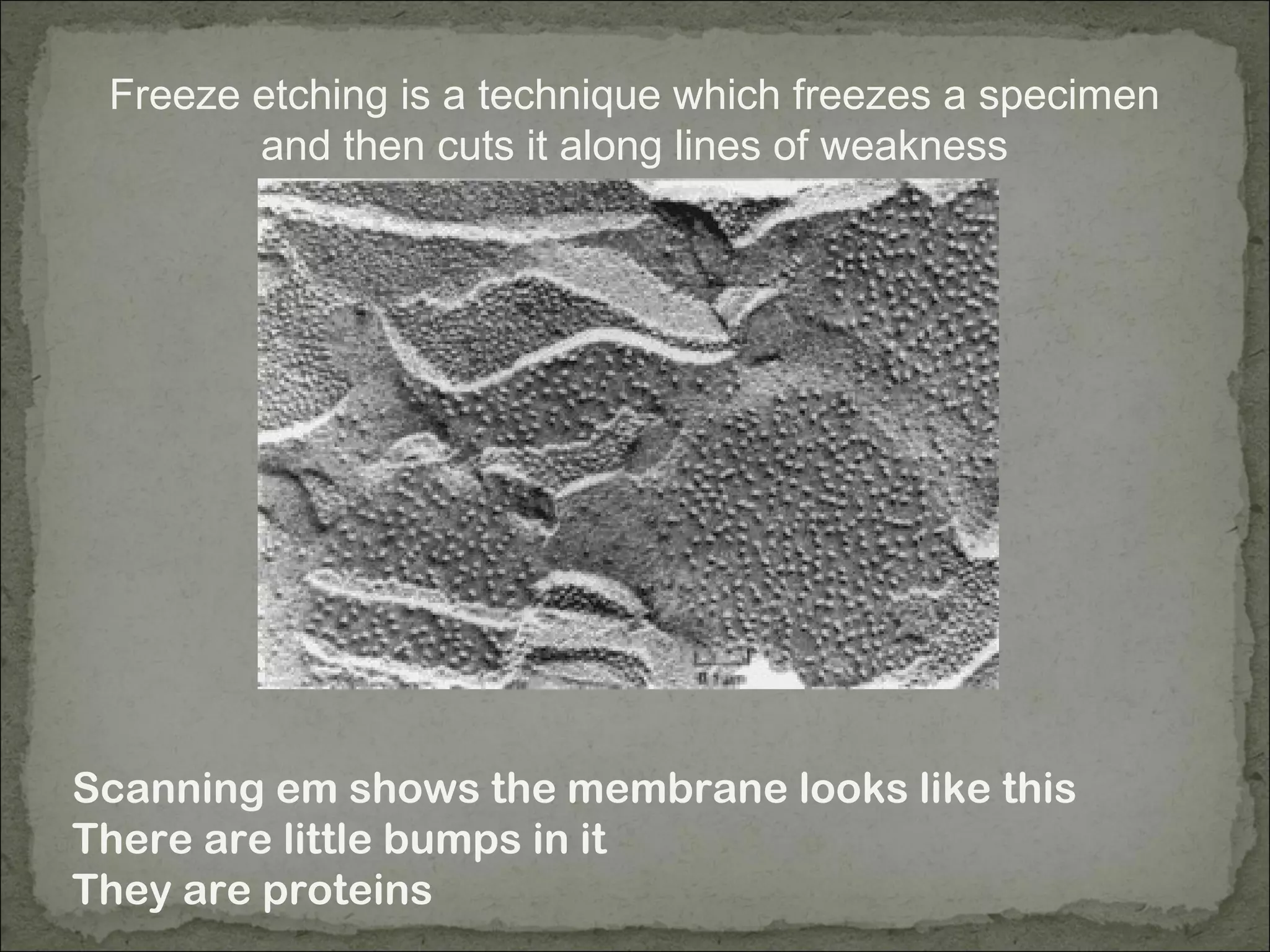
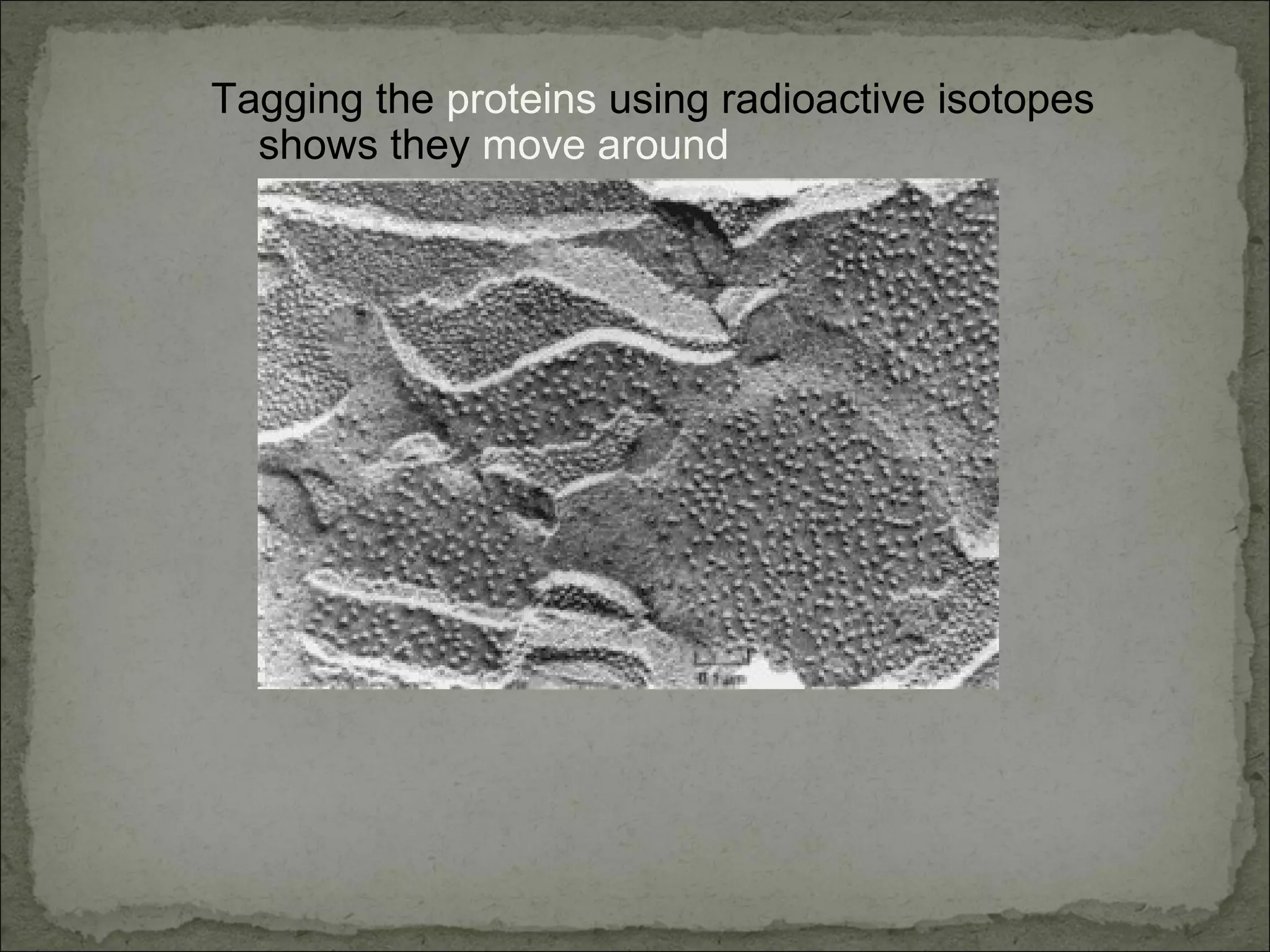
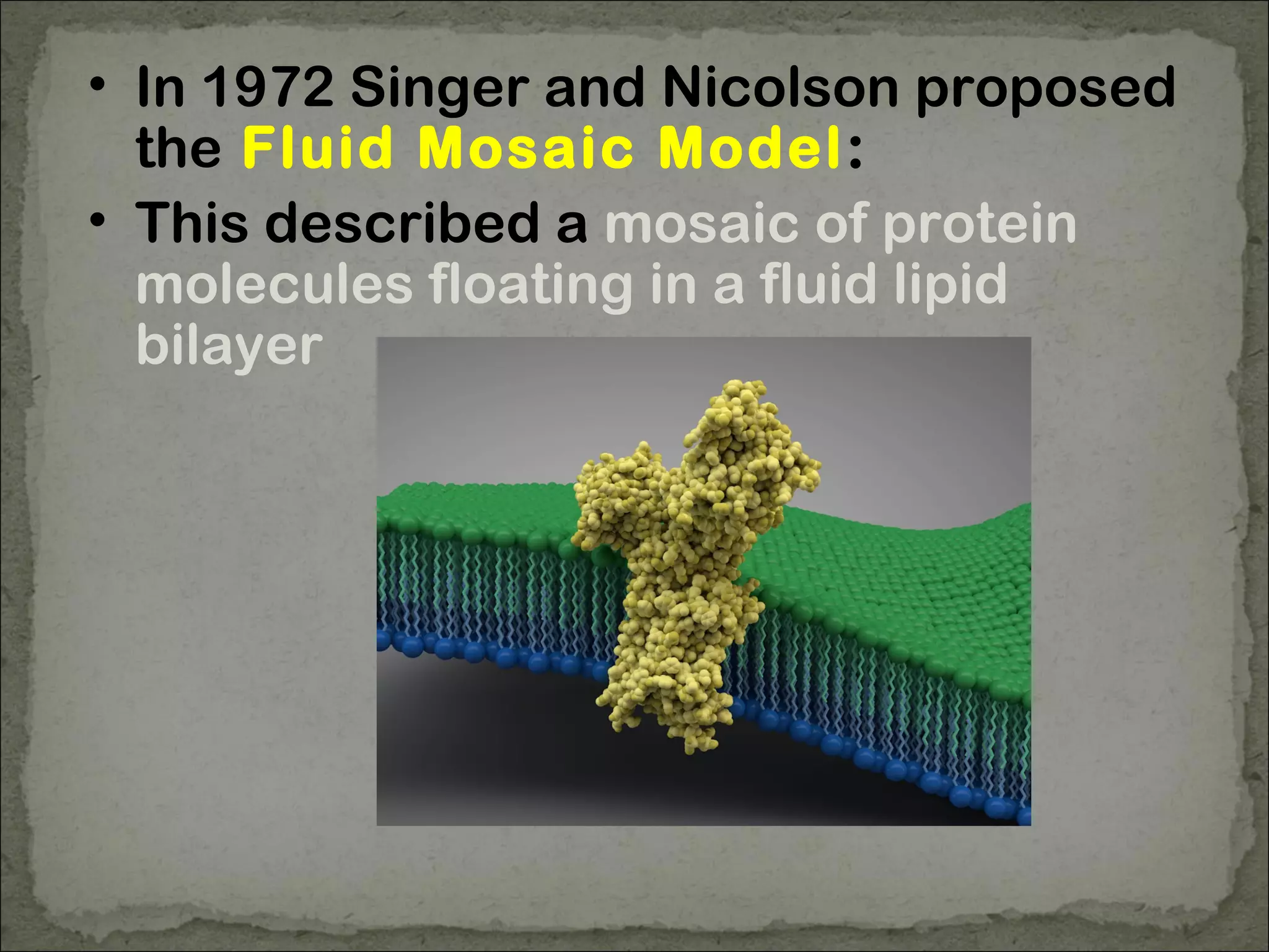
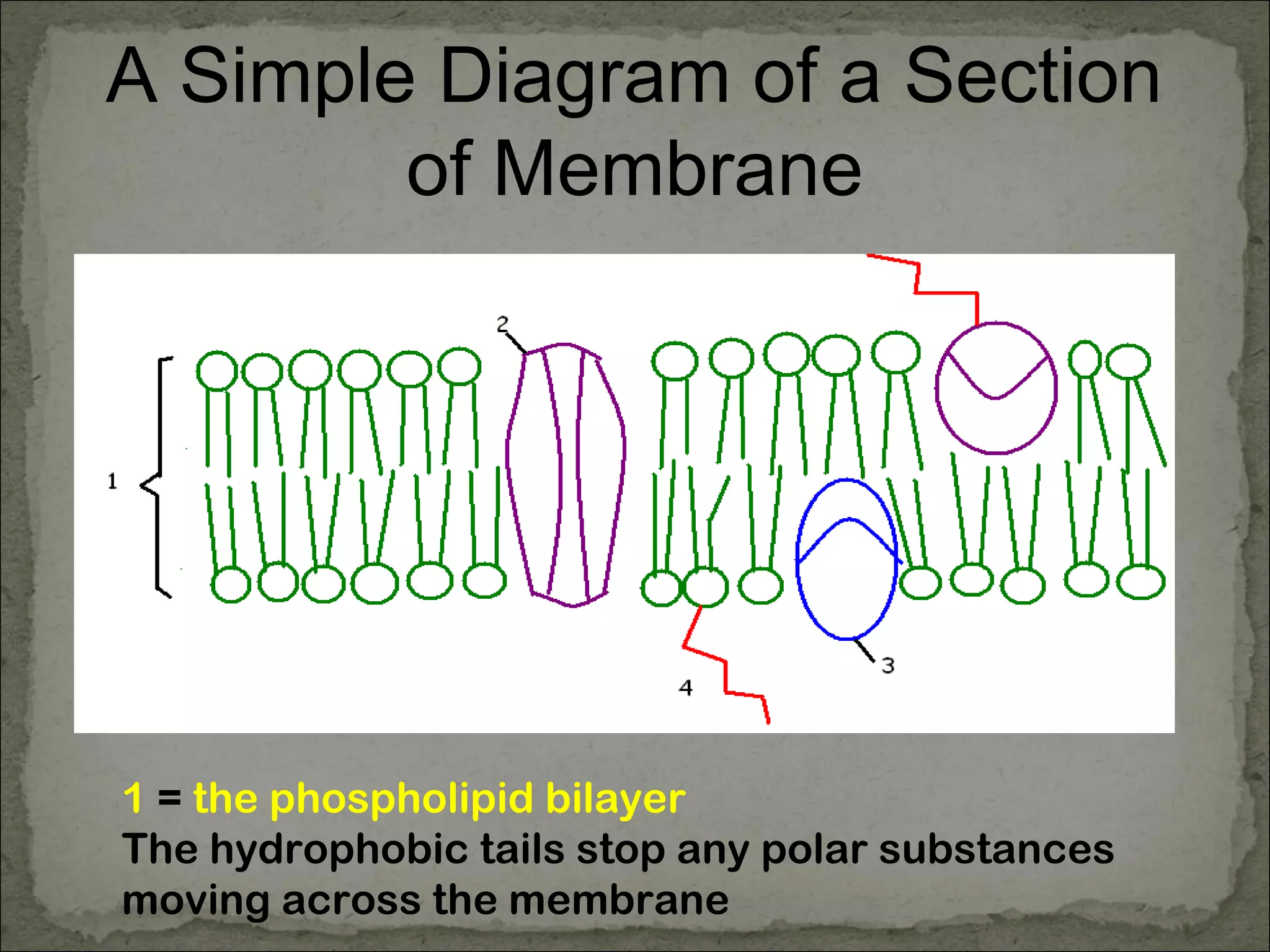
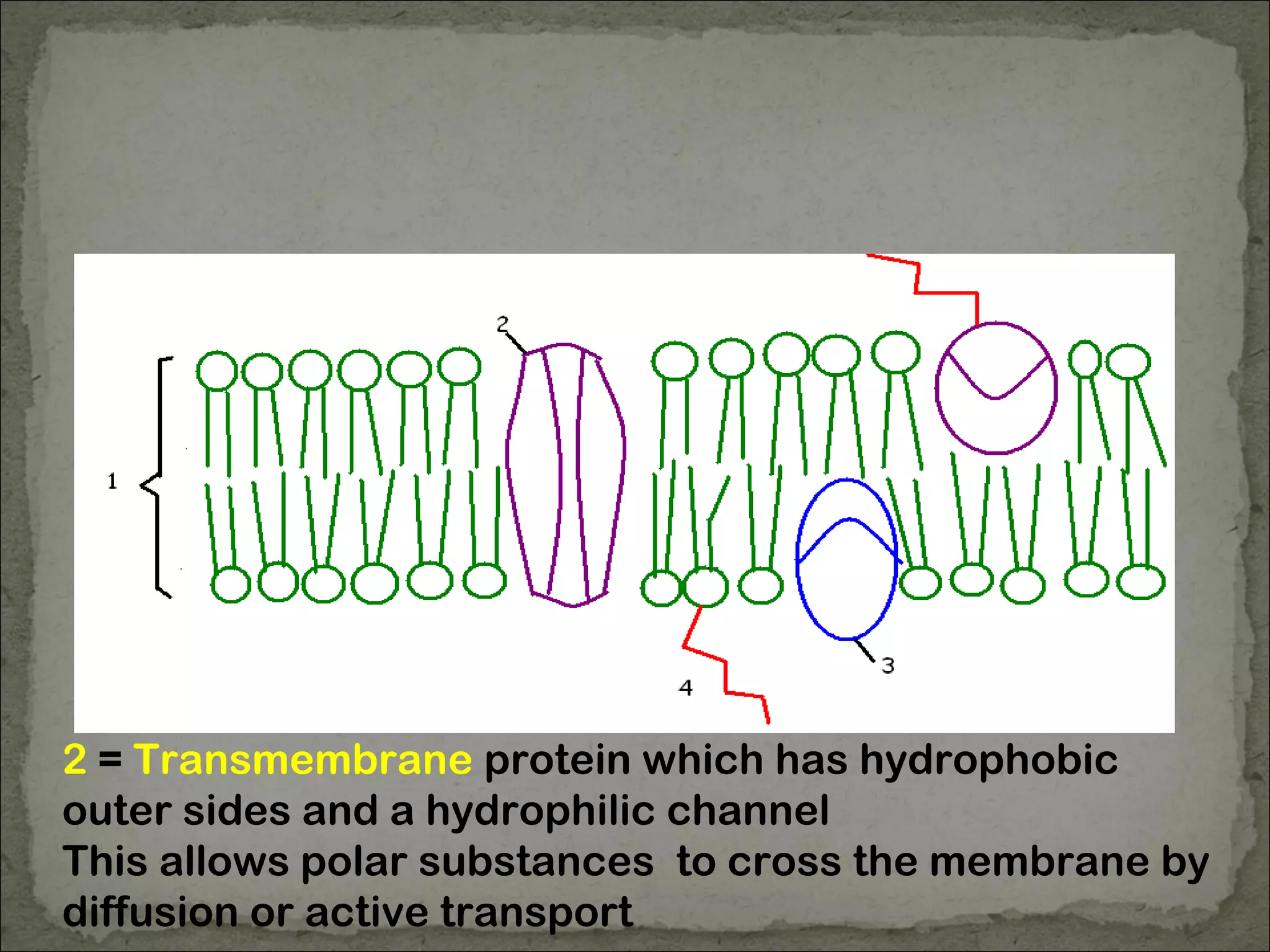
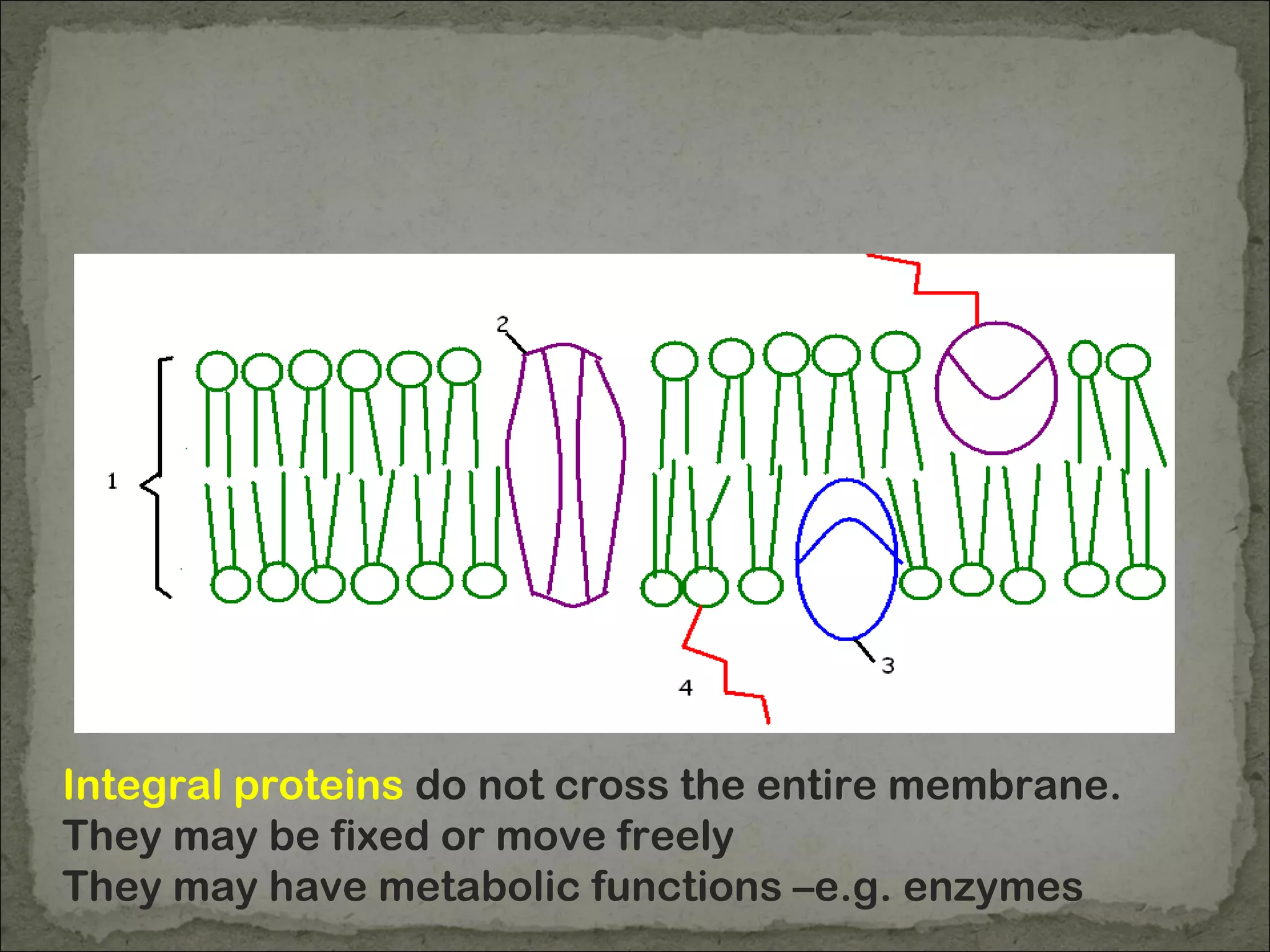
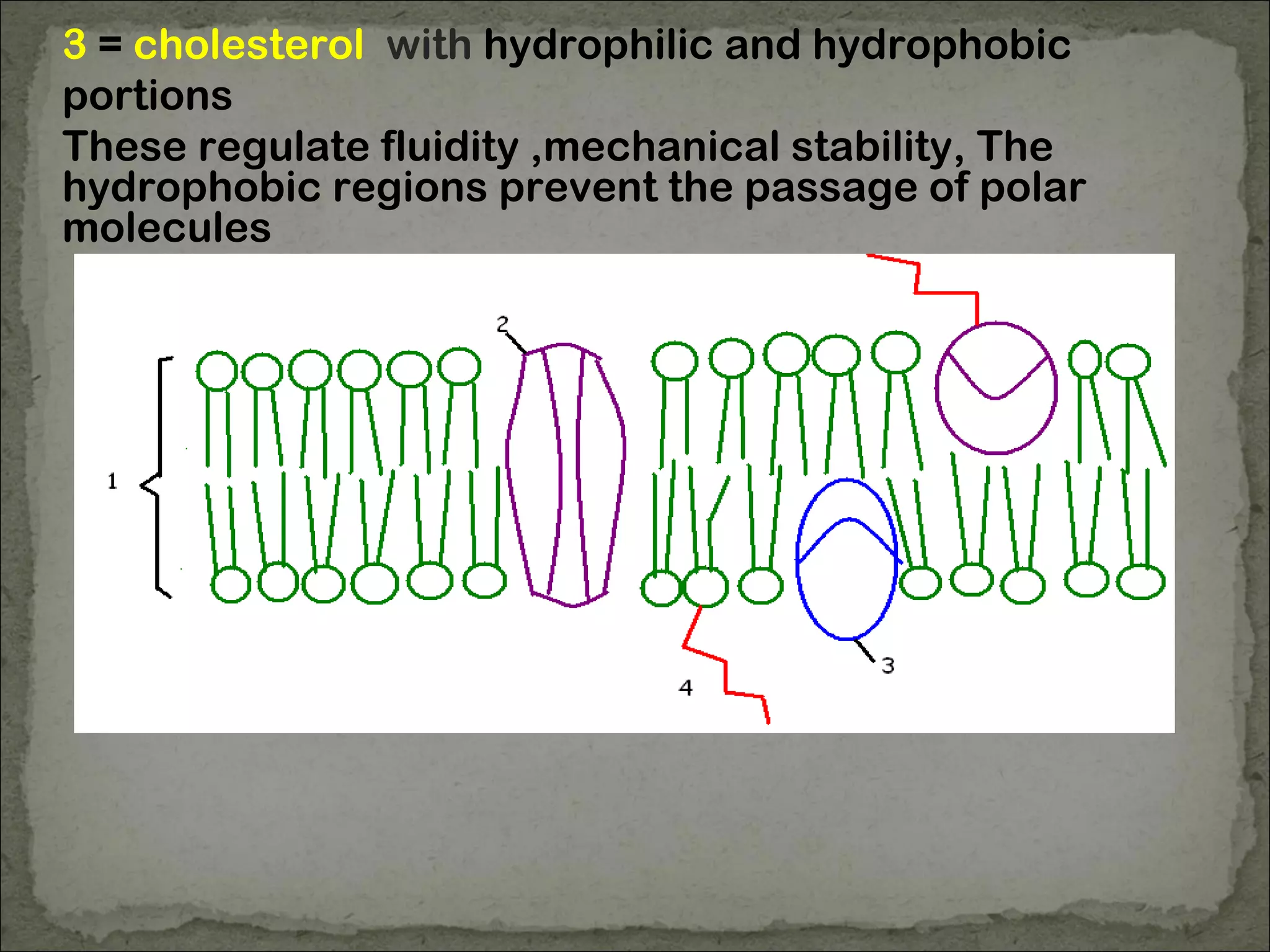
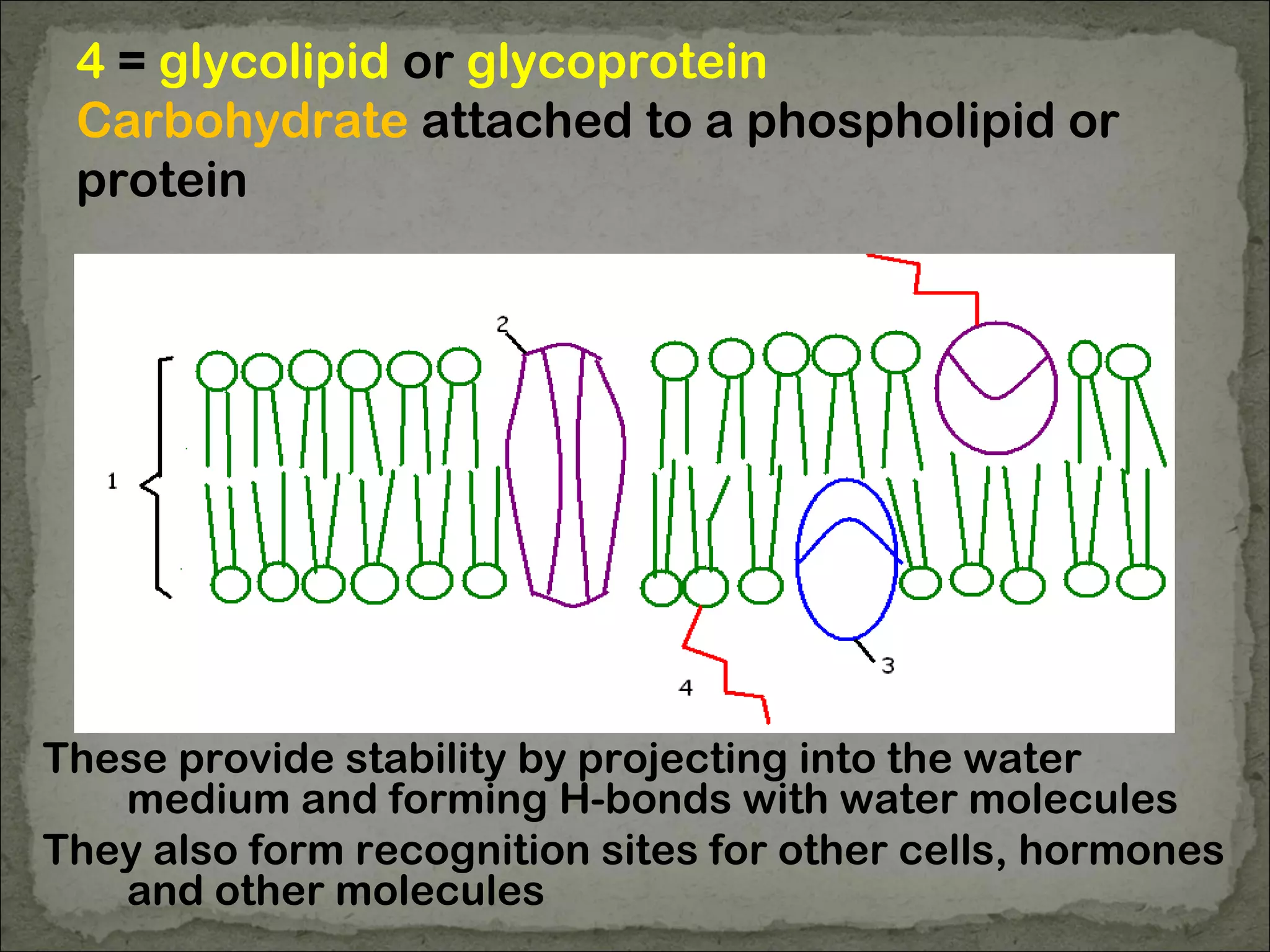
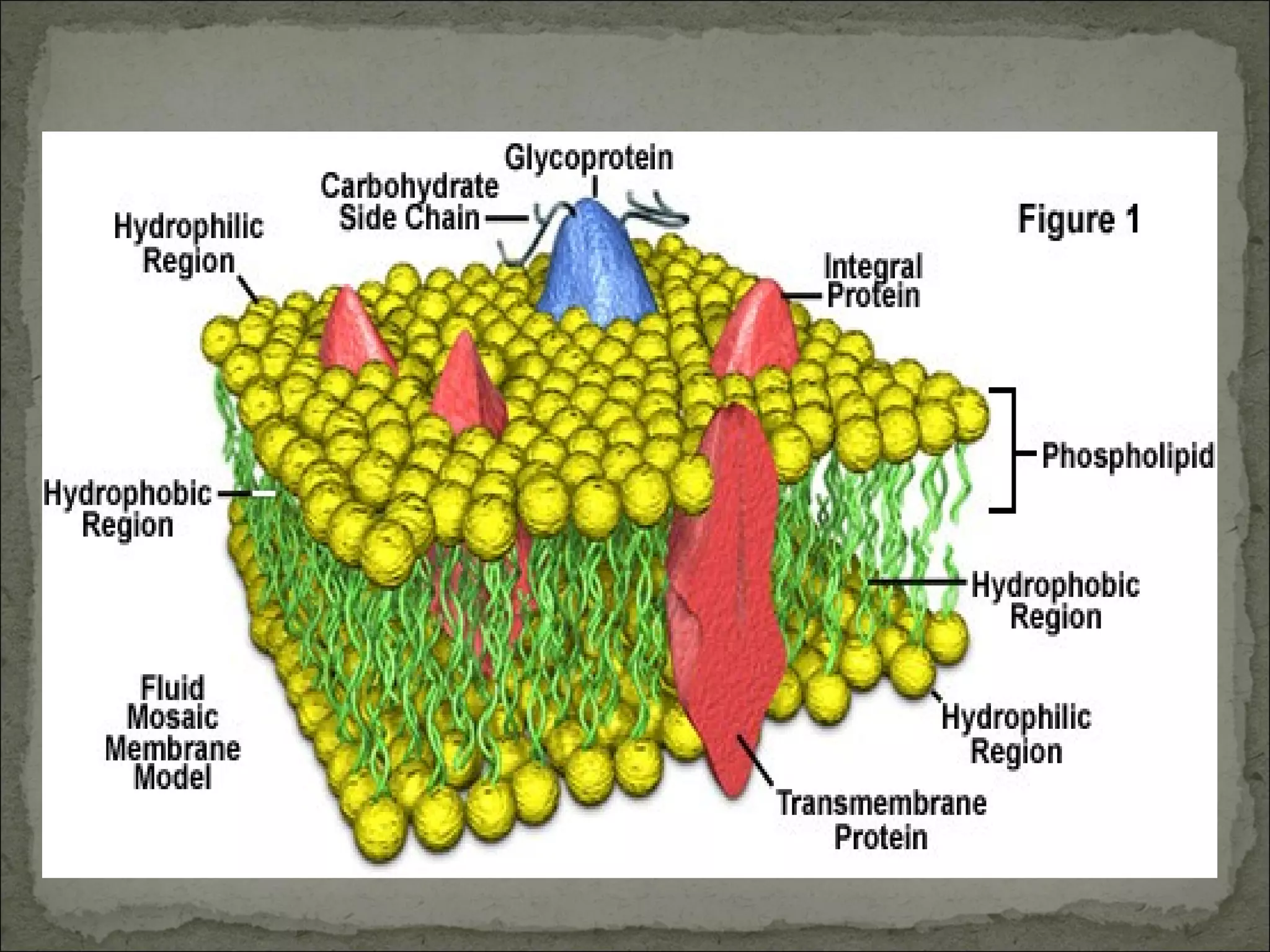
Cell Membranes
• Under the light microscope these are
seen as a single layer
• the electron microscope shows two
layers, 7nm wide
• Chemical Analysis shows the
membrane is made of
Phospholipids and
Proteins (more than lipids by
mass)
Phospholipids
Phosphate head – hydrophillic or
“water loving”
Two fatty acid tails –
hydrophobic or “water hating”
Phospholipids
In water phospholipids'
tails will be repelled and
the water loving heads
will align next to the
water molecules
This forms a micells
Phospholipids
Extend this micelle and
you form a bilayer of
phospholipids
The heads are in the aqueous environment or
water, the tails are away from the water
EM image of a membrane
This shows the
membrane is made
of what appears to
be 3 layers
The total width is
7nm
EM image of a membrane
Chemical analysis
showed the
membrane was
made of
Phospholipids
And
Proteins
Davson Danielli Hypothesis –
which was wrong!
They suggested the
outer layers were
proteins and the inner
layer was a
phospholipid bilayer
proteins
Davson Danielli Hypothesis –
which was wrong!
But this made the
membrane too wide –
The width is the same as a
phospholipid bilayer
7nm
Freeze etching is a technique which freezes a specimen
and then cuts it along lines of weakness
Scanning em shows the membrane looks like this
There are little bumps in it
They are proteins
Tagging the proteins using radioactive isotopes
shows they move around
• In 1972 Singer and Nicolson proposed
the Fluid Mosaic Model:
• This described a mosaic of protein
molecules floating in a fluid lipid
bilayer
A Simple Diagram of a Section
of Membrane
1 = the phospholipid bilayer
The hydrophobic tails stop any polar substances
moving across the membrane
2 = Transmembrane protein which has hydrophobic
outer sides and a hydrophilic channel
This allows polar substances to cross the membrane by
diffusion or active transport
Integral proteins do not cross the entire membrane.
They may be fixed or move freely
They may have metabolic functions –e.g. enzymes
3 = cholesterol with hydrophilic and hydrophobic
portions
These regulate fluidity ,mechanical stability, The
hydrophobic regions prevent the passage of polar
molecules
4 = glycolipid or glycoprotein
Carbohydrate attached to a phospholipid or
protein
These provide stability by projecting into the water
medium and forming H-bonds with water molecules
They also form recognition sites for other cells, hormones
and other molecules
Video

B. membrane structure

  • 1.
  • 2.
    • Under thelight microscope these are seen as a single layer • the electron microscope shows two layers, 7nm wide • Chemical Analysis shows the membrane is made of Phospholipids and Proteins (more than lipids by mass)
  • 3.
    Phospholipids Phosphate head –hydrophillic or “water loving” Two fatty acid tails – hydrophobic or “water hating”
  • 4.
    Phospholipids In water phospholipids' tailswill be repelled and the water loving heads will align next to the water molecules This forms a micells
  • 5.
    Phospholipids Extend this micelleand you form a bilayer of phospholipids
  • 6.
    The heads arein the aqueous environment or water, the tails are away from the water
  • 7.
    EM image ofa membrane This shows the membrane is made of what appears to be 3 layers The total width is 7nm
  • 8.
    EM image ofa membrane Chemical analysis showed the membrane was made of Phospholipids And Proteins
  • 9.
    Davson Danielli Hypothesis– which was wrong! They suggested the outer layers were proteins and the inner layer was a phospholipid bilayer proteins
  • 10.
    Davson Danielli Hypothesis– which was wrong! But this made the membrane too wide – The width is the same as a phospholipid bilayer 7nm
  • 11.
    Freeze etching isa technique which freezes a specimen and then cuts it along lines of weakness Scanning em shows the membrane looks like this There are little bumps in it They are proteins
  • 12.
    Tagging the proteinsusing radioactive isotopes shows they move around
  • 13.
    • In 1972Singer and Nicolson proposed the Fluid Mosaic Model: • This described a mosaic of protein molecules floating in a fluid lipid bilayer
  • 14.
    A Simple Diagramof a Section of Membrane 1 = the phospholipid bilayer The hydrophobic tails stop any polar substances moving across the membrane
  • 15.
    2 = Transmembraneprotein which has hydrophobic outer sides and a hydrophilic channel This allows polar substances to cross the membrane by diffusion or active transport
  • 16.
    Integral proteins donot cross the entire membrane. They may be fixed or move freely They may have metabolic functions –e.g. enzymes
  • 17.
    3 = cholesterolwith hydrophilic and hydrophobic portions These regulate fluidity ,mechanical stability, The hydrophobic regions prevent the passage of polar molecules
  • 18.
    4 = glycolipidor glycoprotein Carbohydrate attached to a phospholipid or protein These provide stability by projecting into the water medium and forming H-bonds with water molecules They also form recognition sites for other cells, hormones and other molecules
  • 20.