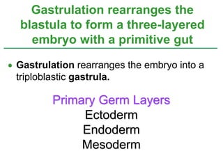
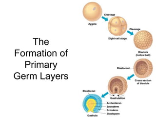
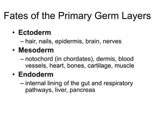
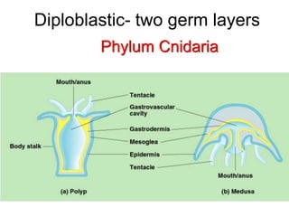
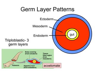
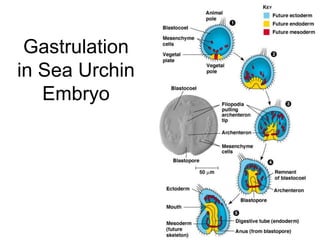
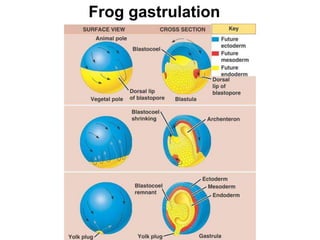
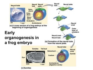
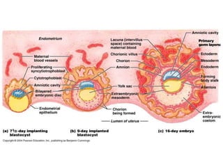
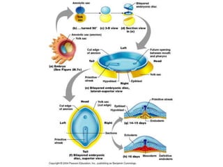
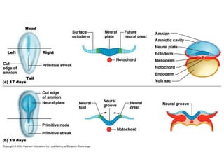
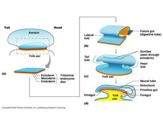
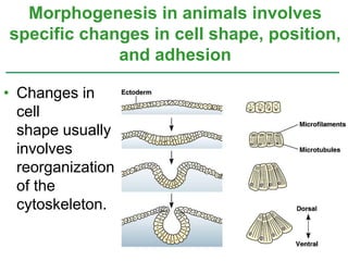
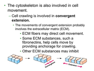
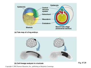
 Gastrulation rearranges the embryo into a
triploblastic gastrula.
Gastrulation rearranges the
blastula to form a three-layered
embryo with a primitive gut
Primary Germ Layers
Ectoderm
Endoderm
Mesoderm
The
Formation of
Primary
Germ Layers
Fates of the Primary Germ Layers
• Ectoderm
– hair, nails, epidermis, brain, nerves
• Mesoderm
– notochord (in chordates), dermis, blood
vessels, heart, bones, cartilage, muscle
• Endoderm
– internal lining of the gut and respiratory
pathways, liver, pancreas
Germ Layer Patterns
Diploblastic
gutEndoderm
Ectoderm
Phylum Cnidaria
Diploblastic- two germ layers
Germ Layer Patterns
Triploblastic- 3
germ layers
acoelomate
gutEndoderm
Ectoderm
Mesoderm
Gastrulation
in Sea Urchin
Embryo
Frog gastrulation
Early
organogenesis in
a frog embryo
• The amniote embryo is the solution to
reproduction in a dry environment.
Amniote embryos develop in a fluid-
filled sac within a shell or uterus
chorion
amnion
embryo
allantois
yolk sac
Extraembryonic membrane
Fetal portion of placenta
Maternal portion of placenta
reptile & bird mammal
• The four extraembryonic membranes are
the yolk sac, amnion, chorion, and
allantois.
–Cells of the yolk sac digest yolk
providing nutrients to the embryo.
–The amnion encloses the embryo in a
fluid-filled amniotic sac which protects
the embryo from drying out.
–The chorion cushions the embryo
against mechanical shocks.
–The allantois functions as a disposal sac
for uric acid.
Mammalian Development.
–Recall:
• The egg and zygote do not exhibit any
obvious polarity.
• Holoblastic cleavage occurs in the zygote.
–Gastrulation and organogenesis follows a
pattern similar to that seen in birds and
reptiles.
Organogenesis
Differentiation of primary
germ layers into tissues
and organs.
• Changes in
cell
shape usually
involves
reorganization
of the
cytoskeleton.
Morphogenesis in animals involves
specific changes in cell shape, position,
and adhesion
• The cytoskeleton is also involved in cell
movement.
– Cell crawling is involved in convergent
extension.
• The movements of convergent extension probably
involves the extracellular matrix (ECM).
• ECM fibers may direct cell movement.
• Some ECM substances, such a
fibronectins, help cells move by
providing anchorage for crawling.
• Other ECM substances may inhibit
movement in certain directions.
•Cell adhesion molecules
(CAMs): located on cell surfaces
bind to CAMs on other cells.
–Differences in CAMs regulate
morphogenetic movement and tissue
binding.
Copyright © 2002 Pearson Education, Inc., publishing as Benjamin Cummings
•Cadherins are also involved in
cell-to-cell adhesion.
–Require the presence of calcium for
proper function.
Copyright © 2002 Pearson Education, Inc., publishing as Benjamin Cummings
• In many animal species (mammals may be a
major exception), the heterogeneous
distribution of cytoplasmic determinants in
the unfertilized egg leads to regional
differences in the early embryo
The developmental fate of cells
depends on cytoplasmic determinants
and cell-cell induction: a review
Copyright © 2002 Pearson Education, Inc., publishing as Benjamin Cummings
• Subsequently, in induction,
interactions among the embryonic
cells themselves induce changes in
gene expression.
–These interactions eventually bring about
the differentiation of the many
specialized cell types making up a new
animal.
Copyright © 2002 Pearson Education, Inc., publishing as Benjamin Cummings
• Fate maps illustrate the developmental
history of cells.
• “Founder cells” give rise to specific tissues in
older embryos.
• As development proceeds a cell’s
developmental potential becomes
restricted.
Fate mapping can reveal cell
genealogies in chordate embryos
Copyright © 2002 Pearson Education, Inc., publishing as Benjamin Cummings
Fig. 47.20
• Polarity and the Basic Body Plan.
– In mammals, polarity may be established by the
entry of the sperm into the egg.
– In frogs, the animal and vegetal pole determine
the anterior-posterior body axis.
The eggs of most vertebrates have
cytoplasmic determinants that help
establish the body axes and differences
among cells of the early embryo
Copyright © 2002 Pearson Education, Inc., publishing as Benjamin Cummings
• Restriction of Cellular Potency.
– The fate of embryonic
cells is affected by
both the distribution
of cytoplasmic
determinants and
by cleavage pattern.
Copyright © 2002 Pearson Education, Inc., publishing as Benjamin Cummings
Fig. 47.21
• Induction: the influence of one set of cells on
a neighboring group of cells.
–Functions by affecting gene expression.
• Results in the differentiation of cells into a
specific type of tissue.
Inductive signals drive differentiation
and pattern formation invertebrates
Copyright © 2002 Pearson Education, Inc., publishing as Benjamin Cummings
• The “Organizer” of Spemann and Mangold.
• Grafting the dorsal lip
of one embryo onto
the ventral surface of
another embryo
results in the develop-
ment of a second
notochord and neural
tube at the site
of the graft.
– Spemann referred
to the dorsal lip
as a primary
organizer.
Copyright © 2002 Pearson Education, Inc., publishing as Benjamin Cummings
Fig. 47.22
 An example of the molecular basis of
induction:
 Bone morphogenetic protein 4 (BMP-4) is
a growth factor promoting promote the
formation of bone and the skeleton
– In amphibians, organizer cells inactivate BMP-4
on the dorsal side of the embryo.
– In humans it’s a critical signaling molecule
required for the early differentiation of the embryo
and establishing of a dorsal-ventral axis
Copyright © 2002 Pearson Education, Inc., publishing as Benjamin Cummings
BMP in neural tube
formation
1. Inhibition of BMP
signaling
2. At end of neurulation the
lateral edges of the
neural plate fuse
3. They segregate from the
non-neural epithelia to
form a neural tube
4. Roof plate of neural tube
now produces BMP. BMP
stimulates neural crest
cell formation
• Pattern Formation in the
Vertebrate Limb.
–Induction plays a major role in
pattern formation.
• Positional information, supplied by
molecular cues, tells a cell where it is
relative to the animals body axes.
Copyright © 2002 Pearson Education, Inc., publishing as Benjamin Cummings
• Limb development in chicks as a model of
pattern formation.
• Wings and legs begin as limb buds.
– Each component
of the limb is
oriented with
regard to
three axes:
– Proximal-distal
– Anterior-posterior
– Dorsal-ventra.
Copyright © 2002 Pearson Education, Inc., publishing as Benjamin Cummings
Fig. 47.23b
Organizer regions.
Copyright © 2002 Pearson Education, Inc., publishing as Benjamin Cummings
• Apical ectodermal ridge (AER).
– Secretes fibroblast growth factor (FGF)
proteins.
– Required for limb growth and patterning along
the proximal-distal axis.
– Required for
pattern formation
along the
dorsal-ventral
axis.
Copyright © 2002 Pearson Education, Inc., publishing as Benjamin Cummings
Fig. 47.23a
• Zone of polarizing activity (ZPA).
–Secretes Sonic hedgehog, a protein
growth factor.
–Required for pattern formation of the limb
along the anterior-posterior axis.
Copyright © 2002 Pearson Education, Inc., publishing as Benjamin Cummings
• Homeobox-containing (Hox) genes
play a role in specifying the identity of
regions of the limb, as well as the body
as a whole.
–In summary, pattern formation is a
chain of events involving cell
signaling and differentiation.
Copyright © 2002 Pearson Education, Inc., publishing as Benjamin Cummings
Guess whom the following grew up to be:
C. D.B.A.
E.
F. G.
H.
C. cat D. humanB. fishA. dolphin
Phylum Chordata
E. Mouse
F. Elephant G. Snake
H. bat

Ch 47 development

  • 2.
     Gastrulation rearrangesthe embryo into a triploblastic gastrula. Gastrulation rearranges the blastula to form a three-layered embryo with a primitive gut Primary Germ Layers Ectoderm Endoderm Mesoderm
  • 3.
  • 4.
    Fates of thePrimary Germ Layers • Ectoderm – hair, nails, epidermis, brain, nerves • Mesoderm – notochord (in chordates), dermis, blood vessels, heart, bones, cartilage, muscle • Endoderm – internal lining of the gut and respiratory pathways, liver, pancreas
  • 5.
  • 6.
  • 7.
    Germ Layer Patterns Triploblastic-3 germ layers acoelomate gutEndoderm Ectoderm Mesoderm
  • 8.
  • 9.
  • 10.
  • 11.
    • The amnioteembryo is the solution to reproduction in a dry environment. Amniote embryos develop in a fluid- filled sac within a shell or uterus chorion amnion embryo allantois yolk sac Extraembryonic membrane Fetal portion of placenta Maternal portion of placenta reptile & bird mammal
  • 12.
    • The fourextraembryonic membranes are the yolk sac, amnion, chorion, and allantois. –Cells of the yolk sac digest yolk providing nutrients to the embryo. –The amnion encloses the embryo in a fluid-filled amniotic sac which protects the embryo from drying out. –The chorion cushions the embryo against mechanical shocks. –The allantois functions as a disposal sac for uric acid.
  • 13.
    Mammalian Development. –Recall: • Theegg and zygote do not exhibit any obvious polarity. • Holoblastic cleavage occurs in the zygote. –Gastrulation and organogenesis follows a pattern similar to that seen in birds and reptiles.
  • 14.
    Organogenesis Differentiation of primary germlayers into tissues and organs.
  • 20.
    • Changes in cell shapeusually involves reorganization of the cytoskeleton. Morphogenesis in animals involves specific changes in cell shape, position, and adhesion
  • 21.
    • The cytoskeletonis also involved in cell movement. – Cell crawling is involved in convergent extension. • The movements of convergent extension probably involves the extracellular matrix (ECM). • ECM fibers may direct cell movement. • Some ECM substances, such a fibronectins, help cells move by providing anchorage for crawling. • Other ECM substances may inhibit movement in certain directions.
  • 22.
    •Cell adhesion molecules (CAMs):located on cell surfaces bind to CAMs on other cells. –Differences in CAMs regulate morphogenetic movement and tissue binding. Copyright © 2002 Pearson Education, Inc., publishing as Benjamin Cummings
  • 23.
    •Cadherins are alsoinvolved in cell-to-cell adhesion. –Require the presence of calcium for proper function. Copyright © 2002 Pearson Education, Inc., publishing as Benjamin Cummings
  • 24.
    • In manyanimal species (mammals may be a major exception), the heterogeneous distribution of cytoplasmic determinants in the unfertilized egg leads to regional differences in the early embryo The developmental fate of cells depends on cytoplasmic determinants and cell-cell induction: a review Copyright © 2002 Pearson Education, Inc., publishing as Benjamin Cummings
  • 25.
    • Subsequently, ininduction, interactions among the embryonic cells themselves induce changes in gene expression. –These interactions eventually bring about the differentiation of the many specialized cell types making up a new animal. Copyright © 2002 Pearson Education, Inc., publishing as Benjamin Cummings
  • 26.
    • Fate mapsillustrate the developmental history of cells. • “Founder cells” give rise to specific tissues in older embryos. • As development proceeds a cell’s developmental potential becomes restricted. Fate mapping can reveal cell genealogies in chordate embryos Copyright © 2002 Pearson Education, Inc., publishing as Benjamin Cummings
  • 27.
    Copyright © 2002Pearson Education, Inc., publishing as Benjamin Cummings Fig. 47.20
  • 28.
    • Polarity andthe Basic Body Plan. – In mammals, polarity may be established by the entry of the sperm into the egg. – In frogs, the animal and vegetal pole determine the anterior-posterior body axis. The eggs of most vertebrates have cytoplasmic determinants that help establish the body axes and differences among cells of the early embryo Copyright © 2002 Pearson Education, Inc., publishing as Benjamin Cummings
  • 29.
    • Restriction ofCellular Potency. – The fate of embryonic cells is affected by both the distribution of cytoplasmic determinants and by cleavage pattern. Copyright © 2002 Pearson Education, Inc., publishing as Benjamin Cummings Fig. 47.21
  • 30.
    • Induction: theinfluence of one set of cells on a neighboring group of cells. –Functions by affecting gene expression. • Results in the differentiation of cells into a specific type of tissue. Inductive signals drive differentiation and pattern formation invertebrates Copyright © 2002 Pearson Education, Inc., publishing as Benjamin Cummings
  • 31.
    • The “Organizer”of Spemann and Mangold. • Grafting the dorsal lip of one embryo onto the ventral surface of another embryo results in the develop- ment of a second notochord and neural tube at the site of the graft. – Spemann referred to the dorsal lip as a primary organizer. Copyright © 2002 Pearson Education, Inc., publishing as Benjamin Cummings Fig. 47.22
  • 32.
     An exampleof the molecular basis of induction:  Bone morphogenetic protein 4 (BMP-4) is a growth factor promoting promote the formation of bone and the skeleton – In amphibians, organizer cells inactivate BMP-4 on the dorsal side of the embryo. – In humans it’s a critical signaling molecule required for the early differentiation of the embryo and establishing of a dorsal-ventral axis Copyright © 2002 Pearson Education, Inc., publishing as Benjamin Cummings
  • 33.
    BMP in neuraltube formation 1. Inhibition of BMP signaling 2. At end of neurulation the lateral edges of the neural plate fuse 3. They segregate from the non-neural epithelia to form a neural tube 4. Roof plate of neural tube now produces BMP. BMP stimulates neural crest cell formation
  • 34.
    • Pattern Formationin the Vertebrate Limb. –Induction plays a major role in pattern formation. • Positional information, supplied by molecular cues, tells a cell where it is relative to the animals body axes. Copyright © 2002 Pearson Education, Inc., publishing as Benjamin Cummings
  • 35.
    • Limb developmentin chicks as a model of pattern formation. • Wings and legs begin as limb buds. – Each component of the limb is oriented with regard to three axes: – Proximal-distal – Anterior-posterior – Dorsal-ventra. Copyright © 2002 Pearson Education, Inc., publishing as Benjamin Cummings Fig. 47.23b
  • 36.
    Organizer regions. Copyright ©2002 Pearson Education, Inc., publishing as Benjamin Cummings
  • 37.
    • Apical ectodermalridge (AER). – Secretes fibroblast growth factor (FGF) proteins. – Required for limb growth and patterning along the proximal-distal axis. – Required for pattern formation along the dorsal-ventral axis. Copyright © 2002 Pearson Education, Inc., publishing as Benjamin Cummings Fig. 47.23a
  • 38.
    • Zone ofpolarizing activity (ZPA). –Secretes Sonic hedgehog, a protein growth factor. –Required for pattern formation of the limb along the anterior-posterior axis. Copyright © 2002 Pearson Education, Inc., publishing as Benjamin Cummings
  • 39.
    • Homeobox-containing (Hox)genes play a role in specifying the identity of regions of the limb, as well as the body as a whole. –In summary, pattern formation is a chain of events involving cell signaling and differentiation. Copyright © 2002 Pearson Education, Inc., publishing as Benjamin Cummings
  • 40.
    Guess whom thefollowing grew up to be: C. D.B.A. E. F. G. H.
  • 41.
    C. cat D.humanB. fishA. dolphin Phylum Chordata E. Mouse F. Elephant G. Snake H. bat