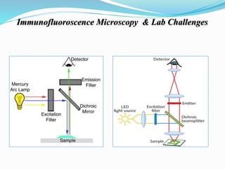
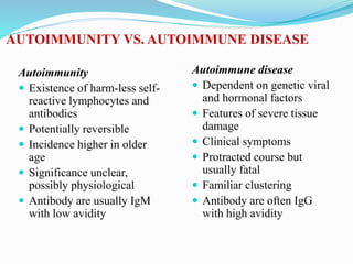
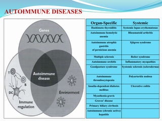
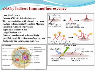
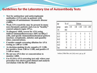
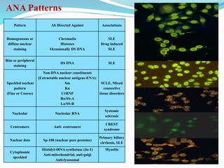
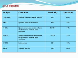
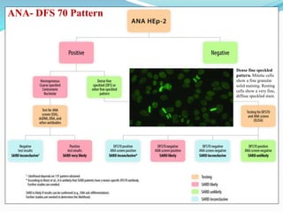
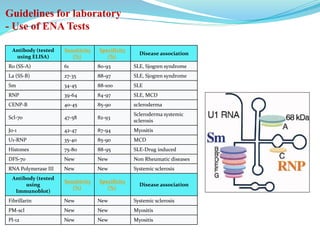
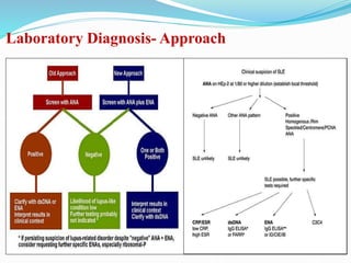
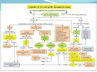
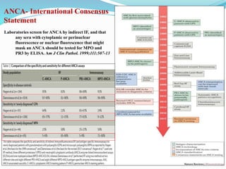
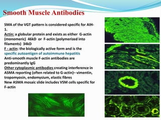
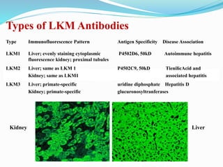
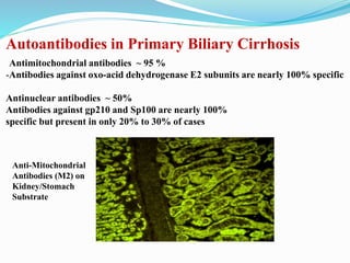
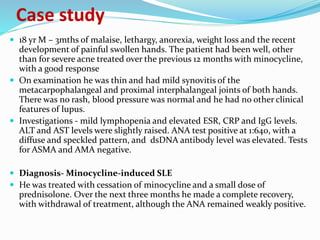
Autoimmune Diseases-
Diagnostic Challenges & Reporting
Dr Rajesh V Bendre
MD(PATH), DNB(PATH), DPB
Chief Pathologist & Head of Laboratory & Blood Bank Services,
Jaslok Hospital & Research Centre, Mumbai
Immunofluoroscence Microscopy & Lab Challenges
AUTOIMMUNITY VS. AUTOIMMUNE DISEASE
Autoimmunity
 Existence of harm-less self-
reactive lymphocytes and
antibodies
 Potentially reversible
 Incidence higher in older
age
 Significance unclear,
possibly physiological
 Antibody are usually IgM
with low avidity
Autoimmune disease
 Dependent on genetic viral
and hormonal factors
 Features of severe tissue
damage
 Clinical symptoms
 Protracted course but
usually fatal
 Familiar clustering
 Antibody are often IgG
with high avidity
AUTOIMMUNE DISEASES
Organ-Specific Systemic
Hashimoto thyroiditis Systemic lupus erythematosus
Autoimmune hemolytic
anemia
Rheumatoid arthritis
Autoimmune atrophic
gastritis
of pernicious anemia
Sjögren syndrome
Multiple sclerosis Reiter syndrome
Autoimmune orchitis Inflammatory myopathies
Goodpasture syndrome Systemic sclerosis (scleroderma)
Autoimmune
thrombocytopenia
Polyarteritis nodosa
Insulin-dependent diabetes
mellitus
Ulcerative colitis
Myasthenia gravis
Graves' disease
Primary biliary cirrhosis
Autoimmune (chronic active)
hepatitis
2. Specific Autoantibody Reactivity
– ENA-
Test methods- ELISA, CLIA,
RIA, Immunoelectrophoresis
and Immunoblotting.
ELISA (monospecific with
purified antigen) &
Immunoblotting are used for
antibodies to DNA, Sm Ag,
U1RNP, SS-A, SS-B, and Scl-70.
Antithyroid antibodies are
usually done by CLIA.
Acetylcholine receptor & MUSK
antibodies are diagnosed using
immunoprecipitation reaction by
RIA.
Laboratory Diagnosis
1. Screening Tests for detection of
systemic autoimmnune diseases for the
last four decades has been the
detection of autoantibodies using
Indirect Immunoflorescence(IIF)
technique with whole cell substrates
eg. ANA, DNA, ANCA
Antinuclear antibodies (ANA) using
HEP-2 cells includes most nuclear and
cytoplasmic antigens, this test can
provide much information regarding
the possible autoantibody system(s)
present, given the pattern of reactivity
seen in the positive ANA.
This can then be utilized to direct
which additional tests the clinician
should order.
ANA by Indirect Immunofluorescence
Uses Hep2 cells –
Detects ANA of clinical relevance
Titers asssociation with clinical relevance
Reduce fading,special Mounting Medium
Optimum Antigen Expression
Significant Mitotic Cells
Large Nuclear size
Pattern correlates with the antibody
specificity and direct immunofluorescence
findings in the skin biopsy material.
Guidelines for the Laboratory Use of Autoantibody Tests
 Test for antinuclear and anticytoplasmic
antibodies (ANA) only in patients with
symptoms of autoimmune rheumatic disease
(ARD),
 Weak ANA reactivity may be present in many
non-rheumatic patients and even in “healthy”
control subjects (upto 15%).
 To diagnose ARD, screen for ANA using
indirect immunofluorescence (IIF) on HEp-2
cells, and specify immunohistochemical pattern
(nuclear, cytoplasmic, mitotic) and quantity
(titer, concentration).
 Consider sample screening dilution for ANA
testing as 1:80 or 1:100
 As decision-making levels: negative if <1:80,
low positive from 1:80 to 1:100, and positive if
1:160 or more.
 Do not use ANA titer or concentration to
monitor ARD.
 Use ELISA-ANA screening test only when your
procedure has shown good clinical and analytic
correlation with the IIF method.
Indian J Med Res 126, July 2007, pp 34-38
ANA Patterns
Pattern Ab Directed Against Associations
Homogeneous or
diffuse nuclear
staining
Chromatin
Histones
Occasionally DS DNA
SLE
Drug induced
SLE
Rim or peripheral
staining
DS DNA SLE
Speckled nuclear
pattern
(Fine or Coarse)
Non-DNA nuclear constituents
(Extractable nuclear antigens-ENA):
Sm
Ku
U1RNP
Ro/SS-A
La/SS-B
SCLE, Mixed
connective
tissue disorders
Nucleolar Nucleolar RNA
Systemic
sclerosis
Centromere Anti- centromere
CREST
syndrome
Nuclear dots Sp-100 (nuclear pore proteins)
Primary biliary
cirrhosis, SLE
Cytoplasmic
speckled
Histidyl-tRNA synthetase (Jo-1)
Anti-mitochondrial, anti-golgi
Anti-lysosomal
Myositis
ANA Patterns
Antigen Condition Sensitivity Specificity
Centromere Limited cutaneous systemic sclerosis 65% 99.9%
dsDNA Systemic lupus erythematosus 57% 97%
SS-B/La Sjogren’s, subacute cutaneous lupus
erythematosus, neonatal lupus
syndrome
16-40% 94%
SS-A/Ro Sjogren’s, subacute cutaneous lupus
erythematosus, neonatal lupus
syndrome
8-70% 87%
U3-RNP Scleroderma 12% 96%
Scl-70 Systemic sclerosis 20% 100%
Dense fine speckled
pattern. Mitotic cells
show a fine granular
solid staining. Resting
cells show a very fine,
diffuse speckled stain.
ANA- DFS 70 Pattern
Guidelines for laboratory
- Use of DsDNA Tests
 Two Types of Antibodies
 dSDNA
 Specific for SLE, react with both ssDNA and dsDNA
 occur in up to 70% of SLE patients, Pathogenic antibodies
 ssDNA
 not specific for SLE , recognize purine and pyrimidine basis
 occur in other autoimmune rheumatic disorders
 Test for anti-dsDNA antibody only in ANA-positive patients in whom
SLE is suspected clinically, using the IIF on Crithidia luciliae or ELISA
methods.
 Use the ELISA to monitor anti dsDNA–positive SLE patients
Am J Clin Pathol 2002;117:316-324
Method Antibody
avidity
Sensitivity &
Specificity
Farr assay (RIA) ++++ 44%
96%
CLIFT ++ 27.4%
95.9%
ELISA
-Conventional dsDNA
-- NcX-dsDNA
+++
41.8%, 97.0%
59.8% , 97.0%
Antibody (tested
using ELISA)
Sensitivity
(%)
Specificity
(%)
Disease association
Ro (SS-A) 61 80-93 SLE, Sjogren syndrome
La (SS-B) 27-35 88-97 SLE, Sjogren syndrome
Sm 34-45 88-100 SLE
RNP 39-64 84-97 SLE, MCD
CENP-B 40-45 85-90 scleroderma
Scl-70 47-58 82-93
Scleroderma systemic
sclerosis
Jo-1 42-47 87-94 Myositis
U1-RNP 35-40 85-90 MCD
Histones 75-80 88-95 SLE-Drug induced
DFS-70 New New Non Rheumatic diseases
RNA Polymerase III New New Systemic sclerosis
Antibody (tested
using
Immunoblot)
Sensitivity
(%)
Specificity
(%)
Disease association
Fibrillarin New New Systemic sclerosis
PM-scl New New Myositis
Pl-12 New New Myositis
Guidelines for laboratory
- Use of ENA Tests
ENA
Major clinical
associations
CIEP ELISA IB
CommentsSens Spec Sens Spec Sens Spec
SS-A Sjogren’s syn 85-95 50-60 90-97 45-50 70-85 40-50 Conformational epitopes on 52- and 60-kDa SS-A
antigen best detected by CIEP; IB is unreliable
because of denaturation of epitopes by SDS-PAGESLE 25-30 50-60 35-60 45-50 10-15 40-50
SS-B Sjogren’s syn 70-80 60-70 75-85 50-60 90-95 55-65 Linear epitopes on 48-kDa SS-B best detected by
IB; CIEP less sensitive but more specificSLE 10-15 50-55 20-30 45-50 30-35 40-50
Sm SLE 30-35 98-100 35-50 95-99 30-35 95-99
ELISA specificity may be improved by use of
highly purified or recombinant antigens;
U1 RNP Mixed CTD 90-95 60-75 95-98 50-60 80-85 55-65 ELISA specificity may be improved by use of
highly purified or recombinant antigens; IB is more
specific than ELISASLE 15-35 55-75 50-60 50-55 30-40 55-70
Scl-70 Scleroderma 25-35 95-99 30-45 80-90 40-55 90-95
CIEP is less sensitive because of the low negative
charge on Scl-70 antigen at pH 8.0-8.4
SLE 0-5 0-5 20-25 15-25 10-20 5-10
Anti-Scl-70 antibodies detected by ELISA in SLE
may not be “false positives”; rather, may identify a
subgroup of patients at high risk of pulmonary
hypertension and renal disease
Jo-1 PM/DM 25-40 95-99 35-45 90-95 60-90 95-99
Anti-Jo-1 antibodies stain the cytoplasm of HEp-2
cells and may be reported as“ANA negative”
Guidelines for laboratory
- Use of ENA Tests
Laboratory Diagnosis- Approach
Classification of Vasculitis
Medium-vessels
• Polyarteritis nodosa
• Kawasaki’s disease
ANCA-Associated Vasculitis
• Wegener’s granulomatosis
• Microscopic polyangiitis
• Churg-Strauss syndrome
• Drug induced
Non-ANCAAssociated Vasculitis
• Immune-complex small-vessel
vasculitis
• Henoch-Schönlein purpura
• Cryoglobulinemic vasculitis
• Cutaneous leukocytoclastic
vasculitis
• Goodpasture’s syndrome
Vasculitis:
Small-vessels Large-vessels
• Giant cell arteritis
• Takayasu’s disease
ANCAAntigens/Fixation
(Atypical)
p-ANCA
(Classical)
p-ANCAc-ANCA
ANCA Patterns
Wegener‘s granulomatosis
Microscopic polyangiitis
Wegener‘s granulomatosis
Microscopic polyangiitis
Ulcerative colitis
Primay sclerosing cholangitis
Autoimmune hepatitis
ANCA- International Consensus
Statement
Laboratories screen for ANCA by indirect IF, and that
any sera with cytoplamic or perinuclear
fluorescence or nuclear fluorescence that might
mask an ANCA should be tested for MPO and
PR3 by ELISA. Am J Clin Pathol. 1999;111:507-13
When an autoimmune liver disease is suspected based on clinical, biochemical or histological pattern, autoantibody
testing of ANA, ASMA, LKM-1 and AMA can clearly help with the diagnosis. Titres can vary throughout the course of
the disease and, therefore, a negative test or a low titre should not exclude a diagnosis; repeat testing may be appropriate
during the initial workup. In adults, a titre of 1:40 or greater is considered positive for pathology, whereas in children, a
cut-off of 1:20 or greater for ANA and ASMA, and 1:10 or greater for LKM-1 are considered positive
Autoimmune Hepatitis
Smooth Muscle Antibodies
SMA of the VGT pattern is considered specific for AIH-
1.
Actin: a globular protein and exists as either G-actin
(monomeric) 46kD or F-actin (polymerized into
filaments) 34kD
F - actin: the biologically active form and is the
specific autoantigen of autoimmune hepatitis
Anti-smooth muscle F-actin antibodies are
predominantly IgG
Other cytoplasmic antibodies creating interference in
ASMA reporting (often related to G-actin)– vimentin,
tropomyosin, endomysium, elastic fibres
New ASMA mosaic slide includes VSM cells specific for
F-actin
Types of LKM Antibodies
Type Immunofluorescence Pattern Antigen Specificity Disease Association
LKM1 Liver; evenly staining cytoplasmic P4502D6, 50kD Autoimmune hepatitis
fluorescence kidney; proximal tubules
LKM2 Liver; same as LKM 1 P4502C9, 50kD TienilicAcid and
Kidney; same as LKM1 associated hepatitis
LKM3 Liver; primate-specific uridine diphosphate Hepatitis D
Kidney; primate-specific glucuronosyltranferases
Kidney Liver
Autoantibodies in Primary Biliary Cirrhosis
Antimitochondrial antibodies ~ 95 %
-Antibodies against oxo-acid dehydrogenase E2 subunits are nearly 100% specific
Antinuclear antibodies ~ 50%
Antibodies against gp210 and Sp100 are nearly 100%
specific but present in only 20% to 30% of cases
Anti-Mitochondrial
Antibodies (M2) on
Kidney/Stomach
Substrate
Case study
 45-yr F presented to GP with 9 mths H/o mild arthralgia predominantly
affecting the proximal interphalangeal joints. She had presented 12 months
earlier with an erythematous rash over her cheeks and nose. The rash was
slightly oily and It seemed to respond to a topical antibiotic. The
development of joint symptoms raised some doubt about the diagnosis of
the rash, which appeared to be confirmed when an ANA test was reported
as positive at a titre of 1:80 with speckled pattern.
 The residual rash was consistent with acne rosacea and the ANA was
thought to be insignificant and coincidental finding.
 The patient was referred to a rheumatologist. O/E- No other clinical
features of lupus. On further Radiological investigations- Early nodal
changes seen on some of the asymptomatic distal interphalangeal joints.
Also patient had strong family history of hand osteoarthritis.
 18 yr M – 3mths of malaise, lethargy, anorexia, weight loss and the recent
development of painful swollen hands. The patient had been well, other
than for severe acne treated over the previous 12 months with minocycline,
with a good response
 On examination he was thin and had mild synovitis of the
metacarpophalangeal and proximal interphalangeal joints of both hands.
There was no rash, blood pressure was normal and he had no other clinical
features of lupus.
 Investigations - mild lymphopenia and elevated ESR, CRP and IgG levels.
ALT and AST levels were slightly raised. ANA test positive at 1:640, with a
diffuse and speckled pattern, and dsDNA antibody level was elevated. Tests
for ASMA and AMA negative.
 Diagnosis- Minocycline-induced SLE
 He was treated with cessation of minocycline and a small dose of
prednisolone. Over the next three months he made a complete recovery,
with withdrawal of treatment, although the ANA remained weakly positive.
Case study
CONCLUSION
 ANA includes many autoantibodies directed towards many nuclear (DNA &
nucleoplasm) & cytoplasmic antigens, which are maximally screened & detected
by using Hep-2 cells in indirect immunofluorescence method, but, not all of these
are always clinically relevant antibodies.
 Thus, there is a possibility of patients having ANA positive by indirect
immunofluorescence method but negative results on immunoblot.
 ANA Immunoblot assays are more sensitive for Ro52/SSb , Scl 70,while poorly
sensitive for dsDNA, hence such patients require further follow-up with
Monspecific Nuclear Antigen ELISAs based on clinical correlation for exact
categorization & diagnosis.
 All weak Positive ANA results- are recommended to be correlated clinically &
repeated after 4-6 weeks OR followed up for further testing with Monospecific
Nuclear Antigen ELISA or Panels for confirmation of specific Autoantibodies.
CONCLUSION
• dsDNA- shift to ELISA along with antinucleosomal antibody
• ANCA- shift to ELISA with purified antigens for MPO, PR3
• ASMA- Report on IIF with F-actin specificity
• AMA,LKM1- Report on IIF using multiple tissue substrate & pattern
recognition
Please remember it is important ‘to treat the
patient and not the serology’.
Autoimmune diseases diagnostic challenges

Autoimmune diseases diagnostic challenges

  • 1.
    Autoimmune Diseases- Diagnostic Challenges& Reporting Dr Rajesh V Bendre MD(PATH), DNB(PATH), DPB Chief Pathologist & Head of Laboratory & Blood Bank Services, Jaslok Hospital & Research Centre, Mumbai
  • 2.
  • 3.
    AUTOIMMUNITY VS. AUTOIMMUNEDISEASE Autoimmunity  Existence of harm-less self- reactive lymphocytes and antibodies  Potentially reversible  Incidence higher in older age  Significance unclear, possibly physiological  Antibody are usually IgM with low avidity Autoimmune disease  Dependent on genetic viral and hormonal factors  Features of severe tissue damage  Clinical symptoms  Protracted course but usually fatal  Familiar clustering  Antibody are often IgG with high avidity
  • 4.
    AUTOIMMUNE DISEASES Organ-Specific Systemic Hashimotothyroiditis Systemic lupus erythematosus Autoimmune hemolytic anemia Rheumatoid arthritis Autoimmune atrophic gastritis of pernicious anemia Sjögren syndrome Multiple sclerosis Reiter syndrome Autoimmune orchitis Inflammatory myopathies Goodpasture syndrome Systemic sclerosis (scleroderma) Autoimmune thrombocytopenia Polyarteritis nodosa Insulin-dependent diabetes mellitus Ulcerative colitis Myasthenia gravis Graves' disease Primary biliary cirrhosis Autoimmune (chronic active) hepatitis
  • 5.
    2. Specific AutoantibodyReactivity – ENA- Test methods- ELISA, CLIA, RIA, Immunoelectrophoresis and Immunoblotting. ELISA (monospecific with purified antigen) & Immunoblotting are used for antibodies to DNA, Sm Ag, U1RNP, SS-A, SS-B, and Scl-70. Antithyroid antibodies are usually done by CLIA. Acetylcholine receptor & MUSK antibodies are diagnosed using immunoprecipitation reaction by RIA. Laboratory Diagnosis 1. Screening Tests for detection of systemic autoimmnune diseases for the last four decades has been the detection of autoantibodies using Indirect Immunoflorescence(IIF) technique with whole cell substrates eg. ANA, DNA, ANCA Antinuclear antibodies (ANA) using HEP-2 cells includes most nuclear and cytoplasmic antigens, this test can provide much information regarding the possible autoantibody system(s) present, given the pattern of reactivity seen in the positive ANA. This can then be utilized to direct which additional tests the clinician should order.
  • 6.
    ANA by IndirectImmunofluorescence Uses Hep2 cells – Detects ANA of clinical relevance Titers asssociation with clinical relevance Reduce fading,special Mounting Medium Optimum Antigen Expression Significant Mitotic Cells Large Nuclear size Pattern correlates with the antibody specificity and direct immunofluorescence findings in the skin biopsy material.
  • 7.
    Guidelines for theLaboratory Use of Autoantibody Tests  Test for antinuclear and anticytoplasmic antibodies (ANA) only in patients with symptoms of autoimmune rheumatic disease (ARD),  Weak ANA reactivity may be present in many non-rheumatic patients and even in “healthy” control subjects (upto 15%).  To diagnose ARD, screen for ANA using indirect immunofluorescence (IIF) on HEp-2 cells, and specify immunohistochemical pattern (nuclear, cytoplasmic, mitotic) and quantity (titer, concentration).  Consider sample screening dilution for ANA testing as 1:80 or 1:100  As decision-making levels: negative if <1:80, low positive from 1:80 to 1:100, and positive if 1:160 or more.  Do not use ANA titer or concentration to monitor ARD.  Use ELISA-ANA screening test only when your procedure has shown good clinical and analytic correlation with the IIF method. Indian J Med Res 126, July 2007, pp 34-38
  • 8.
    ANA Patterns Pattern AbDirected Against Associations Homogeneous or diffuse nuclear staining Chromatin Histones Occasionally DS DNA SLE Drug induced SLE Rim or peripheral staining DS DNA SLE Speckled nuclear pattern (Fine or Coarse) Non-DNA nuclear constituents (Extractable nuclear antigens-ENA): Sm Ku U1RNP Ro/SS-A La/SS-B SCLE, Mixed connective tissue disorders Nucleolar Nucleolar RNA Systemic sclerosis Centromere Anti- centromere CREST syndrome Nuclear dots Sp-100 (nuclear pore proteins) Primary biliary cirrhosis, SLE Cytoplasmic speckled Histidyl-tRNA synthetase (Jo-1) Anti-mitochondrial, anti-golgi Anti-lysosomal Myositis
  • 9.
    ANA Patterns Antigen ConditionSensitivity Specificity Centromere Limited cutaneous systemic sclerosis 65% 99.9% dsDNA Systemic lupus erythematosus 57% 97% SS-B/La Sjogren’s, subacute cutaneous lupus erythematosus, neonatal lupus syndrome 16-40% 94% SS-A/Ro Sjogren’s, subacute cutaneous lupus erythematosus, neonatal lupus syndrome 8-70% 87% U3-RNP Scleroderma 12% 96% Scl-70 Systemic sclerosis 20% 100%
  • 10.
    Dense fine speckled pattern.Mitotic cells show a fine granular solid staining. Resting cells show a very fine, diffuse speckled stain. ANA- DFS 70 Pattern
  • 11.
    Guidelines for laboratory -Use of DsDNA Tests  Two Types of Antibodies  dSDNA  Specific for SLE, react with both ssDNA and dsDNA  occur in up to 70% of SLE patients, Pathogenic antibodies  ssDNA  not specific for SLE , recognize purine and pyrimidine basis  occur in other autoimmune rheumatic disorders  Test for anti-dsDNA antibody only in ANA-positive patients in whom SLE is suspected clinically, using the IIF on Crithidia luciliae or ELISA methods.  Use the ELISA to monitor anti dsDNA–positive SLE patients Am J Clin Pathol 2002;117:316-324 Method Antibody avidity Sensitivity & Specificity Farr assay (RIA) ++++ 44% 96% CLIFT ++ 27.4% 95.9% ELISA -Conventional dsDNA -- NcX-dsDNA +++ 41.8%, 97.0% 59.8% , 97.0%
  • 12.
    Antibody (tested using ELISA) Sensitivity (%) Specificity (%) Diseaseassociation Ro (SS-A) 61 80-93 SLE, Sjogren syndrome La (SS-B) 27-35 88-97 SLE, Sjogren syndrome Sm 34-45 88-100 SLE RNP 39-64 84-97 SLE, MCD CENP-B 40-45 85-90 scleroderma Scl-70 47-58 82-93 Scleroderma systemic sclerosis Jo-1 42-47 87-94 Myositis U1-RNP 35-40 85-90 MCD Histones 75-80 88-95 SLE-Drug induced DFS-70 New New Non Rheumatic diseases RNA Polymerase III New New Systemic sclerosis Antibody (tested using Immunoblot) Sensitivity (%) Specificity (%) Disease association Fibrillarin New New Systemic sclerosis PM-scl New New Myositis Pl-12 New New Myositis Guidelines for laboratory - Use of ENA Tests
  • 13.
    ENA Major clinical associations CIEP ELISAIB CommentsSens Spec Sens Spec Sens Spec SS-A Sjogren’s syn 85-95 50-60 90-97 45-50 70-85 40-50 Conformational epitopes on 52- and 60-kDa SS-A antigen best detected by CIEP; IB is unreliable because of denaturation of epitopes by SDS-PAGESLE 25-30 50-60 35-60 45-50 10-15 40-50 SS-B Sjogren’s syn 70-80 60-70 75-85 50-60 90-95 55-65 Linear epitopes on 48-kDa SS-B best detected by IB; CIEP less sensitive but more specificSLE 10-15 50-55 20-30 45-50 30-35 40-50 Sm SLE 30-35 98-100 35-50 95-99 30-35 95-99 ELISA specificity may be improved by use of highly purified or recombinant antigens; U1 RNP Mixed CTD 90-95 60-75 95-98 50-60 80-85 55-65 ELISA specificity may be improved by use of highly purified or recombinant antigens; IB is more specific than ELISASLE 15-35 55-75 50-60 50-55 30-40 55-70 Scl-70 Scleroderma 25-35 95-99 30-45 80-90 40-55 90-95 CIEP is less sensitive because of the low negative charge on Scl-70 antigen at pH 8.0-8.4 SLE 0-5 0-5 20-25 15-25 10-20 5-10 Anti-Scl-70 antibodies detected by ELISA in SLE may not be “false positives”; rather, may identify a subgroup of patients at high risk of pulmonary hypertension and renal disease Jo-1 PM/DM 25-40 95-99 35-45 90-95 60-90 95-99 Anti-Jo-1 antibodies stain the cytoplasm of HEp-2 cells and may be reported as“ANA negative” Guidelines for laboratory - Use of ENA Tests
  • 14.
  • 16.
    Classification of Vasculitis Medium-vessels •Polyarteritis nodosa • Kawasaki’s disease ANCA-Associated Vasculitis • Wegener’s granulomatosis • Microscopic polyangiitis • Churg-Strauss syndrome • Drug induced Non-ANCAAssociated Vasculitis • Immune-complex small-vessel vasculitis • Henoch-Schönlein purpura • Cryoglobulinemic vasculitis • Cutaneous leukocytoclastic vasculitis • Goodpasture’s syndrome Vasculitis: Small-vessels Large-vessels • Giant cell arteritis • Takayasu’s disease
  • 17.
  • 18.
    (Atypical) p-ANCA (Classical) p-ANCAc-ANCA ANCA Patterns Wegener‘s granulomatosis Microscopicpolyangiitis Wegener‘s granulomatosis Microscopic polyangiitis Ulcerative colitis Primay sclerosing cholangitis Autoimmune hepatitis
  • 19.
    ANCA- International Consensus Statement Laboratoriesscreen for ANCA by indirect IF, and that any sera with cytoplamic or perinuclear fluorescence or nuclear fluorescence that might mask an ANCA should be tested for MPO and PR3 by ELISA. Am J Clin Pathol. 1999;111:507-13
  • 20.
    When an autoimmuneliver disease is suspected based on clinical, biochemical or histological pattern, autoantibody testing of ANA, ASMA, LKM-1 and AMA can clearly help with the diagnosis. Titres can vary throughout the course of the disease and, therefore, a negative test or a low titre should not exclude a diagnosis; repeat testing may be appropriate during the initial workup. In adults, a titre of 1:40 or greater is considered positive for pathology, whereas in children, a cut-off of 1:20 or greater for ANA and ASMA, and 1:10 or greater for LKM-1 are considered positive Autoimmune Hepatitis
  • 21.
    Smooth Muscle Antibodies SMAof the VGT pattern is considered specific for AIH- 1. Actin: a globular protein and exists as either G-actin (monomeric) 46kD or F-actin (polymerized into filaments) 34kD F - actin: the biologically active form and is the specific autoantigen of autoimmune hepatitis Anti-smooth muscle F-actin antibodies are predominantly IgG Other cytoplasmic antibodies creating interference in ASMA reporting (often related to G-actin)– vimentin, tropomyosin, endomysium, elastic fibres New ASMA mosaic slide includes VSM cells specific for F-actin
  • 22.
    Types of LKMAntibodies Type Immunofluorescence Pattern Antigen Specificity Disease Association LKM1 Liver; evenly staining cytoplasmic P4502D6, 50kD Autoimmune hepatitis fluorescence kidney; proximal tubules LKM2 Liver; same as LKM 1 P4502C9, 50kD TienilicAcid and Kidney; same as LKM1 associated hepatitis LKM3 Liver; primate-specific uridine diphosphate Hepatitis D Kidney; primate-specific glucuronosyltranferases Kidney Liver
  • 23.
    Autoantibodies in PrimaryBiliary Cirrhosis Antimitochondrial antibodies ~ 95 % -Antibodies against oxo-acid dehydrogenase E2 subunits are nearly 100% specific Antinuclear antibodies ~ 50% Antibodies against gp210 and Sp100 are nearly 100% specific but present in only 20% to 30% of cases Anti-Mitochondrial Antibodies (M2) on Kidney/Stomach Substrate
  • 24.
    Case study  45-yrF presented to GP with 9 mths H/o mild arthralgia predominantly affecting the proximal interphalangeal joints. She had presented 12 months earlier with an erythematous rash over her cheeks and nose. The rash was slightly oily and It seemed to respond to a topical antibiotic. The development of joint symptoms raised some doubt about the diagnosis of the rash, which appeared to be confirmed when an ANA test was reported as positive at a titre of 1:80 with speckled pattern.  The residual rash was consistent with acne rosacea and the ANA was thought to be insignificant and coincidental finding.  The patient was referred to a rheumatologist. O/E- No other clinical features of lupus. On further Radiological investigations- Early nodal changes seen on some of the asymptomatic distal interphalangeal joints. Also patient had strong family history of hand osteoarthritis.
  • 25.
     18 yrM – 3mths of malaise, lethargy, anorexia, weight loss and the recent development of painful swollen hands. The patient had been well, other than for severe acne treated over the previous 12 months with minocycline, with a good response  On examination he was thin and had mild synovitis of the metacarpophalangeal and proximal interphalangeal joints of both hands. There was no rash, blood pressure was normal and he had no other clinical features of lupus.  Investigations - mild lymphopenia and elevated ESR, CRP and IgG levels. ALT and AST levels were slightly raised. ANA test positive at 1:640, with a diffuse and speckled pattern, and dsDNA antibody level was elevated. Tests for ASMA and AMA negative.  Diagnosis- Minocycline-induced SLE  He was treated with cessation of minocycline and a small dose of prednisolone. Over the next three months he made a complete recovery, with withdrawal of treatment, although the ANA remained weakly positive. Case study
  • 26.
    CONCLUSION  ANA includesmany autoantibodies directed towards many nuclear (DNA & nucleoplasm) & cytoplasmic antigens, which are maximally screened & detected by using Hep-2 cells in indirect immunofluorescence method, but, not all of these are always clinically relevant antibodies.  Thus, there is a possibility of patients having ANA positive by indirect immunofluorescence method but negative results on immunoblot.  ANA Immunoblot assays are more sensitive for Ro52/SSb , Scl 70,while poorly sensitive for dsDNA, hence such patients require further follow-up with Monspecific Nuclear Antigen ELISAs based on clinical correlation for exact categorization & diagnosis.  All weak Positive ANA results- are recommended to be correlated clinically & repeated after 4-6 weeks OR followed up for further testing with Monospecific Nuclear Antigen ELISA or Panels for confirmation of specific Autoantibodies.
  • 27.
    CONCLUSION • dsDNA- shiftto ELISA along with antinucleosomal antibody • ANCA- shift to ELISA with purified antigens for MPO, PR3 • ASMA- Report on IIF with F-actin specificity • AMA,LKM1- Report on IIF using multiple tissue substrate & pattern recognition Please remember it is important ‘to treat the patient and not the serology’.