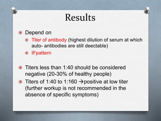
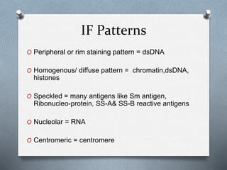
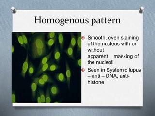
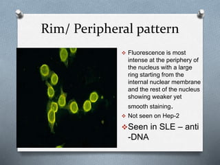
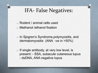
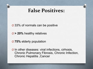
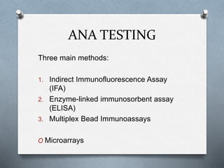
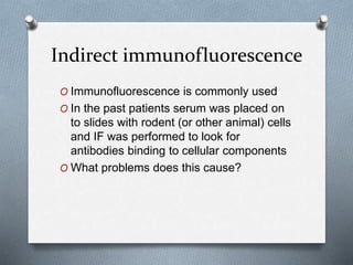
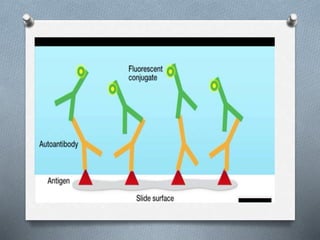
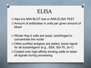
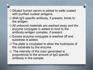
ANA IN
AUTOIMMUNITY
- Dr. Anamika.
D
Autoantibodies
O Directed against
O Components of the cell surface,
O Cytoplasm components
O Nuclear components
 Antibodies to cell nucleus component
ANA, anti-dsDNA, antibodies to extractable
nuclear antigen (ENA), (anti-Sm, anti-RNP)
 Antibodies to cytoplasmic antigens
anti-SSA, ANCA
 Cell-specific autoantibodies
lymphocytotoxic antibodies, anti-neurone
antibodies, anti-erythrocyte antibodies, anti-
platelet antibodies
 Antibodies to serum components
antiphospholipid antibody,antiglobulin,
rheumatoid factor
Anti-nuclear Antibodies
O Also known as antinuclear factor or ANF
O 4 types:
O Anti- nuclear antibodies to DNA
O ANA to histones
O ANA to non-histone proteins bound to
RNA
O ANA to nucleolar antigens
Many sub-types
 anti-Ro/SS-A antibodies Sjogrens syndrome
 anti-La/SS-B antibodies
 anti-dsDNA antibodies
 anti-Sm antibodies SLE
 anti-nRNP antibodies – Mixed connective
tissue disease
 anti-Scl-70 antibodies - scleroderma
 Anti-Jo-1 antibodies – Polymyositis and
dermatomyositis
 anti-histone antibodies – drug induced lupus
 anti-centromere antibodies – CREST syndrome
 anti-sp100 antibodies – primary biliary
cirrhosis
ANA TESTING
Three main methods:
1. Indirect Immunofluorescence Assay
(IFA)
2. Enzyme-linked immunosorbent assay
(ELISA)
3. Multiplex Bead Immunoassays
O Microarrays
Sample preparation
O Collect blood specimens, usually from arm
of pt , at any time.
O No special instructions given
O Separate the serum.
O Specimens may be refrigerated at 2–8°C
for up to seven days or frozen for up to six
months. Avoid repetitive freezing and
thawing.
Indirect immunofluorescence
O Immunofluorescence is commonly used
O In the past patients serum was placed on
to slides with rodent (or other animal) cells
and IF was performed to look for
antibodies binding to cellular components
O What problems does this cause?
O Human and rodent cells differ (slightly),
and so some people with obvious
rheumatic disease would be negative on
this test. “ANA-negative lupus”
O Now there are human tumor cell lines that
are used (HEp-2 are preferred)
How is the test done?
O Whole HEp- 2 cells containing these antigens are attached to
microscope slide (cells fixed into separate dots on the slide)
O Patient serum is diluted and dropped onto HEp-2 slides
O If antibody is resent , it binds to antigen on Hep -2 cells
O Incubate for 20 mins at room temerature.
O Wash to remove unreacted antibody.
O Add secondary antibody -anti-human globulin labeled with
fluorescent tag or enzyme bind to pts antibody
characteristic fluorescent pattern
O Read using an IF scope
O Another source of false negatives includes
how the tissues are fixed onto the slides
O Ethanol and methanol fixation may
remove Ro/SSA antigens from cells, so
the cells are fixed with acetone
Results
 Depend on
 Titer of antibody (highest dilution of serum at which
auto- antibodies are still deectable)
 IFpattern
 Titers less than 1:40 should be considered
negative (20-30% of healthy people)
 Titers of 1:40 to 1:160 positive at low titer
(further workup is not recommended in the
absence of specific symptoms)
Results
O Titers equal to or greater than 1:160
positive
IF Patterns
O Peripheral or rim staining pattern = dsDNA
O Homogenous/ diffuse pattern = chromatin,dsDNA,
histones
O Speckled = many antigens like Sm antigen,
Ribonucleo-protein, SS-A& SS-B reactive antigens
O Nucleolar = RNA
O Centromeric = centromere
Ro
La
Smith
RNP
Jo-1
Scl-70
Ro
Nucleolar
dsDNA
Rim
Speckled
Homogenous
Nucleosomes
Ro
La
Smith
RNP
Jo-1
Scl-70
Ro
dsDNA
Nucleolar proteins
Homogenous pattern
 Smooth, even staining
of the nucleus with or
without
apparent masking of
the nucleoli
 Seen in Systemic lupus
– anti – DNA, anti-
histone
Rim/ Peripheral pattern
 Fluorescence is most
intense at the periphery of
the nucleus with a large
ring starting from the
internal nuclear membrane
and the rest of the nucleus
showing weaker yet
smooth staining.
 Not seen on Hep-2
Seen in SLE – anti
-DNA
Nucleolar pattern
O 23 or 46 (or some
multiple of 46) bright
speckles or ovoid
granules spread over
the nucleus of
interphase cells
O Seen in Diffuse
Scleroderma
O Scl-70
Speckled pattern
O Large speckles
covering the whole
nucleoplasm,
interconnected by a
fine fluorescent
network.
O m/c observed
O Least specific
O Seen in sjogrens
syndrome, SLE,
polymyositis,
dermtomyositis
Patterns
IFA- False Negatives:
o Rodent / animal cells used
o Methanol /ethanol fixation
o In Sjogren’s Syndrome,polymyositis, and
dermatomyositis (ANA -ve in >50%)
o If single antibody, at very low level, is
present: - SSA, subacute cutaneous lupus
- dsDNA, ANA negative lupus
False Positives:
O 33% of normals can be positive
O > 20% healthy relatives
O 75% elderly population
O In other diseases: viral infections, cirhosis,
Chronic Pulmonary Fibrosis, Chronic Infection,
Chronic Hepatitis ,Cancer
ELISA
O Also k/a ANA BLOT test or ANA ELISA TEST
O Amount of antibodies in units per given amount of
blood
O Whole Hep-2 cells are lysed, centrifuged to
concentrate the nuclei
O Other purified antigens are added, boost signal
for all autoantigens (e.g., SSA, Scl-70, Jo-1)
O Coated onto high-affinity binding wells to retain
all signals during processing
O Diluted human serum is added to wells coated
with purified nuclear antigens.
O ANA IgG specific antibody, if present, binds to
the antigen.
O All unbound materials are washed away and the
enzyme conjugate is added to bind to the
antibody-antigen complex, if present.
O Excess enzyme conjugate is washed off and
substrate is added.
O The plate is incubated to allow the hydrolysis of
the substrate by the enzyme.
O The intensity of the color generated is
proportional to the amount of IgG specific
antibody in the sample.
O ELISA detects the same ANA antibodies
as IFA, with improved sensitivity/specificity
O Results in one determination (no
repeating to titrate)
O Objective, automatable, requires no
specialized training
O ELISA reports out Index Values
Multiplex Bead Immunoassay
O Individual antigens, cell components are
coated onto multiple beads
O All activities are detected and identified in
parallel
O High cost of test and automation
To summarize…
O You screen for ANAs using IF on slides
with HEp-2 cells
O If it’s positive look for the specific
antigen using ELISA
O We don’t screen for ANAs using ELISA
because it’s hard to get all the various
antigens (40+) onto the well walls
ANA in autoimmunity by DR. ANAMIKA DEV

ANA in autoimmunity by DR. ANAMIKA DEV

  • 1.
  • 2.
    Autoantibodies O Directed against OComponents of the cell surface, O Cytoplasm components O Nuclear components
  • 3.
     Antibodies tocell nucleus component ANA, anti-dsDNA, antibodies to extractable nuclear antigen (ENA), (anti-Sm, anti-RNP)  Antibodies to cytoplasmic antigens anti-SSA, ANCA  Cell-specific autoantibodies lymphocytotoxic antibodies, anti-neurone antibodies, anti-erythrocyte antibodies, anti- platelet antibodies  Antibodies to serum components antiphospholipid antibody,antiglobulin, rheumatoid factor
  • 4.
    Anti-nuclear Antibodies O Alsoknown as antinuclear factor or ANF O 4 types: O Anti- nuclear antibodies to DNA O ANA to histones O ANA to non-histone proteins bound to RNA O ANA to nucleolar antigens
  • 5.
    Many sub-types  anti-Ro/SS-Aantibodies Sjogrens syndrome  anti-La/SS-B antibodies  anti-dsDNA antibodies  anti-Sm antibodies SLE  anti-nRNP antibodies – Mixed connective tissue disease  anti-Scl-70 antibodies - scleroderma  Anti-Jo-1 antibodies – Polymyositis and dermatomyositis  anti-histone antibodies – drug induced lupus  anti-centromere antibodies – CREST syndrome  anti-sp100 antibodies – primary biliary cirrhosis
  • 6.
    ANA TESTING Three mainmethods: 1. Indirect Immunofluorescence Assay (IFA) 2. Enzyme-linked immunosorbent assay (ELISA) 3. Multiplex Bead Immunoassays O Microarrays
  • 7.
    Sample preparation O Collectblood specimens, usually from arm of pt , at any time. O No special instructions given O Separate the serum. O Specimens may be refrigerated at 2–8°C for up to seven days or frozen for up to six months. Avoid repetitive freezing and thawing.
  • 8.
    Indirect immunofluorescence O Immunofluorescenceis commonly used O In the past patients serum was placed on to slides with rodent (or other animal) cells and IF was performed to look for antibodies binding to cellular components O What problems does this cause?
  • 9.
    O Human androdent cells differ (slightly), and so some people with obvious rheumatic disease would be negative on this test. “ANA-negative lupus” O Now there are human tumor cell lines that are used (HEp-2 are preferred)
  • 10.
    How is thetest done? O Whole HEp- 2 cells containing these antigens are attached to microscope slide (cells fixed into separate dots on the slide) O Patient serum is diluted and dropped onto HEp-2 slides O If antibody is resent , it binds to antigen on Hep -2 cells O Incubate for 20 mins at room temerature. O Wash to remove unreacted antibody. O Add secondary antibody -anti-human globulin labeled with fluorescent tag or enzyme bind to pts antibody characteristic fluorescent pattern O Read using an IF scope
  • 14.
    O Another sourceof false negatives includes how the tissues are fixed onto the slides O Ethanol and methanol fixation may remove Ro/SSA antigens from cells, so the cells are fixed with acetone
  • 15.
    Results  Depend on Titer of antibody (highest dilution of serum at which auto- antibodies are still deectable)  IFpattern  Titers less than 1:40 should be considered negative (20-30% of healthy people)  Titers of 1:40 to 1:160 positive at low titer (further workup is not recommended in the absence of specific symptoms)
  • 16.
    Results O Titers equalto or greater than 1:160 positive
  • 17.
    IF Patterns O Peripheralor rim staining pattern = dsDNA O Homogenous/ diffuse pattern = chromatin,dsDNA, histones O Speckled = many antigens like Sm antigen, Ribonucleo-protein, SS-A& SS-B reactive antigens O Nucleolar = RNA O Centromeric = centromere
  • 18.
  • 19.
    Homogenous pattern  Smooth,even staining of the nucleus with or without apparent masking of the nucleoli  Seen in Systemic lupus – anti – DNA, anti- histone
  • 20.
    Rim/ Peripheral pattern Fluorescence is most intense at the periphery of the nucleus with a large ring starting from the internal nuclear membrane and the rest of the nucleus showing weaker yet smooth staining.  Not seen on Hep-2 Seen in SLE – anti -DNA
  • 21.
    Nucleolar pattern O 23or 46 (or some multiple of 46) bright speckles or ovoid granules spread over the nucleus of interphase cells O Seen in Diffuse Scleroderma O Scl-70
  • 23.
    Speckled pattern O Largespeckles covering the whole nucleoplasm, interconnected by a fine fluorescent network. O m/c observed O Least specific O Seen in sjogrens syndrome, SLE, polymyositis, dermtomyositis
  • 25.
  • 26.
    IFA- False Negatives: oRodent / animal cells used o Methanol /ethanol fixation o In Sjogren’s Syndrome,polymyositis, and dermatomyositis (ANA -ve in >50%) o If single antibody, at very low level, is present: - SSA, subacute cutaneous lupus - dsDNA, ANA negative lupus
  • 27.
    False Positives: O 33%of normals can be positive O > 20% healthy relatives O 75% elderly population O In other diseases: viral infections, cirhosis, Chronic Pulmonary Fibrosis, Chronic Infection, Chronic Hepatitis ,Cancer
  • 28.
    ELISA O Also k/aANA BLOT test or ANA ELISA TEST O Amount of antibodies in units per given amount of blood O Whole Hep-2 cells are lysed, centrifuged to concentrate the nuclei O Other purified antigens are added, boost signal for all autoantigens (e.g., SSA, Scl-70, Jo-1) O Coated onto high-affinity binding wells to retain all signals during processing
  • 29.
    O Diluted humanserum is added to wells coated with purified nuclear antigens. O ANA IgG specific antibody, if present, binds to the antigen. O All unbound materials are washed away and the enzyme conjugate is added to bind to the antibody-antigen complex, if present. O Excess enzyme conjugate is washed off and substrate is added. O The plate is incubated to allow the hydrolysis of the substrate by the enzyme. O The intensity of the color generated is proportional to the amount of IgG specific antibody in the sample.
  • 30.
    O ELISA detectsthe same ANA antibodies as IFA, with improved sensitivity/specificity O Results in one determination (no repeating to titrate) O Objective, automatable, requires no specialized training O ELISA reports out Index Values
  • 31.
    Multiplex Bead Immunoassay OIndividual antigens, cell components are coated onto multiple beads O All activities are detected and identified in parallel O High cost of test and automation
  • 32.
    To summarize… O Youscreen for ANAs using IF on slides with HEp-2 cells O If it’s positive look for the specific antigen using ELISA O We don’t screen for ANAs using ELISA because it’s hard to get all the various antigens (40+) onto the well walls