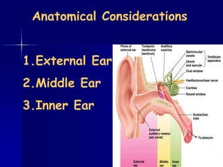
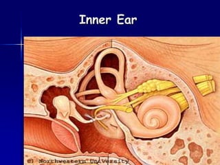
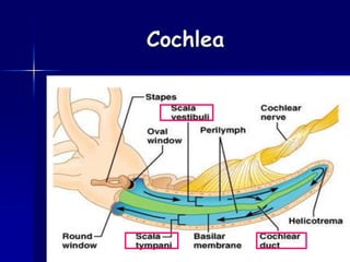
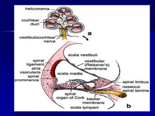
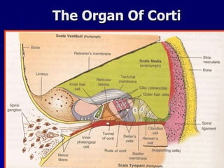
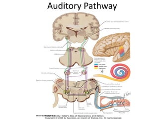
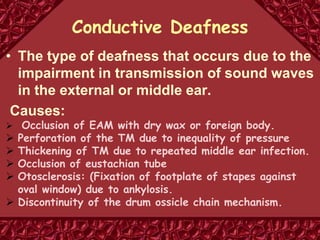
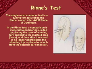
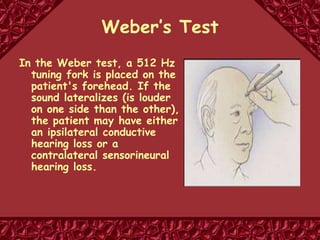
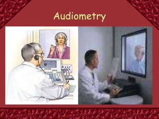
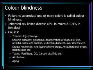
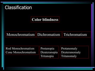
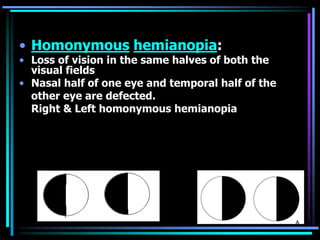
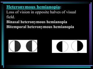
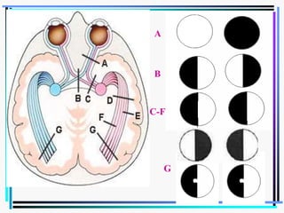
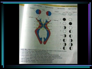
receptor organ
for
hearing and
equilibrium
Anatomical Considerations
1.External Ear
2.Middle Ear
3.Inner Ear
EXTERNAL EAR
 Auricle or Pinna -
Collection and funnelling sound
Localization of sound
 External auditory canal –
Amplification of sound
Protection to tympanic membrane
Middle Ear
Comprises of
 Tympanic membrane
 2 Muscles-Tensor tympani
Stapedius
 3 Ossicles – Malleus
Incus
Stapes
 Eustachian tube
 Nerves,blood vessels,
 lymphatics
Middle Ear Functions
 Impedance matching
 Protection of inner ear
structures
 Static pressure equilibration
 Preferential route of
conduction
 Acts as physiological filter
The Eustachian tube functions
to equalize pressure
Inner Ear
Inner Ear
Cochlea
The Organ Of Corti
Hair Cells
Types of Hair Cells
 Outer hair cell
 Inner hair cell
Inner hair Cell Outer Hair Cell
Number 3,500 20,000
Rows One Three
Shape Flask shaped Cylindrical
Nerve supply Primarily
afferent,very
few efferent
Mainly
efferent,very
few afferent
Function Transmit
auditory
stimuli
Modulate
function of IHC
Auditory pathway
Comprise:
 Spiral ganglion
 Superior olivary nucleus, trapezoid nucleus
and nucleus of lateral lemniscus
 Inferior colliculus
 Medial geniculate body
 Auditory cortex
Spiral ganglion
FIRST ORDER NEURONS
Bipolar cells of the spiral ganglion
Situated in Rosenthal’s canal (Canal running along
osseous spiral lamina)
 Dendrites:
Afferent fibres innervating the hair cells
 Axons:
Cochlear division of VIIIth cranial nerve
Ends in cochlear nuclei in medulla
Cochlear nuclei
SECOND ORDER NEURONS
Two cochlear nuclei:
Dorsal cochlear nuclei
Ventral cochlear nuclei
Axons of 2nd order neurons pass medially in the dorsal part of
pons
Most of them cross to opposite side, but some remain
uncrossed
• Trapezoid body-The crossing fibres of two sides form a
conspicuous mass of fibres
• Some crossing fibres run separately and do not form
trapezoid body
Superior olivary complex, Trapezoid
nucleus and nucleus of lateral lemniscus
THIRD ORDER NEURONS
 Superior olivary nuclear complex
Receive the fibres from the cochlear nuclei
Axons arising from the superior olivary complex- Lateral
lemniscus
 Trapezoid nucleus
Some cochlear fibres that donot relay in superior olivary
nucleus join lateral lemniscus after relaying in trapezoid
nucleus
 Nucleus of lateral lemniscus
Collections of cell that lie within the lemniscus itself
Some cochlear fibres relay in these cells
The fibres of lateral lemniscus ascend to the mid brain
and terminate in the nferior nucleus
Inferior colliculus
FOURTH ORDER NEURONS
Fibres of lateral lemniscus terminate here
Fibres arising in the inferior colliculus enter the
inferior brachium to reach the medial
geniculate body
Medial geniculate body
FIFTH ORDER NEURONS
Fibres from inferior colliculus terminate
Some fibres from the lateral lemniscus reach
this body without relay in inferior colliculus
Fibres arising in MGB form acoustic radiation
which ends in acoustic area of cerebral cortex
Auditory cortex
Present in temporal bone
 Primary auditory cortex (areas 41 and 42)
 Auditory association areas (areas 22, 21 and
20)
Auditory Pathway
Types
 Conductive Deafness
Sensorineural or Nerve Deafness
Deafness
Conductive Deafness
• The type of deafness that occurs due to the
impairment in transmission of sound waves
in the external or middle ear.
Causes:
 Occlusion of EAM with dry wax or foreign body.
 Perforation of the TM due to inequality of pressure
 Thickening of TM due to repeated middle ear infection.
 Occlusion of eustachian tube
 Otosclerosis: (Fixation of footplate of stapes against
oval window) due to ankylosis.
 Discontinuity of the drum ossicle chain mechanism.
Nerve deafness
• Deafness caused by damage of any structure
in cochlea, such as hair cell, organ of Corti,
basilar membrane, cochlear duct or the lesion
in the auditory pathway.
• Causes:
– Degeneration of hair cells due to some antibiotics
like streptomycin & gentamicin.
– Damage of cochlea by prolonged exposure to loud
noise.
– Tumor affecting VIIIth cranial nerve.
Tests For Hearing
• Tuning Fork Tests
1) Rinne’s Test
2) Weber’s Test
• Audiometry
Rinne’s Test
The single most common test is a
tuning fork test called the
Rinne, named after Adolf Rinne
of Gottingen,
In the Rinne test, a comparison is
made between hearing elicited
by placing the base of a tuning
fork applied to the mastoid area
(bone), and then after the sound
is no longer appreciated, the
vibrating top is placed one inch
from the external ear canal (air).
Weber’s Test
In the Weber test, a 512 Hz
tuning fork is placed on the
patient's forehead. If the
sound lateralizes (is louder
on one side than the other),
the patient may have either
an ipsilateral conductive
hearing loss or a
contralateral sensorineural
hearing loss.
Audiometry
Colour blindness
• Failure to appreciate one or more colors is called colour
blindness.
• Inherited sex linked disease (8% in males & 0.4% in
females)
• Causes:
– Trauma: Injury to eye
– Chronic diseases: glaucoma, degeneration of macula of eye,
retinitis, sickle cell anemia, leukemia, diabetes, liver disease etc.
– Drugs: Antibiotics, Anti hypertensive drugs, Antituberulosis drugs,
Barbiturates etc
– Toxins: Fertilizers, CO, Carbon disulfide etc.
– Alcoholism
– Aging
Classification
Color blindness
Monochromatism Dichromatism Trichromatism
Rod Monochromatism
Cone Monochromatism
Protanopia
Deuteranopia
Tritanopia
Protanomaly
Deuteranomaly
Tritanomaly
• Monochromatism: Condition characterized by total inability to
perceive colour. Also called total blindness
– Rod monochromatism: Cones are functionless
– Cone monochromatism: Vision depends upon one single type of cone.
• Dichromatism: Colour blindness in which the subject can
appreciate only two colours.
– Protonopia: defect in receptor of primary color. i.e, Red.
– Deuteranopia: defect in receptor of primary color. i.e, Green
– Tritanopia: defect in receptor of primary color. i.e, Blue
• Trichromatism: Intensity of one of the primary color cannot be
appreciated correctly though the affected persons are able to
perceive all the three colors.
– Protonomaly: Red is weak
– Deuteranomaly: Green is weak
– Tritanomaly: Blue is weak
TESTS FOR COLOR BLINDNESS
• Lantern Test –Edridge-Green’s Lantern
• Holmgren’s Wool Test
• Isochromatic charts – Ishihara chart
Presbyopia
• Age related loss of accommodation
due to decreased elasticity of the
crystalline lens capsule and hardness
of lens
• Corrected by convex lens
Refractive Errors -Ametropia
The inability of the eye to accurately focus
the parallel rays of light coming from
distance (with accomodation at rest) on the
retina
Are focussed either in front or behind the
retina
May be due to imbalance between the
refractive power and axial length of the
eyeball
Refractive Errors
 Myopia
 Hypermetropia
 Astigmatism
Myopia- Nearsightedness
Cause:
 Refractive power is normal.
Antero-posterior diameter of
the eyeball is abnormally long.
Correction:
Biconcave Lens- diverging
lens or minus lens
Nearsightedness (Myopia)
Cause:
• Shortening of eyeball
•Light rays are not
converged to form
clear image.
Correction:
Biconvex lens or
Converging lensor
Plus lens
Farsightedness (Hyperopia)
Astigmatism is a condition in which an abnormal
curvature of the cornea can cause two focal
points to fall in two different locations -
making objects up close and at a distance
appear blurry.
• Visual acuity uneven , part of image blurry
• Cause- cornea or lens has uneven surfaces
• Horizontal and vertical rays focused at different points
• Distorted or blurred vision for far and near
Types of astigmatism
Curvature astigmatism
Index astigmatism
Correction
• By Cylindrical Lenses
Lesions of visual pathway
• Anopia: Loss of vision in one visual field
• Hemianopia: Loss of vision in one half
of visual field
– Homonymous hemianopia
– Heteronymous hemianopia
• Homonymous hemianopia:
• Loss of vision in the same halves of both the
visual fields
• Nasal half of one eye and temporal half of the
other eye are defected.
Right & Left homonymous hemianopia
Heteronymous hemianopia:
Loss of vision in opposite halves of visual
field.
Binasal heteronymous hemianopia
Bitemporal heteronymous hemianopia
A
B
C-F
G
AUDITORY PATHWAY anatomy and physiologys

AUDITORY PATHWAY anatomy and physiologys

  • 1.
  • 2.
  • 4.
    EXTERNAL EAR  Auricleor Pinna - Collection and funnelling sound Localization of sound  External auditory canal – Amplification of sound Protection to tympanic membrane
  • 5.
    Middle Ear Comprises of Tympanic membrane  2 Muscles-Tensor tympani Stapedius  3 Ossicles – Malleus Incus Stapes  Eustachian tube  Nerves,blood vessels,  lymphatics
  • 7.
    Middle Ear Functions Impedance matching  Protection of inner ear structures  Static pressure equilibration  Preferential route of conduction  Acts as physiological filter
  • 8.
    The Eustachian tubefunctions to equalize pressure
  • 9.
  • 10.
  • 11.
  • 13.
  • 14.
    Hair Cells Types ofHair Cells  Outer hair cell  Inner hair cell
  • 15.
    Inner hair CellOuter Hair Cell Number 3,500 20,000 Rows One Three Shape Flask shaped Cylindrical Nerve supply Primarily afferent,very few efferent Mainly efferent,very few afferent Function Transmit auditory stimuli Modulate function of IHC
  • 17.
    Auditory pathway Comprise:  Spiralganglion  Superior olivary nucleus, trapezoid nucleus and nucleus of lateral lemniscus  Inferior colliculus  Medial geniculate body  Auditory cortex
  • 18.
    Spiral ganglion FIRST ORDERNEURONS Bipolar cells of the spiral ganglion Situated in Rosenthal’s canal (Canal running along osseous spiral lamina)  Dendrites: Afferent fibres innervating the hair cells  Axons: Cochlear division of VIIIth cranial nerve Ends in cochlear nuclei in medulla
  • 19.
    Cochlear nuclei SECOND ORDERNEURONS Two cochlear nuclei: Dorsal cochlear nuclei Ventral cochlear nuclei Axons of 2nd order neurons pass medially in the dorsal part of pons Most of them cross to opposite side, but some remain uncrossed • Trapezoid body-The crossing fibres of two sides form a conspicuous mass of fibres • Some crossing fibres run separately and do not form trapezoid body
  • 20.
    Superior olivary complex,Trapezoid nucleus and nucleus of lateral lemniscus THIRD ORDER NEURONS  Superior olivary nuclear complex Receive the fibres from the cochlear nuclei Axons arising from the superior olivary complex- Lateral lemniscus  Trapezoid nucleus Some cochlear fibres that donot relay in superior olivary nucleus join lateral lemniscus after relaying in trapezoid nucleus  Nucleus of lateral lemniscus Collections of cell that lie within the lemniscus itself Some cochlear fibres relay in these cells The fibres of lateral lemniscus ascend to the mid brain and terminate in the nferior nucleus
  • 21.
    Inferior colliculus FOURTH ORDERNEURONS Fibres of lateral lemniscus terminate here Fibres arising in the inferior colliculus enter the inferior brachium to reach the medial geniculate body
  • 22.
    Medial geniculate body FIFTHORDER NEURONS Fibres from inferior colliculus terminate Some fibres from the lateral lemniscus reach this body without relay in inferior colliculus Fibres arising in MGB form acoustic radiation which ends in acoustic area of cerebral cortex
  • 23.
    Auditory cortex Present intemporal bone  Primary auditory cortex (areas 41 and 42)  Auditory association areas (areas 22, 21 and 20)
  • 24.
  • 25.
  • 26.
    Conductive Deafness • Thetype of deafness that occurs due to the impairment in transmission of sound waves in the external or middle ear. Causes:  Occlusion of EAM with dry wax or foreign body.  Perforation of the TM due to inequality of pressure  Thickening of TM due to repeated middle ear infection.  Occlusion of eustachian tube  Otosclerosis: (Fixation of footplate of stapes against oval window) due to ankylosis.  Discontinuity of the drum ossicle chain mechanism.
  • 27.
    Nerve deafness • Deafnesscaused by damage of any structure in cochlea, such as hair cell, organ of Corti, basilar membrane, cochlear duct or the lesion in the auditory pathway. • Causes: – Degeneration of hair cells due to some antibiotics like streptomycin & gentamicin. – Damage of cochlea by prolonged exposure to loud noise. – Tumor affecting VIIIth cranial nerve.
  • 28.
    Tests For Hearing •Tuning Fork Tests 1) Rinne’s Test 2) Weber’s Test • Audiometry
  • 29.
    Rinne’s Test The singlemost common test is a tuning fork test called the Rinne, named after Adolf Rinne of Gottingen, In the Rinne test, a comparison is made between hearing elicited by placing the base of a tuning fork applied to the mastoid area (bone), and then after the sound is no longer appreciated, the vibrating top is placed one inch from the external ear canal (air).
  • 30.
    Weber’s Test In theWeber test, a 512 Hz tuning fork is placed on the patient's forehead. If the sound lateralizes (is louder on one side than the other), the patient may have either an ipsilateral conductive hearing loss or a contralateral sensorineural hearing loss.
  • 31.
  • 32.
    Colour blindness • Failureto appreciate one or more colors is called colour blindness. • Inherited sex linked disease (8% in males & 0.4% in females) • Causes: – Trauma: Injury to eye – Chronic diseases: glaucoma, degeneration of macula of eye, retinitis, sickle cell anemia, leukemia, diabetes, liver disease etc. – Drugs: Antibiotics, Anti hypertensive drugs, Antituberulosis drugs, Barbiturates etc – Toxins: Fertilizers, CO, Carbon disulfide etc. – Alcoholism – Aging
  • 33.
    Classification Color blindness Monochromatism DichromatismTrichromatism Rod Monochromatism Cone Monochromatism Protanopia Deuteranopia Tritanopia Protanomaly Deuteranomaly Tritanomaly
  • 34.
    • Monochromatism: Conditioncharacterized by total inability to perceive colour. Also called total blindness – Rod monochromatism: Cones are functionless – Cone monochromatism: Vision depends upon one single type of cone. • Dichromatism: Colour blindness in which the subject can appreciate only two colours. – Protonopia: defect in receptor of primary color. i.e, Red. – Deuteranopia: defect in receptor of primary color. i.e, Green – Tritanopia: defect in receptor of primary color. i.e, Blue • Trichromatism: Intensity of one of the primary color cannot be appreciated correctly though the affected persons are able to perceive all the three colors. – Protonomaly: Red is weak – Deuteranomaly: Green is weak – Tritanomaly: Blue is weak
  • 35.
    TESTS FOR COLORBLINDNESS • Lantern Test –Edridge-Green’s Lantern • Holmgren’s Wool Test • Isochromatic charts – Ishihara chart
  • 36.
    Presbyopia • Age relatedloss of accommodation due to decreased elasticity of the crystalline lens capsule and hardness of lens • Corrected by convex lens
  • 37.
    Refractive Errors -Ametropia Theinability of the eye to accurately focus the parallel rays of light coming from distance (with accomodation at rest) on the retina Are focussed either in front or behind the retina May be due to imbalance between the refractive power and axial length of the eyeball
  • 38.
    Refractive Errors  Myopia Hypermetropia  Astigmatism
  • 40.
    Myopia- Nearsightedness Cause:  Refractivepower is normal. Antero-posterior diameter of the eyeball is abnormally long. Correction: Biconcave Lens- diverging lens or minus lens
  • 42.
  • 44.
    Cause: • Shortening ofeyeball •Light rays are not converged to form clear image. Correction: Biconvex lens or Converging lensor Plus lens
  • 45.
  • 47.
    Astigmatism is acondition in which an abnormal curvature of the cornea can cause two focal points to fall in two different locations - making objects up close and at a distance appear blurry.
  • 48.
    • Visual acuityuneven , part of image blurry • Cause- cornea or lens has uneven surfaces • Horizontal and vertical rays focused at different points • Distorted or blurred vision for far and near
  • 49.
    Types of astigmatism Curvatureastigmatism Index astigmatism Correction • By Cylindrical Lenses
  • 50.
    Lesions of visualpathway • Anopia: Loss of vision in one visual field • Hemianopia: Loss of vision in one half of visual field – Homonymous hemianopia – Heteronymous hemianopia
  • 51.
    • Homonymous hemianopia: •Loss of vision in the same halves of both the visual fields • Nasal half of one eye and temporal half of the other eye are defected. Right & Left homonymous hemianopia
  • 52.
    Heteronymous hemianopia: Loss ofvision in opposite halves of visual field. Binasal heteronymous hemianopia Bitemporal heteronymous hemianopia
  • 53.