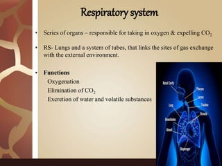
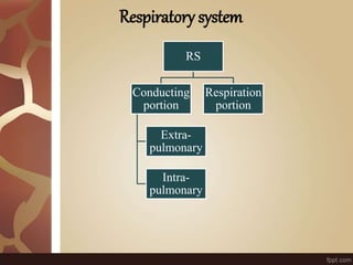
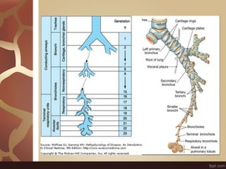
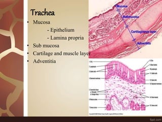
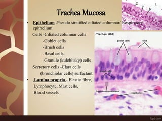
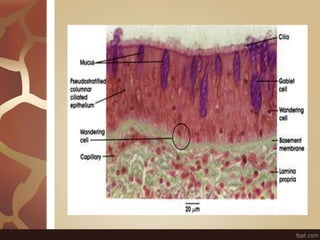
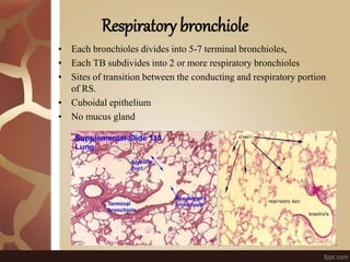
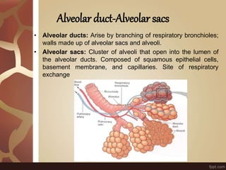
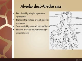
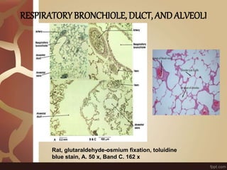
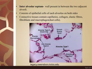
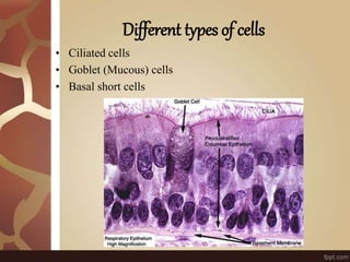
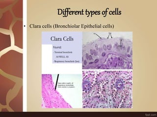
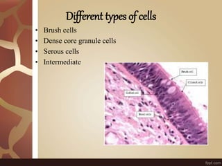
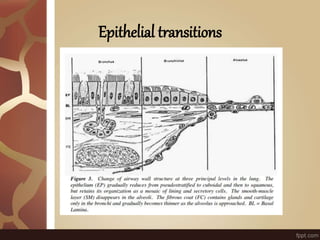
LUNGS-
HISTOLOGY
Gurvinder kaur
P hD
GADVASU
Respiratory system
• Series of organs – responsible for taking in oxygen & expelling CO2
• RS- Lungs and a system of tubes, that links the sites of gas exchange
with the external environment.
• Functions
Oxygenation
Elimination of CO2
Excretion of water and volatile substances
Respiratory system
RS
Conducting
portion
Extra-
pulmonary
Intra-
pulmonary
Respiration
portion
Respiratory systemHistology
• Trachea
• Bronchus
-Primary bronchus
-Secondary bronchus
-Tertiary bronchus
• Bronchiole
• Lung
Trachea
• Mucosa
- Epithelium
- Lamina propria
• Sub mucosa
• Cartilage and muscle layer
• Adventitia
Trachea Mucosa
• Epithelium -Pseudo stratified ciliated columnar/ Respiratory
epithelium
Cells -Ciliated columnar cells
-Goblet cells
-Brush cells
-Basal cells
-Granule (kulchitsky) cells
Secretory cells -Clara cells
(bronchiolar cells) surfactant.
• Lamina propria - Elastic fibre,
Lymphocyte, Mast cells,
Blood vessels
• Sub mucosa- Loose connective tissue
Tracheal glands-Mixed (serous &mucus) glands
Blood vessels and ducts
• Cartilage &smooth muscle layer- ”C” Shaped hyaline cartilage
having perichondrium and chondrocytes
Ends of cartilage connected by smooth muscles
• Adventitia-fibro elastic tissue
BRONCHUS
• Principal bronchus -same as trachea
• Secondary /Lobar bronchus -Irregular hyaline cartilage, Pseudo
stratified ciliated columnar
• Tertiary /Segmental bronchus -Columnar epithelium, Patches of
cartilage
Tertiary/segmental Bronchus
•The number of goblet cells reduced.
•The number of glands reduced.
•The cartilage is present in the irregular plates.
Bronchiole
• The tertiary brochi repeatedly divide, at minimal 10 times and
bronchioles arise, conduct air into pulmonary lobule.
• Bronchioles > 5mm diameter
• No cartilage or glands in mucosa
• Epithelium changes from pseudo-
stratified to cuboidal epithelium.
• Scattered goblet cells initially.
• Clara cells secrete protective proteins.
• Lamina propria smooth muscle
and elastic fibers.Vagus nerve,
sympathetic neuron
Respiratory bronchiole
• Each bronchioles divides into 5-7 terminal bronchioles,
• Each TB subdivides into 2 or more respiratory bronchioles
• Sites of transition between the conducting and respiratory portion
of RS.
• Cuboidal epithelium
• No mucus gland
Alveolar duct-Alveolar sacs
• Alveolar ducts: Arise by branching of respiratory bronchioles;
walls made up of alveolar sacs and alveoli.
• Alveolar sacs: Cluster of alveoli that open into the lumen of
the alveolar ducts. Composed of squamous epithelial cells,
basement membrane, and capillaries. Site of respiratory
exchange
Alveolar duct-Alveolar sacs
• Duct lined by simple squamous
epithelium
• Increase the surface area of gaseous
exchange
• Surrounded by network of capillaries.
• Smooth muscles only at opening of
alveolar ducts
RESPIRATORY BRONCHIOLE, DUCT, ANDALVEOLI
Rat, glutaraldehyde-osmium fixation, toluidine
blue stain, A. 50 x, Band C. 162 x
• Inter alveolar septum- wall present in between the two adjacent
alveoli.
• Consists of epithelial cells of each alveolus on both sides
• Connective tissues contain capillaries, collagen, elastic fibres,
fibroblasts and macrophages(dust cells).
legacy.owensboro.kctcs.edu
Alveolar cells
• Alveoli are thin walled outpouchings , lined by a single layer of
cells.
• Type I alveolar cells.
- Simple squamous cells
adjacent cells joined by tight junctions.
• Type II cells
- Surfactant - Cuboidal cells, scattered among type 1 cells.
Secrete Pulmonary surfactant which lowers the surface tension and
prevents alveoli from collapsing.
• Alveolar macrophages or Dust cells – Remove debris.
Different types of cells
• In progressing from trachea to bronchioles, eight different cell
types are found within the epithelium
Different types of cells
• Ciliated cells
• Goblet (Mucous) cells
• Basal short cells
Different types of cells
• Clara cells (Bronchiolar Epithelial cells)
Different types of cells
• Brush cells
• Dense core granule cells
• Serous cells
• Intermediate
Epithelial transitions
Thank you

Lungs -Histology

  • 1.
  • 2.
    Respiratory system • Seriesof organs – responsible for taking in oxygen & expelling CO2 • RS- Lungs and a system of tubes, that links the sites of gas exchange with the external environment. • Functions Oxygenation Elimination of CO2 Excretion of water and volatile substances
  • 3.
  • 5.
    Respiratory systemHistology • Trachea •Bronchus -Primary bronchus -Secondary bronchus -Tertiary bronchus • Bronchiole • Lung
  • 6.
    Trachea • Mucosa - Epithelium -Lamina propria • Sub mucosa • Cartilage and muscle layer • Adventitia
  • 7.
    Trachea Mucosa • Epithelium-Pseudo stratified ciliated columnar/ Respiratory epithelium Cells -Ciliated columnar cells -Goblet cells -Brush cells -Basal cells -Granule (kulchitsky) cells Secretory cells -Clara cells (bronchiolar cells) surfactant. • Lamina propria - Elastic fibre, Lymphocyte, Mast cells, Blood vessels
  • 9.
    • Sub mucosa-Loose connective tissue Tracheal glands-Mixed (serous &mucus) glands Blood vessels and ducts • Cartilage &smooth muscle layer- ”C” Shaped hyaline cartilage having perichondrium and chondrocytes Ends of cartilage connected by smooth muscles • Adventitia-fibro elastic tissue
  • 10.
    BRONCHUS • Principal bronchus-same as trachea • Secondary /Lobar bronchus -Irregular hyaline cartilage, Pseudo stratified ciliated columnar • Tertiary /Segmental bronchus -Columnar epithelium, Patches of cartilage
  • 11.
    Tertiary/segmental Bronchus •The numberof goblet cells reduced. •The number of glands reduced. •The cartilage is present in the irregular plates.
  • 12.
    Bronchiole • The tertiarybrochi repeatedly divide, at minimal 10 times and bronchioles arise, conduct air into pulmonary lobule. • Bronchioles > 5mm diameter • No cartilage or glands in mucosa • Epithelium changes from pseudo- stratified to cuboidal epithelium. • Scattered goblet cells initially. • Clara cells secrete protective proteins. • Lamina propria smooth muscle and elastic fibers.Vagus nerve, sympathetic neuron
  • 14.
    Respiratory bronchiole • Eachbronchioles divides into 5-7 terminal bronchioles, • Each TB subdivides into 2 or more respiratory bronchioles • Sites of transition between the conducting and respiratory portion of RS. • Cuboidal epithelium • No mucus gland
  • 15.
    Alveolar duct-Alveolar sacs •Alveolar ducts: Arise by branching of respiratory bronchioles; walls made up of alveolar sacs and alveoli. • Alveolar sacs: Cluster of alveoli that open into the lumen of the alveolar ducts. Composed of squamous epithelial cells, basement membrane, and capillaries. Site of respiratory exchange
  • 16.
    Alveolar duct-Alveolar sacs •Duct lined by simple squamous epithelium • Increase the surface area of gaseous exchange • Surrounded by network of capillaries. • Smooth muscles only at opening of alveolar ducts
  • 17.
    RESPIRATORY BRONCHIOLE, DUCT,ANDALVEOLI Rat, glutaraldehyde-osmium fixation, toluidine blue stain, A. 50 x, Band C. 162 x
  • 18.
    • Inter alveolarseptum- wall present in between the two adjacent alveoli. • Consists of epithelial cells of each alveolus on both sides • Connective tissues contain capillaries, collagen, elastic fibres, fibroblasts and macrophages(dust cells). legacy.owensboro.kctcs.edu
  • 19.
    Alveolar cells • Alveoliare thin walled outpouchings , lined by a single layer of cells. • Type I alveolar cells. - Simple squamous cells adjacent cells joined by tight junctions. • Type II cells - Surfactant - Cuboidal cells, scattered among type 1 cells. Secrete Pulmonary surfactant which lowers the surface tension and prevents alveoli from collapsing. • Alveolar macrophages or Dust cells – Remove debris.
  • 20.
    Different types ofcells • In progressing from trachea to bronchioles, eight different cell types are found within the epithelium
  • 21.
    Different types ofcells • Ciliated cells • Goblet (Mucous) cells • Basal short cells
  • 22.
    Different types ofcells • Clara cells (Bronchiolar Epithelial cells)
  • 23.
    Different types ofcells • Brush cells • Dense core granule cells • Serous cells • Intermediate
  • 24.
  • 25.