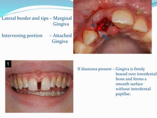
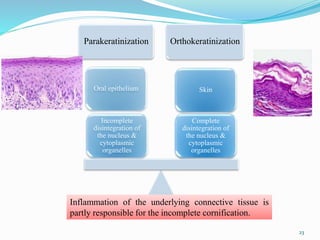
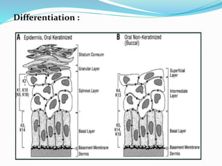
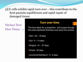
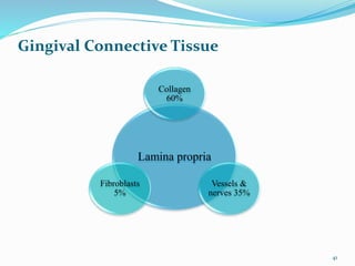
SlideShare a Scribd company logo
1 of 51
By DR . Ayesha
TABLE OF CONTENT
Introduction Changes in
Oral mucosa Older adults
Gingiva Diabetes Mellitus
Macroscopic anatomy Conclusion
Microscopic anatomy References
Epithelium
Lamina Propria
Fiber Apparatus
Introduction
 “Architecture of Periodontal Protection”- Schroeder
INTRODUCTION
Masticatory Mucosa – Gingiva and covering of
the hard palate
Oral Mucosa Specialized Mucosa – Dorsum of the tongue.
Lining Mucosa - Gingiva Part of oral
mucosa that covers the
alveolar processes of the
jaws and surrounds the
necks of teeth.
DEFINITION
Common term – Gums
It is a combination of epithelium and connective tissue and is defined as
that portion of oral mucosa which in complete post eruptive dentition of a
healthy young individual, surrounds and is attached to the teeth and alvoelar
processes.
SCHROEDER
Part of masticatory mucosa covering the alvoelar process and the cervical
portions of the teeth.
LINDHE
The mucous membrane, with supporting fibrous tissue, covering the tooth-
bearing border of the jaw are called as gingiva.
MEDICAL DICTIONARY
ANATOMY OF GINGIVA
Marginal Gingiva
Attached Gingiva
Interdental Gingiva
Marginal Gingiva
Unattached Gingiva
Terminal edge or border surrounding to the collar like
fashion.
Free Gingival Groove
Marginal gingiva demarcated from attached gingiva
by a shallow depression.
“ Line of demarcation between free and attached
gingiva ( Orban 1948 )”
Gingival Zenith
Apical point of marginal gingival scallop.
Apicoronal – 0.06 mm
Mesiodistal – 0.96 mm
Gingival Sulcus
 Shallow crevice or space around the
tooth.
 Bounded by surface of tooth on one
side
 Epithelium lining the free margin of
gingiva.
Depth Of Sulcus
Normal or ideal conditions : 0
( Gottelib and Orban )
Histologically – 1.8 mm
Ranges from 0-6mm
1.5mm (Saito et al)
0.69mm (Eveerts et al)
Histologic Depth
Depth of
penetration
probe
Attached Gingiva
 Continuous with marginal gingiva.
 Firm, resilient and tightly bound to periosteum.
Mucogingival Junction : Facial aspect of attached gingiva
extends to relatively loose and
movable alveolar mucosa.
Width of Attached Gingiva
Distance between mucogingival junction and projection on the external
surface of bottom of gingival sulcus:
Facial aspect: Maxilla Mandible
Incisor Premolar Incisor Premolar
3.5-4.5mm 1.9mm 3.3-3.9mm 1.8mm
Methods to measure gingiva
1)Measurement approach
2)Tension test
3)Roll test
4)By using potassium iodide solution
Width increases with age
in supraerupted teeth
Lingual Aspect of Mandible - terminates at the junction of lingual alveolar
mucosa.
Palatal Surface - in maxilla it blends imperceptibly with equally
firm and resilient palatal mucosa.
Interdental Gingiva
Shape Pyramidal or Col non keratinised
Facial and lingual surfaces tapered
towards interproximal contact area
Mesial and distal – CONCAVE
 Contact relationships between the teeth & the course
of CEJ
 Anterior regions - pyramidal form
 Molar regions - flattened in the buccolingual direction
(sagging pup tent)
Col
 In the histological section, is covered by a thin non-
keratinized epithelium.
Lateral border and tips – Marginal
Gingiva
Intervening portion – Attached
Gingiva
If diastema present – Gingiva is firmly
bound over interdental
bone and forms a
smooth surface
without interdental
papillae.
Tissue Biotypes
 Ochsenbein & Ross
 Scalloped thin & flat thick gingiva
 Claffey & Shanley
 Thin tissue biotype - gingival thickness of <1.5 mm
 Thick tissue biotype - tissue thickness >/+2 mm
 Vertical distance between the interdental bone crest & buccal
bone
 0.9-1.2mm – ideal for predictive periodontal prognosis
18
MICROSCOPIC FEATURES
Overlying - Straited and squamous epithelium Predominantly Cellular
Underlying - Central core connectivity tissue. Less cellular and primarily
collagen fibres.
Gingival Epithelium
Historically Epithelial Component act as physical barrier to
infection.
Recent Epithelial cells play an active role in innate host
defense
Functions and Features of Gingival Epithelium
Architectural Integrity : Cell – cell attachments
Basal Lamina
Keratin Cytoskeleton
Major cell type - Keratinocyte
Other cell type - Langerhans cells,
Melanocytes,
Merkel cells
Constant Renewal - Replacement of
damaged cells.
Cell – cell attachments - Desmosomes,
adherens junctions,
Tight junctions,
gap junctions
Cell Basal Lamina - Synthesis of basal
lamina components
Hemidesmosome
Modified from Dale BA Peridontal 2000
Keratinocyte :
 Principal cell type
 Main function of gingival epithelium is to protect the deep
structures.
 Proliferation of keratinocytes takes place by mitosis in basal
layer and less frequently in suprabasal layers.
 Small portion of cells Proliferative Component
Larger portion of cells Migrate to surface
Parakeratinization Orthokeratinization
Complete
disintegration of
the nucleus &
cytoplasmic
organelles
Skin
Incomplete
disintegration of
the nucleus &
cytoplasmic
organelles
Oral epithelium
Inflammation of the underlying connective tissue is
partly responsible for the incomplete cornification.
23
Differentiation :
Process of Keratinization from the basal layer
 Flatening of the cell with increased
Prevalance of tonofilaments.
 Intercellular junctions coupled to the
production of keratohyalin granules.
 Disappearance of the nucleus
SCHROEDER
Gingival Epithelium
Some areas ( outer ) orthokeratinized
other areas Para or non-keratinized
Intermediate stages of keratinization
Progress to maturity under physiologic or
pathologic condition
 Parakeratinized epithelia The stratum corneum retains pyknotic nuclei
and the keratohyaline granules are dispersed
not giving rise to a stratum granulosum.
 Non–Keratinized epithelium Although cytokeratins are major components,
as in all epithelia
Structural and Metabolic Characteristics of Gingival Epithelium
 Oral Epithelium
Protective in function
 Sulcular Epithelium
 Junctional Epithelium To maintain tissue health
Epithelial cells are not PASSIVE BYSTANDERS
Metabolically active
Capable of reacting to external stimuli
Oral (Outer ) Epithelium : Covers the crest and outer surface of
marginal and attached gingiva.
Prevalence Parakeratinized
1. Stratum Basale
2. Stratum Spinosum
3. Stratum Granulosum
4. Stratum Corneum
Degree of Gingival Keratinization with age and onset of menopause
Most Keratinized : Palate
Least : Gingiva and Ventral aspect of tongue, cheek.
Concentration of glycogen in normal gingiva is inversely related to
degree Of keratinization and inflamation.
Sulcular Epithelium : lines gingival sulcus
thin, non-keratinized stratified squamous epithelium
 No rete pegs
 Lacks stratum granulosum and corneum
 Contains K4, K13 ( Oesophageal type cytokeratin ) and K19.
 Lower degree of enzymes than in outer epithelium.
 Acid phosphate staining negative
 Has the potential to keratinize
If
1. It is reflected and exposed in
oral cavity.
2. Bacterial flora of sulcus is totally
eliminated
 Local irritation of sulcus prevents
sulcular keratinization.
 Semi Permeable
JUNCTIONAL EPITHELIUM
Collar like band of stratified squamous epithelium
0.25mm-1.35mm
Early life 3-4 layers thick
 with age 10 or 20 layers thick
In healthy tissue
tapers from coronal end 10-29 cells
TO
Apical termination 1-2 cells
Two layers
1. Basal layer facing connective
tissue.
2. Suprabasal layer extending to
tooth surface.
J.E = O.E + R.E.E
 Exhibits lower glycolytic enzyme
activity than outer epithelium.
 Lacks acid phosphatase activity.
 Attached to the tooth surface by
Internal basal lamina.
 Attached to gingival connective
tissue by external basal lamina.
Lamina Densa
Internal Basal Lamina
Lamina Lucida ( to which hemidesmosomes
are attached )
Hemidesmosomes :
Firm attachment of cells to internal basal lamina.
Specific sides for signal transduction
Gene expression, cell proliferation, cell differentiation
Organic strands from enamel appear to extend into lamina densa.
 J.E attaches to afibrillar cementum present on the
crown.
 Neutral polysacchrides in zone of epethelial
attachment.
 Dentogingival Unit: J.E + gingival fibers
functional unit
Functions:
1) Epithelial barrier against plaque bacteria.
2) It allows access of gingival fluid, inflammatory cells
components of immunologic host defense to gigival
margin.
3)J.E cells exhibit rapid turn over – this contribute to the
host parasite equilibrium and rapid repair of
damaged tissue.
Various Turn
Over Times:
Development of gingival sulcus: After enamel formation
is complete, enamel is covered by REE.
When tooth penetrates oral mucosa REE unites
with oral epithelium transforms into JEE.
Transformation of REE into JEE proceeds in an apical
direction without interrupting the attachments to the
tooth ------- occurs at 1 to 2 yrs.
SCHROEDER , LISTGARTEN
Continually self renewing with mitotic activity occurring in
all cell layers.
Renewal of gingival epthelium:
 Mitotic activity exhibits 24hrs periodicity.
Highest  morning
(Nonkeratinized areas, gingivitis)
Lowest Evening
Mitotic Rate Buccal mucosa
Hard Palate
Sulcular Epithelium
JE
Outer surface of marginal gingiva
Attached gingiva
Cuticular structures on tooth
 Cuticle: Thin acellular structure.
 Cuticular structures into coatings of developmental
origin and acquired coatings.
LISTGARTEN
 Acquired coatings: Saliva, bacteria, calculus and surface
stains.
 Coatings of developmental origin: REE, coronal
cementum and dental
cuticle.
CUTICLE ---------PRESENT BETWEEN JE & TOOTH
Gingival fluid:
 Transudate or Exudate
 Dignostic or Prognostic Biomaker
 Contents: Connective tissue epithelium, inflammatory cells, serum,
microbial flora inhabiting gingival margin or sulcus.
Healthy---------------------- GCF
Inflammation-------------- GCF
 Functions:
1. Cleanse material from sulcus.
2. Contain plasma proteins.
3. Possess anti microbial properties.
4. Exert antibody activity to defend gingiva.
Lamina propria
Collagen
60%
Vessels &
nerves 35%
Fibroblasts
5%
Gingival Connective Tissue
41
A papillary layer subjacent to the
epithelium  Retepegs.
 Lamina Propria:
Reticular layer  periosteum of
alveolarbone.
 Ground substance fills space between fibers and cells & high
content of water.
composed of proteoglycans, hyaluronic acid, chondrotin sulphate.
Fibers
Collagen
fibers
Reticulum
fibers
Oxytalan
fibers
Elastic fibers
43
 3 types of fibers: Type I (bulk of lamina
propria)
 1) Collagen
 Type IV (branches b/n
collagen Type I)
 2) Reticular
 3) Elastic
 Oxytalan Elaunin Elastin
Clinical features
 Color: coral pink
 The attached gingiva is demarcated from the adjacent
alveolar mucosa on the buccal aspect by a clearly
defined mucogingival line.
Blood Supply:
Lymphatic supply
Nerve Supply:
Gingiva.

More Related Content

What's hot

Smoking and periodontal disease
Smoking and periodontal diseaseSmoking and periodontal disease
Smoking and periodontal diseaseNavneet Randhawa
 
Necrotising periodontal diseases
Necrotising periodontal diseasesNecrotising periodontal diseases
Necrotising periodontal diseasesRitam Kundu
 
Aggressive periodontitis
Aggressive periodontitisAggressive periodontitis
Aggressive periodontitisBinaya Subedi
 
Dento gingival unit
Dento gingival unitDento gingival unit
Dento gingival unitsangeeta roy
 
Aging and the periodontium
Aging and the periodontiumAging and the periodontium
Aging and the periodontiumPartha Singha
 
Defence mechanism of gingiva
Defence mechanism of gingivaDefence mechanism of gingiva
Defence mechanism of gingivaParth Thakkar
 
Development of periodontium
Development of periodontiumDevelopment of periodontium
Development of periodontiumFatima Gilani
 
039.splints in periodontal therapy
039.splints in periodontal therapy039.splints in periodontal therapy
039.splints in periodontal therapyDr.Jaffar Raza BDS
 
Development of tooth and periodontium
Development of tooth and periodontiumDevelopment of tooth and periodontium
Development of tooth and periodontiumSoni Bista
 
Phase 1 periodontal therapy
Phase 1 periodontal therapyPhase 1 periodontal therapy
Phase 1 periodontal therapyDr.Shraddha Kode
 
Microbiology of periodontal diseases
Microbiology of periodontal diseasesMicrobiology of periodontal diseases
Microbiology of periodontal diseasesAishwarya Hajare
 

What's hot (20)

Smoking and periodontal disease
Smoking and periodontal diseaseSmoking and periodontal disease
Smoking and periodontal disease
 
Necrotising periodontal diseases
Necrotising periodontal diseasesNecrotising periodontal diseases
Necrotising periodontal diseases
 
Chronic periodontitis (1)
Chronic periodontitis (1)Chronic periodontitis (1)
Chronic periodontitis (1)
 
Trauma from occlusion
Trauma from occlusionTrauma from occlusion
Trauma from occlusion
 
Gingival crevicular fluid
Gingival crevicular fluidGingival crevicular fluid
Gingival crevicular fluid
 
Aggressive periodontitis
Aggressive periodontitisAggressive periodontitis
Aggressive periodontitis
 
Dento gingival unit
Dento gingival unitDento gingival unit
Dento gingival unit
 
Junctional epithelium
Junctional epitheliumJunctional epithelium
Junctional epithelium
 
Aging and the periodontium
Aging and the periodontiumAging and the periodontium
Aging and the periodontium
 
Defence mechanism of gingiva
Defence mechanism of gingivaDefence mechanism of gingiva
Defence mechanism of gingiva
 
Development of periodontium
Development of periodontiumDevelopment of periodontium
Development of periodontium
 
Defense mechanisms of gingiva
Defense mechanisms of gingivaDefense mechanisms of gingiva
Defense mechanisms of gingiva
 
039.splints in periodontal therapy
039.splints in periodontal therapy039.splints in periodontal therapy
039.splints in periodontal therapy
 
Modified widman flap
Modified widman flapModified widman flap
Modified widman flap
 
"GINGIVAL-ENLARGEMENT"
"GINGIVAL-ENLARGEMENT""GINGIVAL-ENLARGEMENT"
"GINGIVAL-ENLARGEMENT"
 
Peridontal pocket
Peridontal pocketPeridontal pocket
Peridontal pocket
 
Development of tooth and periodontium
Development of tooth and periodontiumDevelopment of tooth and periodontium
Development of tooth and periodontium
 
Phase 1 periodontal therapy
Phase 1 periodontal therapyPhase 1 periodontal therapy
Phase 1 periodontal therapy
 
Microbiology of periodontal diseases
Microbiology of periodontal diseasesMicrobiology of periodontal diseases
Microbiology of periodontal diseases
 
Dental calculus
Dental calculusDental calculus
Dental calculus
 

Similar to Gingiva. (20)

GINGIVA IN HEALTH.ppt
GINGIVA IN HEALTH.pptGINGIVA IN HEALTH.ppt
GINGIVA IN HEALTH.ppt
 
Gingiva
GingivaGingiva
Gingiva
 
Gingiva (2)
Gingiva (2)Gingiva (2)
Gingiva (2)
 
Gingiva
GingivaGingiva
Gingiva
 
Gingiva
Gingiva Gingiva
Gingiva
 
GINGIVA SEMINAR .pptx
GINGIVA SEMINAR .pptxGINGIVA SEMINAR .pptx
GINGIVA SEMINAR .pptx
 
Applied anatomy
Applied anatomyApplied anatomy
Applied anatomy
 
gigiva2ndseminar- 2.pdf
gigiva2ndseminar- 2.pdfgigiva2ndseminar- 2.pdf
gigiva2ndseminar- 2.pdf
 
Gingiva
Gingiva Gingiva
Gingiva
 
Ultrastructure of gingiva
Ultrastructure of gingiva Ultrastructure of gingiva
Ultrastructure of gingiva
 
Gingiva
GingivaGingiva
Gingiva
 
Gingiva seminar final first october 2015
Gingiva seminar final first october 2015Gingiva seminar final first october 2015
Gingiva seminar final first october 2015
 
Gingiva
GingivaGingiva
Gingiva
 
Gingiva. Lilly & Bong.pptx
Gingiva. Lilly & Bong.pptxGingiva. Lilly & Bong.pptx
Gingiva. Lilly & Bong.pptx
 
Gingiva
GingivaGingiva
Gingiva
 
Gingiva
Gingiva Gingiva
Gingiva
 
Histology of oral mucous membrane and gingiva
Histology of oral mucous membrane and gingivaHistology of oral mucous membrane and gingiva
Histology of oral mucous membrane and gingiva
 
Oral mucosa carmi
Oral mucosa carmiOral mucosa carmi
Oral mucosa carmi
 
oral mucous membrane
oral mucous membraneoral mucous membrane
oral mucous membrane
 
Oral Mucosa
Oral MucosaOral Mucosa
Oral Mucosa
 

Recently uploaded

Presiding Officer Training module 2024 lok sabha elections
Presiding Officer Training module 2024 lok sabha electionsPresiding Officer Training module 2024 lok sabha elections
Presiding Officer Training module 2024 lok sabha electionsanshu789521
 
AmericanHighSchoolsprezentacijaoskolama.
AmericanHighSchoolsprezentacijaoskolama.AmericanHighSchoolsprezentacijaoskolama.
AmericanHighSchoolsprezentacijaoskolama.arsicmarija21
 
Types of Journalistic Writing Grade 8.pptx
Types of Journalistic Writing Grade 8.pptxTypes of Journalistic Writing Grade 8.pptx
Types of Journalistic Writing Grade 8.pptxEyham Joco
 
Capitol Tech U Doctoral Presentation - April 2024.pptx
Capitol Tech U Doctoral Presentation - April 2024.pptxCapitol Tech U Doctoral Presentation - April 2024.pptx
Capitol Tech U Doctoral Presentation - April 2024.pptxCapitolTechU
 
MICROBIOLOGY biochemical test detailed.pptx
MICROBIOLOGY biochemical test detailed.pptxMICROBIOLOGY biochemical test detailed.pptx
MICROBIOLOGY biochemical test detailed.pptxabhijeetpadhi001
 
POINT- BIOCHEMISTRY SEM 2 ENZYMES UNIT 5.pptx
POINT- BIOCHEMISTRY SEM 2 ENZYMES UNIT 5.pptxPOINT- BIOCHEMISTRY SEM 2 ENZYMES UNIT 5.pptx
POINT- BIOCHEMISTRY SEM 2 ENZYMES UNIT 5.pptxSayali Powar
 
Pharmacognosy Flower 3. Compositae 2023.pdf
Pharmacognosy Flower 3. Compositae 2023.pdfPharmacognosy Flower 3. Compositae 2023.pdf
Pharmacognosy Flower 3. Compositae 2023.pdfMahmoud M. Sallam
 
Introduction to ArtificiaI Intelligence in Higher Education
Introduction to ArtificiaI Intelligence in Higher EducationIntroduction to ArtificiaI Intelligence in Higher Education
Introduction to ArtificiaI Intelligence in Higher Educationpboyjonauth
 
Full Stack Web Development Course for Beginners
Full Stack Web Development Course  for BeginnersFull Stack Web Development Course  for Beginners
Full Stack Web Development Course for BeginnersSabitha Banu
 
Employee wellbeing at the workplace.pptx
Employee wellbeing at the workplace.pptxEmployee wellbeing at the workplace.pptx
Employee wellbeing at the workplace.pptxNirmalaLoungPoorunde1
 
Solving Puzzles Benefits Everyone (English).pptx
Solving Puzzles Benefits Everyone (English).pptxSolving Puzzles Benefits Everyone (English).pptx
Solving Puzzles Benefits Everyone (English).pptxOH TEIK BIN
 
Roles & Responsibilities in Pharmacovigilance
Roles & Responsibilities in PharmacovigilanceRoles & Responsibilities in Pharmacovigilance
Roles & Responsibilities in PharmacovigilanceSamikshaHamane
 
Introduction to AI in Higher Education_draft.pptx
Introduction to AI in Higher Education_draft.pptxIntroduction to AI in Higher Education_draft.pptx
Introduction to AI in Higher Education_draft.pptxpboyjonauth
 
ENGLISH 7_Q4_LESSON 2_ Employing a Variety of Strategies for Effective Interp...
ENGLISH 7_Q4_LESSON 2_ Employing a Variety of Strategies for Effective Interp...ENGLISH 7_Q4_LESSON 2_ Employing a Variety of Strategies for Effective Interp...
ENGLISH 7_Q4_LESSON 2_ Employing a Variety of Strategies for Effective Interp...JhezDiaz1
 
Difference Between Search & Browse Methods in Odoo 17
Difference Between Search & Browse Methods in Odoo 17Difference Between Search & Browse Methods in Odoo 17
Difference Between Search & Browse Methods in Odoo 17Celine George
 
Enzyme, Pharmaceutical Aids, Miscellaneous Last Part of Chapter no 5th.pdf
Enzyme, Pharmaceutical Aids, Miscellaneous Last Part of Chapter no 5th.pdfEnzyme, Pharmaceutical Aids, Miscellaneous Last Part of Chapter no 5th.pdf
Enzyme, Pharmaceutical Aids, Miscellaneous Last Part of Chapter no 5th.pdfSumit Tiwari
 
ECONOMIC CONTEXT - PAPER 1 Q3: NEWSPAPERS.pptx
ECONOMIC CONTEXT - PAPER 1 Q3: NEWSPAPERS.pptxECONOMIC CONTEXT - PAPER 1 Q3: NEWSPAPERS.pptx
ECONOMIC CONTEXT - PAPER 1 Q3: NEWSPAPERS.pptxiammrhaywood
 
MARGINALIZATION (Different learners in Marginalized Group
MARGINALIZATION (Different learners in Marginalized GroupMARGINALIZATION (Different learners in Marginalized Group
MARGINALIZATION (Different learners in Marginalized GroupJonathanParaisoCruz
 

Recently uploaded (20)

Presiding Officer Training module 2024 lok sabha elections
Presiding Officer Training module 2024 lok sabha electionsPresiding Officer Training module 2024 lok sabha elections
Presiding Officer Training module 2024 lok sabha elections
 
AmericanHighSchoolsprezentacijaoskolama.
AmericanHighSchoolsprezentacijaoskolama.AmericanHighSchoolsprezentacijaoskolama.
AmericanHighSchoolsprezentacijaoskolama.
 
ESSENTIAL of (CS/IT/IS) class 06 (database)
ESSENTIAL of (CS/IT/IS) class 06 (database)ESSENTIAL of (CS/IT/IS) class 06 (database)
ESSENTIAL of (CS/IT/IS) class 06 (database)
 
Types of Journalistic Writing Grade 8.pptx
Types of Journalistic Writing Grade 8.pptxTypes of Journalistic Writing Grade 8.pptx
Types of Journalistic Writing Grade 8.pptx
 
Capitol Tech U Doctoral Presentation - April 2024.pptx
Capitol Tech U Doctoral Presentation - April 2024.pptxCapitol Tech U Doctoral Presentation - April 2024.pptx
Capitol Tech U Doctoral Presentation - April 2024.pptx
 
MICROBIOLOGY biochemical test detailed.pptx
MICROBIOLOGY biochemical test detailed.pptxMICROBIOLOGY biochemical test detailed.pptx
MICROBIOLOGY biochemical test detailed.pptx
 
Model Call Girl in Bikash Puri Delhi reach out to us at 🔝9953056974🔝
Model Call Girl in Bikash Puri  Delhi reach out to us at 🔝9953056974🔝Model Call Girl in Bikash Puri  Delhi reach out to us at 🔝9953056974🔝
Model Call Girl in Bikash Puri Delhi reach out to us at 🔝9953056974🔝
 
POINT- BIOCHEMISTRY SEM 2 ENZYMES UNIT 5.pptx
POINT- BIOCHEMISTRY SEM 2 ENZYMES UNIT 5.pptxPOINT- BIOCHEMISTRY SEM 2 ENZYMES UNIT 5.pptx
POINT- BIOCHEMISTRY SEM 2 ENZYMES UNIT 5.pptx
 
Pharmacognosy Flower 3. Compositae 2023.pdf
Pharmacognosy Flower 3. Compositae 2023.pdfPharmacognosy Flower 3. Compositae 2023.pdf
Pharmacognosy Flower 3. Compositae 2023.pdf
 
Introduction to ArtificiaI Intelligence in Higher Education
Introduction to ArtificiaI Intelligence in Higher EducationIntroduction to ArtificiaI Intelligence in Higher Education
Introduction to ArtificiaI Intelligence in Higher Education
 
Full Stack Web Development Course for Beginners
Full Stack Web Development Course  for BeginnersFull Stack Web Development Course  for Beginners
Full Stack Web Development Course for Beginners
 
Employee wellbeing at the workplace.pptx
Employee wellbeing at the workplace.pptxEmployee wellbeing at the workplace.pptx
Employee wellbeing at the workplace.pptx
 
Solving Puzzles Benefits Everyone (English).pptx
Solving Puzzles Benefits Everyone (English).pptxSolving Puzzles Benefits Everyone (English).pptx
Solving Puzzles Benefits Everyone (English).pptx
 
Roles & Responsibilities in Pharmacovigilance
Roles & Responsibilities in PharmacovigilanceRoles & Responsibilities in Pharmacovigilance
Roles & Responsibilities in Pharmacovigilance
 
Introduction to AI in Higher Education_draft.pptx
Introduction to AI in Higher Education_draft.pptxIntroduction to AI in Higher Education_draft.pptx
Introduction to AI in Higher Education_draft.pptx
 
ENGLISH 7_Q4_LESSON 2_ Employing a Variety of Strategies for Effective Interp...
ENGLISH 7_Q4_LESSON 2_ Employing a Variety of Strategies for Effective Interp...ENGLISH 7_Q4_LESSON 2_ Employing a Variety of Strategies for Effective Interp...
ENGLISH 7_Q4_LESSON 2_ Employing a Variety of Strategies for Effective Interp...
 
Difference Between Search & Browse Methods in Odoo 17
Difference Between Search & Browse Methods in Odoo 17Difference Between Search & Browse Methods in Odoo 17
Difference Between Search & Browse Methods in Odoo 17
 
Enzyme, Pharmaceutical Aids, Miscellaneous Last Part of Chapter no 5th.pdf
Enzyme, Pharmaceutical Aids, Miscellaneous Last Part of Chapter no 5th.pdfEnzyme, Pharmaceutical Aids, Miscellaneous Last Part of Chapter no 5th.pdf
Enzyme, Pharmaceutical Aids, Miscellaneous Last Part of Chapter no 5th.pdf
 
ECONOMIC CONTEXT - PAPER 1 Q3: NEWSPAPERS.pptx
ECONOMIC CONTEXT - PAPER 1 Q3: NEWSPAPERS.pptxECONOMIC CONTEXT - PAPER 1 Q3: NEWSPAPERS.pptx
ECONOMIC CONTEXT - PAPER 1 Q3: NEWSPAPERS.pptx
 
MARGINALIZATION (Different learners in Marginalized Group
MARGINALIZATION (Different learners in Marginalized GroupMARGINALIZATION (Different learners in Marginalized Group
MARGINALIZATION (Different learners in Marginalized Group
 

Gingiva.

  • 1. By DR . Ayesha
  • 2. TABLE OF CONTENT Introduction Changes in Oral mucosa Older adults Gingiva Diabetes Mellitus Macroscopic anatomy Conclusion Microscopic anatomy References Epithelium Lamina Propria Fiber Apparatus
  • 3. Introduction  “Architecture of Periodontal Protection”- Schroeder
  • 4.
  • 5. INTRODUCTION Masticatory Mucosa – Gingiva and covering of the hard palate Oral Mucosa Specialized Mucosa – Dorsum of the tongue. Lining Mucosa - Gingiva Part of oral mucosa that covers the alveolar processes of the jaws and surrounds the necks of teeth.
  • 6. DEFINITION Common term – Gums It is a combination of epithelium and connective tissue and is defined as that portion of oral mucosa which in complete post eruptive dentition of a healthy young individual, surrounds and is attached to the teeth and alvoelar processes. SCHROEDER Part of masticatory mucosa covering the alvoelar process and the cervical portions of the teeth. LINDHE The mucous membrane, with supporting fibrous tissue, covering the tooth- bearing border of the jaw are called as gingiva. MEDICAL DICTIONARY
  • 7. ANATOMY OF GINGIVA Marginal Gingiva Attached Gingiva Interdental Gingiva
  • 8. Marginal Gingiva Unattached Gingiva Terminal edge or border surrounding to the collar like fashion. Free Gingival Groove Marginal gingiva demarcated from attached gingiva by a shallow depression. “ Line of demarcation between free and attached gingiva ( Orban 1948 )” Gingival Zenith Apical point of marginal gingival scallop. Apicoronal – 0.06 mm Mesiodistal – 0.96 mm
  • 9. Gingival Sulcus  Shallow crevice or space around the tooth.  Bounded by surface of tooth on one side  Epithelium lining the free margin of gingiva. Depth Of Sulcus Normal or ideal conditions : 0 ( Gottelib and Orban ) Histologically – 1.8 mm Ranges from 0-6mm 1.5mm (Saito et al) 0.69mm (Eveerts et al) Histologic Depth Depth of penetration probe
  • 10. Attached Gingiva  Continuous with marginal gingiva.  Firm, resilient and tightly bound to periosteum. Mucogingival Junction : Facial aspect of attached gingiva extends to relatively loose and movable alveolar mucosa.
  • 11. Width of Attached Gingiva Distance between mucogingival junction and projection on the external surface of bottom of gingival sulcus: Facial aspect: Maxilla Mandible Incisor Premolar Incisor Premolar 3.5-4.5mm 1.9mm 3.3-3.9mm 1.8mm
  • 12. Methods to measure gingiva 1)Measurement approach 2)Tension test 3)Roll test 4)By using potassium iodide solution
  • 13.
  • 14. Width increases with age in supraerupted teeth Lingual Aspect of Mandible - terminates at the junction of lingual alveolar mucosa. Palatal Surface - in maxilla it blends imperceptibly with equally firm and resilient palatal mucosa.
  • 15. Interdental Gingiva Shape Pyramidal or Col non keratinised Facial and lingual surfaces tapered towards interproximal contact area Mesial and distal – CONCAVE
  • 16.  Contact relationships between the teeth & the course of CEJ  Anterior regions - pyramidal form  Molar regions - flattened in the buccolingual direction (sagging pup tent) Col  In the histological section, is covered by a thin non- keratinized epithelium.
  • 17. Lateral border and tips – Marginal Gingiva Intervening portion – Attached Gingiva If diastema present – Gingiva is firmly bound over interdental bone and forms a smooth surface without interdental papillae.
  • 18. Tissue Biotypes  Ochsenbein & Ross  Scalloped thin & flat thick gingiva  Claffey & Shanley  Thin tissue biotype - gingival thickness of <1.5 mm  Thick tissue biotype - tissue thickness >/+2 mm  Vertical distance between the interdental bone crest & buccal bone  0.9-1.2mm – ideal for predictive periodontal prognosis 18
  • 19. MICROSCOPIC FEATURES Overlying - Straited and squamous epithelium Predominantly Cellular Underlying - Central core connectivity tissue. Less cellular and primarily collagen fibres.
  • 20. Gingival Epithelium Historically Epithelial Component act as physical barrier to infection. Recent Epithelial cells play an active role in innate host defense Functions and Features of Gingival Epithelium Architectural Integrity : Cell – cell attachments Basal Lamina Keratin Cytoskeleton
  • 21. Major cell type - Keratinocyte Other cell type - Langerhans cells, Melanocytes, Merkel cells Constant Renewal - Replacement of damaged cells. Cell – cell attachments - Desmosomes, adherens junctions, Tight junctions, gap junctions Cell Basal Lamina - Synthesis of basal lamina components Hemidesmosome Modified from Dale BA Peridontal 2000
  • 22. Keratinocyte :  Principal cell type  Main function of gingival epithelium is to protect the deep structures.  Proliferation of keratinocytes takes place by mitosis in basal layer and less frequently in suprabasal layers.  Small portion of cells Proliferative Component Larger portion of cells Migrate to surface
  • 23. Parakeratinization Orthokeratinization Complete disintegration of the nucleus & cytoplasmic organelles Skin Incomplete disintegration of the nucleus & cytoplasmic organelles Oral epithelium Inflammation of the underlying connective tissue is partly responsible for the incomplete cornification. 23
  • 25. Process of Keratinization from the basal layer  Flatening of the cell with increased Prevalance of tonofilaments.  Intercellular junctions coupled to the production of keratohyalin granules.  Disappearance of the nucleus SCHROEDER Gingival Epithelium Some areas ( outer ) orthokeratinized other areas Para or non-keratinized Intermediate stages of keratinization Progress to maturity under physiologic or pathologic condition
  • 26.  Parakeratinized epithelia The stratum corneum retains pyknotic nuclei and the keratohyaline granules are dispersed not giving rise to a stratum granulosum.  Non–Keratinized epithelium Although cytokeratins are major components, as in all epithelia
  • 27.
  • 28. Structural and Metabolic Characteristics of Gingival Epithelium  Oral Epithelium Protective in function  Sulcular Epithelium  Junctional Epithelium To maintain tissue health Epithelial cells are not PASSIVE BYSTANDERS Metabolically active Capable of reacting to external stimuli
  • 29. Oral (Outer ) Epithelium : Covers the crest and outer surface of marginal and attached gingiva. Prevalence Parakeratinized 1. Stratum Basale 2. Stratum Spinosum 3. Stratum Granulosum 4. Stratum Corneum
  • 30. Degree of Gingival Keratinization with age and onset of menopause Most Keratinized : Palate Least : Gingiva and Ventral aspect of tongue, cheek. Concentration of glycogen in normal gingiva is inversely related to degree Of keratinization and inflamation. Sulcular Epithelium : lines gingival sulcus thin, non-keratinized stratified squamous epithelium  No rete pegs  Lacks stratum granulosum and corneum
  • 31.  Contains K4, K13 ( Oesophageal type cytokeratin ) and K19.  Lower degree of enzymes than in outer epithelium.  Acid phosphate staining negative  Has the potential to keratinize If 1. It is reflected and exposed in oral cavity. 2. Bacterial flora of sulcus is totally eliminated  Local irritation of sulcus prevents sulcular keratinization.  Semi Permeable
  • 32. JUNCTIONAL EPITHELIUM Collar like band of stratified squamous epithelium 0.25mm-1.35mm Early life 3-4 layers thick  with age 10 or 20 layers thick In healthy tissue tapers from coronal end 10-29 cells TO Apical termination 1-2 cells
  • 33. Two layers 1. Basal layer facing connective tissue. 2. Suprabasal layer extending to tooth surface. J.E = O.E + R.E.E  Exhibits lower glycolytic enzyme activity than outer epithelium.  Lacks acid phosphatase activity.  Attached to the tooth surface by Internal basal lamina.  Attached to gingival connective tissue by external basal lamina.
  • 34. Lamina Densa Internal Basal Lamina Lamina Lucida ( to which hemidesmosomes are attached ) Hemidesmosomes : Firm attachment of cells to internal basal lamina. Specific sides for signal transduction Gene expression, cell proliferation, cell differentiation Organic strands from enamel appear to extend into lamina densa.
  • 35.  J.E attaches to afibrillar cementum present on the crown.  Neutral polysacchrides in zone of epethelial attachment.  Dentogingival Unit: J.E + gingival fibers functional unit Functions: 1) Epithelial barrier against plaque bacteria. 2) It allows access of gingival fluid, inflammatory cells components of immunologic host defense to gigival margin.
  • 36. 3)J.E cells exhibit rapid turn over – this contribute to the host parasite equilibrium and rapid repair of damaged tissue. Various Turn Over Times:
  • 37. Development of gingival sulcus: After enamel formation is complete, enamel is covered by REE. When tooth penetrates oral mucosa REE unites with oral epithelium transforms into JEE. Transformation of REE into JEE proceeds in an apical direction without interrupting the attachments to the tooth ------- occurs at 1 to 2 yrs. SCHROEDER , LISTGARTEN Continually self renewing with mitotic activity occurring in all cell layers.
  • 38. Renewal of gingival epthelium:  Mitotic activity exhibits 24hrs periodicity. Highest  morning (Nonkeratinized areas, gingivitis) Lowest Evening Mitotic Rate Buccal mucosa Hard Palate Sulcular Epithelium JE Outer surface of marginal gingiva Attached gingiva
  • 39. Cuticular structures on tooth  Cuticle: Thin acellular structure.  Cuticular structures into coatings of developmental origin and acquired coatings. LISTGARTEN  Acquired coatings: Saliva, bacteria, calculus and surface stains.  Coatings of developmental origin: REE, coronal cementum and dental cuticle. CUTICLE ---------PRESENT BETWEEN JE & TOOTH
  • 40. Gingival fluid:  Transudate or Exudate  Dignostic or Prognostic Biomaker  Contents: Connective tissue epithelium, inflammatory cells, serum, microbial flora inhabiting gingival margin or sulcus. Healthy---------------------- GCF Inflammation-------------- GCF  Functions: 1. Cleanse material from sulcus. 2. Contain plasma proteins. 3. Possess anti microbial properties. 4. Exert antibody activity to defend gingiva.
  • 41. Lamina propria Collagen 60% Vessels & nerves 35% Fibroblasts 5% Gingival Connective Tissue 41
  • 42. A papillary layer subjacent to the epithelium  Retepegs.  Lamina Propria: Reticular layer  periosteum of alveolarbone.  Ground substance fills space between fibers and cells & high content of water. composed of proteoglycans, hyaluronic acid, chondrotin sulphate.
  • 44.  3 types of fibers: Type I (bulk of lamina propria)  1) Collagen  Type IV (branches b/n collagen Type I)  2) Reticular  3) Elastic  Oxytalan Elaunin Elastin
  • 45.
  • 46. Clinical features  Color: coral pink  The attached gingiva is demarcated from the adjacent alveolar mucosa on the buccal aspect by a clearly defined mucogingival line.
  • 49.

Editor's Notes

  1. I