The document provides an overview of assessing the musculo-skeletal system including describing the types of muscles, tendons, ligaments, bones, and joints. It outlines the steps of physical examination including inspection, palpation, range of motion testing, and evaluating muscle strength. The goal of assessment is to identify any abnormalities, pain, or limitations in movement.
This document provides guidance on conducting a physical examination of the musculoskeletal system. It outlines general principles such as ensuring privacy and comfort for the patient. The objectives are to apply anatomy and physiology knowledge to differentiate normal from abnormal findings through physical assessment. Assessments should be conducted when examining bone, muscle or joint injuries, disorders, or pain. Common disorders are described. The assessment involves subjective history collection and physical examination. Key areas to examine include joints, muscles, nerves and blood vessels. Common examination techniques like inspection, palpation, and assessing range of motion are outlined for each body area. Common abnormalities that may be found and diagnostic tests are also summarized.
Musculoskeletal System Anatomy and AssessmentJofred Martinez
The skeletal system has four components: bones, cartilage, tendons, and ligaments. It provides support, protection, movement, mineral homeostasis, blood cell production, and triglyceride storage. There are three types of muscle tissue: skeletal, smooth, and cardiac. Skeletal muscle is responsible for locomotion and other movements while cardiac muscle contracts the heart and smooth muscle regulates organs and blood vessels. Joints allow movement and are classified as fibrous, cartilaginous, or synovial joints which can be immovable, slightly movable, or freely movable. A variety of diagnostic tests assess the musculoskeleton including arthrocentesis, arthroscopy, bone density tests, bone scans, gallium/thall
The document provides guidance on assessing the musculoskeletal system. It details how to inspect and palpate various joints and structures, including the temporomandibular joint, sternoclavicular joint, cervical, thoracic and lumbar spine, shoulders, arms, elbows, wrists, and tests range of motion and neurological function. Assessment findings considered normal include symmetry, smooth movement, and no pain. Abnormalities include tenderness, swelling, limited range of motion, muscle weakness or atrophy.
The document provides an overview of musculoskeletal assessment including the components of the musculoskeletal system, functions of muscles and bones, types of muscle contractions and classifications of joints. It describes approaches to subjective and objective assessment including inspection, palpation, range of motion assessment and special tests. Common chief complaints, effects on daily living, past medical history and medications are discussed for subjective data collection. The document also outlines various diagnostic studies for musculoskeletal conditions.
Musculoskeletal assessment involves a thorough subjective and objective examination of the muscles, bones, and joints. The subjective examination includes gathering information on the patient's chief complaint, pain characteristics, functional limitations, and relevant medical history. The objective examination consists of inspection and palpation techniques to evaluate the musculoskeletal system, range of motion, limb measurements, and diagnostic tests. Together, the subjective and objective data aim to determine the degree to which the patient's daily activities are affected by any musculoskeletal problems.
Goniometry refers to the measurement of joint angles in the human body. It is an important part of a physical examination to determine range of motion, evaluate progress, and modify treatment. There are different types of goniometers used to measure motion in various planes at joints like the shoulder, elbow, wrist, fingers, hip, and spine. Factors like a person's age, joint health, surrounding soft tissues, and pathological conditions can impact the normal range of motion values. Proper positioning, stabilization, and identification of bony landmarks is required to accurately measure and document a joint's range of motion.
This document discusses the assessment of the cervical spine. It begins with an introduction to the anatomy and biomechanics of the cervical spine. It then describes taking a patient history, including questions about pain and symptoms. The examination involves observation, palpation, range of motion testing, muscle strength testing, sensory testing, and special tests like Spurling's test. Diagnostic imaging options like x-rays, CT scans, and MRI are also discussed.
Bones provide structure and protection for the body and are made mainly of calcium deposits. Joints connect bones and allow movement, with freely movable, partially movable, and immovable types. Muscles are elastic tissues that pull on bones to enable movement, with skeletal muscles controlled voluntarily and smooth muscles working involuntarily.
Physical Assessment of Musculoskeletal SystemAshwini K N
This document provides an overview of musculoskeletal system assessment including the types of bones and joints in the human body, how to collect subjective and objective data, and how to assess different parts of the musculoskeletal system like the spine, shoulders, hips, knees, ankles and feet. Assessment involves inspection, palpation, range of motion testing, and strength testing to evaluate for abnormalities, pain, swelling or limited movement. Common musculoskeletal problems and abnormalities are also outlined.
The document provides an overview of the musculoskeletal system, including its anatomy, physiology, assessment, diagnostic tests, and age-related changes. Key points include:
- The skeletal and muscular systems make up the musculoskeletal system, with bones, cartilage, tendons, ligaments, and joints.
- Assessment involves history, physical exam including gait, posture, range of motion, and related systems. Diagnostic tests include radiography, MRI, lab tests, and invasive procedures.
- Age-related changes consist of decreased bone density, increased prominence, kyphosis, cartilage degeneration, decreased range of motion, muscle atrophy, and slowed movement.
Bone is a composite material formed mostly of calcium phosphate. There are two types of bone tissue: compact bone and spongy bone. Cortical bone accounts for 80% of the total bone mass in the adult skeleton. There are two processes of bone formation: intramembranous ossification which forms flat bones of the skull, and endochondral ossification which forms most other bones through a cartilage model. Bone is constantly remodeled through the actions of osteoblasts which build bone and osteoclasts which break it down.
Pain is an unpleasant sensory experience associated with actual or potential tissue damage. It serves an important protective function but can limit functions. Pain is classified based on location, type, duration and origin, and is assessed through patient reports and observations. Management involves non-drug approaches like repositioning and relaxation as well as drug therapies ranging from over-the-counter drugs for mild pain to opioids for severe pain. Proper pain assessment and ongoing reevaluation are important for effective management.
This document provides details on performing an examination of the foot and ankle. It begins with taking a history including pain characteristics, swelling, deformity, and instability. The physical examination is then described step-by-step including inspection of gait, the feet while standing and sitting, and range of motion testing. Specific tests are outlined to assess the ligaments, tendons, nerves and vasculature. Common foot deformities and injuries are also discussed. The presentation concludes with references for further information.
This document contains information about the human skeletal system including:
- There are 206 bones in the human body which are categorized into long bones, short bones, flat bones, and irregular bones.
- It describes the basic structure and composition of bones including diaphysis, epiphysis, metaphysis, marrow cavity, periosteum, and endosteum.
- There are two processes for bone formation - endochondral and intramembranous ossification.
This document provides an overview of manual therapy and mobilization techniques. It discusses the history of manual therapy, originating from practitioners like Cyriax, Kaltenborn, Travell, and Maitland. Maitland developed specific mobilization grades and techniques. The document defines key concepts like arthrokinematics, osteokinematics, joint play, and provides guidelines for properly applying mobilization forces and determining directions. The goals of mobilization are to restore normal joint motion and function through specific oscillating movements while avoiding pain and resistance.
This document discusses the different types of joints in the human body. It begins by defining a joint as the connection between two or more bones, noting there are 230 joints total. Joints are classified by their movement capabilities and tissue composition. The three main types are fibrous, cartilaginous, and synovial joints. Synovial joints are the most common and movable, with six subtypes described including ball-and-socket, hinge, and gliding joints. Examples are provided of each joint along with their distinguishing structural features and ranges of motion. The document concludes by defining various movements joints can perform such as flexion, extension, and rotation.
This document lists and describes various orthotic braces and devices for the upper body, lower body, and spine. It includes braces for the shoulder, elbow, wrist, hand, back, knee, ankle, and foot as well as cervical collars and braces used for spinal curvature and rehabilitation.
This document discusses musculoskeletal examination techniques used by physical therapists. It covers screening examinations to identify red flags and impairments, as well as detailed examinations of mobility, muscle performance, joints, gait, and pain. Techniques include observation, palpation of bones, muscles, and joints, range of motion and strength testing, and functional assessments. The goal is to comprehensively evaluate patients and guide diagnosis, treatment planning, and outcomes.
The skeletal system consists of 206 bones that support the body and enable movement. The axial skeleton includes the skull, vertebrae, ribs, and sternum, while the appendicular skeleton comprises the shoulder and pelvic girdles and upper and lower limbs. Bones are living tissues composed of compact and spongy bone, and come in long, short, flat, and irregular shapes. Joints like the ball-and-socket hip and shoulder joints provide flexibility, while ligaments and tendons connect bones to muscles to facilitate movement. The three types of muscles - skeletal, cardiac, and smooth - work with the skeletal system to enable both voluntary and involuntary body functions.
This document provides information on examining the musculoskeletal system. It begins by outlining the objectives of reviewing anatomy and physiology, differentiating normal and abnormal findings, and focused history and physical exam. Various techniques for examining muscles, bones, and joints are described, including inspection, palpation, range of motion tests, and assessing for tenderness, swelling, and deformities. Common musculoskeletal disorders that may be identified during the history and physical exam are also listed. The document aims to equip practitioners to properly examine the musculoskeletal system and identify any potential disorders.
The document discusses the process of assessing the musculoskeletal system which includes gathering a health history, conducting a physical exam, and ordering diagnostic tests. The physical exam involves inspecting and palpating bones, joints, muscles, and range of motion. Normal findings and deviations are described for each part of the exam. Common diagnostic tests like imaging, nuclear studies, and lab work are also mentioned. The overall assessment provides information on the client's musculoskeletal system through a detailed history and physical combined with additional objective medical testing.
This document provides an assessment of the musculoskeletal system. It discusses the skeletal system including bone types, structure, and function. It describes the 206 bones in the human body and the types of bone (compact and spongy). It also discusses bone marrow, joints, ligaments, tendons, and muscles (skeletal, smooth, cardiac). The document outlines diagnostic tests for musculoskeletal problems including blood tests, imaging like x-rays, CT scans, and MRIs. It discusses common musculoskeletal complaints like pain, stiffness, swelling, and weakness. It also provides details on examining the musculoskeletal system.
Musculoskeletal System Assessment & DisordersMuhammadasif909
Red bone marrow
Found in flat bones of sternum, ribs, and ileum
Produces blood cells and hemoglobin
Yellow bone marrow
Found in shaft of long bones
Contains fat and connective tissue
The musculoskeletal system comprises the bones, muscles, cartilage, tendons and ligaments in the body. It provides form, support and movement. The muscular system includes voluntary and involuntary muscles that enable movement and maintain posture. The skeletal system comprises bones and joints, including 206 bones that make up the framework of the body. Bones are classified as flat, irregular, long or short. Joints include fibrous, cartilaginous and synovial joints like the ball and socket hip joint that allow movement. Together the musculoskeletal system provides structure, movement and protection to the body.
Hallux limitus is a progressive arthritic condition that limits the upward motion of the big toe (hallux). Over time, it can worsen and lead to hallux rigidus, where there is no motion in the big toe joint. Risk factors include repetitive stress on the big toe, abnormal foot muscle imbalance, flat feet, and inflammatory conditions like rheumatoid arthritis or gout. Common signs are pain, stiffness, swelling in the big toe joint, limping, and decreased range of motion.
This document provides information on various types of splints, including their indications, application techniques, and materials. It describes splints for the upper extremity including figure of eight, sling and swathe, and aeroplane splints for the shoulder/arm; long arm posterior and double sugar-tong splints for the elbow/forearm; volar forearm, cockup, and sugar-tong splints for the forearm/wrist; ulnar gutter, radial gutter, thumb spica, finger splints, and knuckle-bender splints for the hand/fingers. For the lower extremity it covers Von Rosen's, Thomas, Bohler-Braun splints for the hip/
Orthotics are devices used to support or correct deformities and impairments of the foot, ankle, knee, and hip joints. A foot orthotic is customized to fit inside the shoe to correct foot alignment. An ankle-foot orthosis (AFO) consists of a shoe attachment, ankle control, and leg band to support the ankle. A knee-ankle-foot orthosis (KAFO) adds a knee control to an AFO. The most specialized orthosis is a total hip-knee-ankle-foot orthosis (THKAFO) which incorporates a hip joint and trunk band. Orthoses are customized to meet individual functional needs and goals.
The document describes a new B-Flex post system that is patent pending worldwide. The system is intended for use in sports fields, playgrounds, and schools to improve safety and reduce injuries during activities. Initial test results show that the new system reduces shock by about 50% compared to a standard fence. An external certifier conducted laboratory tests to evaluate the new system according to UNI safety norms, and the first results showed the B-Flex post significantly reduces shock.
Neuromuscular coordination allows the nervous system and muscles to work together to enable body movement. It functions on two levels: intra-muscular coordination which controls individual muscle fiber activation, and inter-muscular coordination which coordinates muscle groups. Neuromuscular coordination depends on factors like rate coding of motor unit firing, recruitment of more motor units through resistance training, and synchronization of motor unit discharge. Strength training exercises can improve neuromuscular coordination by increasing rate coding, recruitment, and synchronization.
This document provides guidance on performing a musculoskeletal examination of the joints. It describes the general principles of joint examination, including inspection, palpation, and assessing range of motion. Specific instructions are given for examining each major joint, including hands/wrists, elbows, shoulders, and assessment of surrounding structures like muscles. Inspection involves evaluating for swelling, deformities, and skin changes. Palpation feels for tenderness, swelling, temperature differences. Range of motion testing determines a joint's flexion, extension, abduction, adduction, and other movements.
The musculoskeletal system consists of two main systems - the skeletal system and the muscular system. The three types of muscles are smooth, skeletal, and cardiac muscles. Skeletal muscles are voluntary muscles that produce movement and are attached to bones via tendons. Bones provide structure, protect organs, allow movement, produce blood cells, and store minerals. The skeletal system works with skeletal muscles to provide functions like protection, support, movement, and mineral storage.
1. Drugs can interact with receptors, ion channels, enzymes, and carrier molecules in cells.
2. Receptor-mediated mechanisms involve drugs binding to receptors, forming drug-receptor complexes that trigger biological responses. Non-receptor mechanisms do not involve receptors.
3. There are different types of receptors and signal transduction pathways, including ionotropic receptors, G-protein coupled receptors, enzyme-linked receptors, and receptors regulating gene expression.
The document provides information on assessing the musculo-skeletal system including:
1. It describes the components of the musculo-skeletal system including muscles, tendons, bones, cartilage and joints.
2. It outlines the physical exam process including inspection, palpation, range of motion testing, and assessment of gait, posture, and spinal alignment.
3. Key areas of examination are described for the upper extremities, lower extremities, neck, back, and joints like the knee. Abnormal findings and ranges of motion are defined.
This document provides guidelines for conducting a physical examination of the musculoskeletal system. It outlines the standard protocol, including performing hand hygiene and explaining the procedure to the patient. It describes obtaining subjective information from the patient regarding joints, muscles, bones and functional assessment. It also lists the objective assessment steps including inspecting various areas like the temporomandibular joint, cervical spine, and shoulders while observing for any abnormalities.
The document provides guidance on performing a neurological examination. It outlines assessing the cranial nerves, reflexes, motor system, and sensory system. The cranial nerve examination involves testing each nerve individually. Reflex testing grades reflexes on a scale from 0 to 4. The motor exam evaluates muscle strength on a scale from 0 to 5. Finally, the sensory exam tests sensations like pain, touch, and position sense. The goal is to identify which parts of the neurological system may be affected.
Jose Austine- Orthopaedic evaluation of cerebral palsyJose Austine
This document provides an overview of cerebral palsy (CP), including its history, definition, classification, etiology, assessment, and orthopedic evaluation. CP is caused by non-progressive brain lesions early in development that result in abnormal muscle tone and movement. It is important to correctly classify a patient's CP based on their motor function and movement disorder. A thorough orthopedic evaluation includes assessing medical history, physical exam of muscle tone and strength, contractures, deformities, and gait. Gait analysis through observation and in a lab helps document the patient's movement and plan for surgical interventions. The goal of evaluation and classification is to guide appropriate treatment and management of CP.
The document provides guidance on performing a neurological assessment to identify abnormalities. The assessment involves gathering information on symptoms, medical history, and conducting a mental status exam, cranial nerve assessment, reflex testing, motor and sensory exams, and evaluating coordination and gait. The goal is to screen for neurological disorders and determine the location and components affected. The assessment uses basic equipment and involves systematically testing various reflexes, sensations, strengths, and movements.
The document describes how to examine the motor system, including inspection and palpation of muscles, assessment of tone, testing movement and power, examining reflexes, and testing coordination. Key points covered include how to assess muscle bulk, fasciculation, involuntary movements, tone, power in different joints, deep tendon reflexes, plantar reflexes, abdominal reflexes, and tests of coordination like finger-to-nose. Sensory system examination is also outlined, covering testing of nerves like the median, radial, ulnar, common peroneal and lateral cutaneous nerve of thigh. Meningeal irritation signs and disorders of movement, stance and gait are briefly discussed.
The document provides guidance on assessing a patient's musculoskeletal system and rheumatological symptoms. It describes:
1. Questions to ask about joint pain, stiffness, swelling, deformities, and ability to perform activities.
2. How to perform a focused physical exam using a modified GALS (gait, arms, legs, spine) method to quickly check mobility.
3. Details on examining individual joints like the back, shoulders, hands, hips, and feet to identify areas of pain or reduced movement that could indicate arthritis.
The assessment aims to identify key symptoms, affected joints, onset and progression of issues in order to formulate potential diagnoses.
This document provides an overview of basic human anatomy, including:
1) The organization of the body into anatomical positions, planes, and directions that are used as a reference.
2) Descriptions of the skeletal, muscular, nervous, and other body systems.
3) Explanations of basic joints, bones, muscles, and movements.
Rotator Cuff Evaluation
- The document summarizes evaluation and examination of rotator cuff injuries, including descriptions of common tests like the empty can test, Neer's test, and Hawkins-Kennedy test. It also reviews rotator cuff anatomy and covers potential orders and referrals for primary care providers. Examples of shoulder injuries like SLAP tears, Bankart tears, and Drew Brees' shoulder dislocation are examined.
This document provides guidance on performing a physical exam to assess muscle strength. It describes how to test individual muscle groups, including those in the upper and lower extremities. A 5-point scale is presented for grading muscle strength from 0 (no contraction) to 5 (full strength against resistance). Proximal and distal weakness are defined, and tips are provided for identifying each, such as asking about difficulty with certain movements. The various methods of testing major muscle groups like the biceps, triceps, hips and knees are outlined as well.
This document provides information about performing a motor examination, including assessing muscle tone, reflexes, and strength. It describes how to evaluate tone through passive movement testing, defines types of abnormal tone like spasticity and rigidity, and compares conditions. Reflex testing techniques are outlined for superficial reflexes like plantar and abdominal reflexes as well as deep tendon reflexes at sites like the biceps, triceps, knee and ankle. Clinical scales for grading tone and reflexes are also presented.
Lower limb neurological examination frequently appears in OSCEs. You’ll be expected to pick up the relevant clinical signs using your examination skills. This lower limb neurological examination OSCE guide provides a clear, concise, step-by-step approach to performing a neurological examination of the lower limb
This document provides information about obstetrical brachial plexus palsy (OBPP), including its definition, risk factors, classification, and management through physiotherapy. OBPP is a flaccid paralysis of the upper extremity caused by traumatic stretching of the brachial plexus during childbirth. It has an incidence of 0.19-2.5 per 1000 births. Risk factors include high birth weight, low APGAR scores, and breech position. Physiotherapy management includes initial rest, passive range of motion exercises, positioning, stretching, sensory stimulation, and splinting/bracing. Early intervention and recovery of muscle function by 3 months improves prognosis.
This document provides an outline for examining the motor system, including muscles, reflexes, tone, and coordination. It describes the anatomy and examination of major muscle groups in the upper and lower limbs, including specific tests to assess muscle power on a standardized grading scale. Examination of posture, movement, tone, and reflexes is also outlined. The goal is to provide a thorough yet concise motor examination.
This document discusses body mechanics, mobility, immobility, and range of motion. It defines key terms like kyphosis, lordosis, flexion, extension, supination, and pronation. It describes principles of good body mechanics for moving and lifting patients, including maintaining good posture, keeping weight close to the body, and requesting assistance for heavy loads. Common positions used for patient exams and procedures are explained, as well as range of motion exercises. The effects of immobility on body systems like musculoskeletal, cardiovascular, and integumentary are summarized. Care for immobilized patients focuses on preventing complications through skin assessments, pressure relief, proper positioning and alignment.
Fon body mechanics, positions, rom exerciseP V GREESHMA
Body mechanics involves coordinating muscles, joints, and posture to maintain balance and reduce injury risk. Proper body mechanics principles include maintaining a stable center of gravity over a wide base of support, keeping the back straight, and lifting objects close to the body. Improper body mechanics can lead to muscle strains, injuries to joints or spine, and increased fatigue. Nurses must follow principles of body mechanics when moving or transferring patients to prevent injuries to both patients and staff.
The document discusses flexibility and stretching, defining flexibility as the range of motion in joints and length of muscles. It discusses factors affecting flexibility like joints, ligaments, and age. It describes types of flexibility and stretching exercises including dynamic, static active, and static passive. Benefits of stretching are enhanced fitness and reduced injury risk. Guidelines for safe stretching are provided, like warming up and avoiding bouncing motions. Examples of stretching exercises are given for different muscle groups.
Part 4 examination of motor and sensory systemAtul Saswat
This document summarizes the examination of the motor and sensory systems. It describes how to examine muscle bulk, tone, power, and involuntary movements. It also outlines how to test various sensory modalities like pain, touch, temperature, proprioception, vibration, and cortical sensations. Key points examined include muscle wasting, tone (loss or increase), power grading, reflexes, coordination, dermatomes, and signs for proprioception. Assessment methods are provided for each test with normal and abnormal findings.
1. The neurological examination document outlines the process and components of examining a patient's nervous system, including terminology, indications, and aspects of the exam such as level of consciousness, cranial nerve function, motor function, and reflexes.
2. Nurses play an important role in conducting and documenting the neurological exam. This includes setting up equipment, assessing vital signs, performing tests of mental status, cranial nerves, motor skills, sensation, and reflexes, and communicating findings to doctors.
3. The goal of the neurological exam is to determine if there is any disease or abnormality present in the nervous system by thoroughly assessing multiple domains of neurological function.
Similar to Assessment of the musclo skletal system (20)
micro teaching on communication m.sc nursing.pdfAnurag Sharma
Microteaching is a unique model of practice teaching. It is a viable instrument for the. desired change in the teaching behavior or the behavior potential which, in specified types of real. classroom situations, tends to facilitate the achievement of specified types of objectives.
Here is the updated list of Top Best Ayurvedic medicine for Gas and Indigestion and those are Gas-O-Go Syp for Dyspepsia | Lavizyme Syrup for Acidity | Yumzyme Hepatoprotective Capsules etc
Integrating Ayurveda into Parkinson’s Management: A Holistic ApproachAyurveda ForAll
Explore the benefits of combining Ayurveda with conventional Parkinson's treatments. Learn how a holistic approach can manage symptoms, enhance well-being, and balance body energies. Discover the steps to safely integrate Ayurvedic practices into your Parkinson’s care plan, including expert guidance on diet, herbal remedies, and lifestyle modifications.
Knee anatomy and clinical tests 2024.pdfvimalpl1234
This includes all relevant anatomy and clinical tests compiled from standard textbooks, Campbell,netter etc..It is comprehensive and best suited for orthopaedicians and orthopaedic residents.
ABDOMINAL TRAUMA in pediatrics part one.drhasanrajab
Abdominal trauma in pediatrics refers to injuries or damage to the abdominal organs in children. It can occur due to various causes such as falls, motor vehicle accidents, sports-related injuries, and physical abuse. Children are more vulnerable to abdominal trauma due to their unique anatomical and physiological characteristics. Signs and symptoms include abdominal pain, tenderness, distension, vomiting, and signs of shock. Diagnosis involves physical examination, imaging studies, and laboratory tests. Management depends on the severity and may involve conservative treatment or surgical intervention. Prevention is crucial in reducing the incidence of abdominal trauma in children.
Title: Sense of Smell
Presenter: Dr. Faiza, Assistant Professor of Physiology
Qualifications:
MBBS (Best Graduate, AIMC Lahore)
FCPS Physiology
ICMT, CHPE, DHPE (STMU)
MPH (GC University, Faisalabad)
MBA (Virtual University of Pakistan)
Learning Objectives:
Describe the primary categories of smells and the concept of odor blindness.
Explain the structure and location of the olfactory membrane and mucosa, including the types and roles of cells involved in olfaction.
Describe the pathway and mechanisms of olfactory signal transmission from the olfactory receptors to the brain.
Illustrate the biochemical cascade triggered by odorant binding to olfactory receptors, including the role of G-proteins and second messengers in generating an action potential.
Identify different types of olfactory disorders such as anosmia, hyposmia, hyperosmia, and dysosmia, including their potential causes.
Key Topics:
Olfactory Genes:
3% of the human genome accounts for olfactory genes.
400 genes for odorant receptors.
Olfactory Membrane:
Located in the superior part of the nasal cavity.
Medially: Folds downward along the superior septum.
Laterally: Folds over the superior turbinate and upper surface of the middle turbinate.
Total surface area: 5-10 square centimeters.
Olfactory Mucosa:
Olfactory Cells: Bipolar nerve cells derived from the CNS (100 million), with 4-25 olfactory cilia per cell.
Sustentacular Cells: Produce mucus and maintain ionic and molecular environment.
Basal Cells: Replace worn-out olfactory cells with an average lifespan of 1-2 months.
Bowman’s Gland: Secretes mucus.
Stimulation of Olfactory Cells:
Odorant dissolves in mucus and attaches to receptors on olfactory cilia.
Involves a cascade effect through G-proteins and second messengers, leading to depolarization and action potential generation in the olfactory nerve.
Quality of a Good Odorant:
Small (3-20 Carbon atoms), volatile, water-soluble, and lipid-soluble.
Facilitated by odorant-binding proteins in mucus.
Membrane Potential and Action Potential:
Resting membrane potential: -55mV.
Action potential frequency in the olfactory nerve increases with odorant strength.
Adaptation Towards the Sense of Smell:
Rapid adaptation within the first second, with further slow adaptation.
Psychological adaptation greater than receptor adaptation, involving feedback inhibition from the central nervous system.
Primary Sensations of Smell:
Camphoraceous, Musky, Floral, Pepperminty, Ethereal, Pungent, Putrid.
Odor Detection Threshold:
Examples: Hydrogen sulfide (0.0005 ppm), Methyl-mercaptan (0.002 ppm).
Some toxic substances are odorless at lethal concentrations.
Characteristics of Smell:
Odor blindness for single substances due to lack of appropriate receptor protein.
Behavioral and emotional influences of smell.
Transmission of Olfactory Signals:
From olfactory cells to glomeruli in the olfactory bulb, involving lateral inhibition.
Primitive, less old, and new olfactory systems with different path
- Video recording of this lecture in English language: https://youtu.be/kqbnxVAZs-0
- Video recording of this lecture in Arabic language: https://youtu.be/SINlygW1Mpc
- Link to download the book free: https://nephrotube.blogspot.com/p/nephrotube-nephrology-books.html
- Link to NephroTube website: www.NephroTube.com
- Link to NephroTube social media accounts: https://nephrotube.blogspot.com/p/join-nephrotube-on-social-media.html
Local Advanced Lung Cancer: Artificial Intelligence, Synergetics, Complex Sys...Oleg Kshivets
Overall life span (LS) was 1671.7±1721.6 days and cumulative 5YS reached 62.4%, 10 years – 50.4%, 20 years – 44.6%. 94 LCP lived more than 5 years without cancer (LS=2958.6±1723.6 days), 22 – more than 10 years (LS=5571±1841.8 days). 67 LCP died because of LC (LS=471.9±344 days). AT significantly improved 5YS (68% vs. 53.7%) (P=0.028 by log-rank test). Cox modeling displayed that 5YS of LCP significantly depended on: N0-N12, T3-4, blood cell circuit, cell ratio factors (ratio between cancer cells-CC and blood cells subpopulations), LC cell dynamics, recalcification time, heparin tolerance, prothrombin index, protein, AT, procedure type (P=0.000-0.031). Neural networks, genetic algorithm selection and bootstrap simulation revealed relationships between 5YS and N0-12 (rank=1), thrombocytes/CC (rank=2), segmented neutrophils/CC (3), eosinophils/CC (4), erythrocytes/CC (5), healthy cells/CC (6), lymphocytes/CC (7), stick neutrophils/CC (8), leucocytes/CC (9), monocytes/CC (10). Correct prediction of 5YS was 100% by neural networks computing (error=0.000; area under ROC curve=1.0).
share - Lions, tigers, AI and health misinformation, oh my!.pptxTina Purnat
• Pitfalls and pivots needed to use AI effectively in public health
• Evidence-based strategies to address health misinformation effectively
• Building trust with communities online and offline
• Equipping health professionals to address questions, concerns and health misinformation
• Assessing risk and mitigating harm from adverse health narratives in communities, health workforce and health system
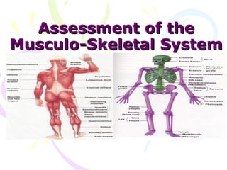
2. Review of Anatomy and Physiology
• The musculo-skeletal system consists of the
muscles, tendons, bones and cartilage
together with the joints
• The primary function of which is to produce
skeletal movements
3. Muscles
Three types of muscles exist in the body
• 1. Skeletal Muscles
– Voluntary and striated
• 2. Cardiac muscles
– Involuntary and striated
• 3. Smooth/Visceral muscles
– Involuntary and NON-striated
6. BONES
• Variously classified according to shape, location and
size
• Functions
1. Locomotion
2. Protection
3. Support and lever
4. Blood production
5. Mineral deposition
10. • CHECK YOUR EQUIPMENT PRIOR
TO ENTERING THE PATIENT’S
ROOM. MAKE SURE YOU HAVE
EVERYTHING YOU NEED TO
COMPLETE YOUR ASSESSMENT
PRIOR TO ENTERING THE
PATIENT’S ROOM
11. Make the Patient Comfortable
• Showing concern for privacy and patient
modesty must become ingrained in your
professional behavior
• Be sure to close nearby doors or
examination room PRIOR to beginning
physical examination
• Your goal is to visualize one area of the
body at a time
12. Make the Patient
Comfortable
• Be sensitive to the patient’s feelings and
physical comfort
• When you have completed the examination,
show your attentiveness, by rearranging the
patients pillows, or adding blankets for
warmth; make sure their immediate
environment is to their satisfaction
• Be sure to lower the bed completely, and
make sure side rails are up and call bell is in
the patient’s reach
• As you leave be sure to WASH YOUR HANDS
16. Outlines
2. Review of Anatomy and physiology
of musculoskeletal system
3. Physical Exam
4. Inspection
5. Palpation
6. ROM (Rang of motion)
17. Objectives
• Apply knowledge of Anatomy and
physiology of musculoskeletal
system
• Differentiate between normal and
abnormal
• Implement physical assessment
18. Musculoskeletal
• Muscle or joint pain
• Stiffness
• Arthritis
• Gout
• Backache
• If present, describe location or affected joints or
muscles, any swelling, redness, pain, tenderness,
stiffness, weakness, or limitation of motion or
activity; include timing of symptoms duration, and
any history of trauma
• Neck or low back pain
• Joint pain with systemic features such as fever,
chills, rash, anorexia, weight loss, or weakness
19. Skin
• Rashes
• Lumps
• Sores
• Itching
• Dryness
• Changes in color
• Changes in hair or nails
20.
21. What do muscles do ?
• Muscles simply move you!
• Without muscles you couldn't open your
mouth, speak, shake hands, walk, talk, or
move your food through your digestive
system.
• There would be no exploring, running,
climbing, smiling, blinking, breathing. You
couldn't move anything inside or outside
you. The fact is, without muscles, you
wouldn't be alive for very long
22.
23. The skeleton is the name given to the
collection of bones that holds our body
up.
Our skeleton is very important to us. It
does three major jobs.
1. It protects our vital organs such as
the brain, the heart, and the lungs.
2. It gives us the shape that we have.
Without our skeleton we would just be
a blob of blood and tissue on the floor.
3. It allows us to move. Because our
muscles are attached to our bones,
when our muscles move, they move
the bones, and we move
24. Physical Exam
1. Inspection
• Observe any lack of symmetry and
any evidence of trauma or disease.
• Look for muscle wasting;
• Inspect the joint contour (shape)
and observe any evidence of
swelling, deformity or inflammation.
25. • Ask the client to point to, or
otherwise identify, any painful areas,
including sites of radiation of
pain.
Screening questions for
musculoskeletal disorders
1. Do you have any pain or
stiffness in your arms, legs or
back?
2. Can you walk up and down
stairs without difficulty?
3. Can you dress yourself in
everyday clothes without any
difficulty?
26. • Assessment of Gait
• Ask the patient to walk back and forth
across the room.
• Observe for equality of arm swing ,
balance and rapidity and ease of heels
turning.
• Next, ask the patient to walk on his
tiptoes, then on heels.
• Ask the patient to tandem walk.
• Test patient's ability to stand with feet tiptoes
together with eyes open and then
closed. (Romberg's test). Reassure
patient that you will support him, in
case he becomes unsteady.
• Normal: Person can walk in balance
with the arms swinging at sides and can
turn smoothly. Person should be able to
stand with feet together without falling
with eyes open or closed.
tandem
27. Upper Extremity Muscles
• Inspect the muscles of
the shoulder, arm,
forearm and hand.
• Note muscle size (bulk).
• Look for asymmetry,
atrophy and
fasciculation.
• Look for tremor and
other abnormal
movement at rest and
with arms outstretched.
28. Determine muscle power
by
• Gently trying to
Abduction
overpower
contraction of each
group of muscles.
– Shoulder:
Abduction
(Deltoid) Adduction
–, Adduction
–, (Trapezius) (Trapezius
30. – Hand: Grip
Grip
– opposition of
thumb and index finger
– opposition of
thumb and little finger
and
– finger abduction and
31. • Determine limb tone
(resistance to
passive stretch).
• With the patient
relaxed
• Gently move the
limb at the shoulder,
elbow and wrist
joints and note
whether tone is
normal, increased or
decreased
32. Normal findings
• Muscles are symmetrical in size with
no involuntary movements.
• In some, muscles may be slightly
larger on the dominant side.
• Muscle power obviously varies. You
should not be able to overpower with
reasonable resistance.
• You have to learn to appreciate the
normal tone from practice.
33. Neck: Range of Motion of
• Fix the head with one hand while you
examine neck
• Inspection
– Note the normal concavity of cervical
spine
– Identify Transverse process of C7
– Observe Trapezius and Sternomastoid
muscles
• Palpation
– Feel each spinous process looking for focal
areas of tenderness
– Joint
• Feel for crepitus during passive motion Touch chin
– Para spinal muscles
• Range of motion
– Active
• Touch chin for flexion
• Throw head back for extension
Throw head back
34. • Touch each shoulder with ears for
lateral flexion
• Touch each shoulder with chin for
lateral rotation
– Passive
• Feel for crepitus during passive
motion
• Normal:
– 30 degree rotation, able to
touch chest with chin, 55
degree extension and 40
degree lateral bend.
– No resistance during the range
of motion.
35. Muscles of Lower Extremity
Inspect the muscles of the hip,
knee and ankle.
• Note muscle size (bulk).
• Look for asymmetry, atrophy
and fasciculation.
Hip flexion
• Look for abnormal movement.
• Determine muscle power by
gently trying to overpower
contraction of each group of
muscles.
– Hip: Flexion (Iliopsoas), Extension
(Gluteus maximus), Abduction,
Adduction.
36. Assessment of the
Musculoskelet al System
Muscle Strength scale
0 No detection of muscular contraction
1 A barely detectable flicker or trace of contraction
with observation or palpation.
2 Active movement of body part with elimination of
gravity.
3 Active movement against gravity only and not
against resistance
4 Active movement against gravity & some
resistance
5 Active movement against full resistance without
evident fatigue (Normal muscle strength)
37. The Knee Exam
• Inspection
• Make sure that both knees are
fully exposed. The patient should
be in either a gown or shorts.
Rolled up pant legs do not
provide good exposure!
• Watch the patient walk.
• Do they limp or appear to be in varus Knee
pain? deormity ,more
• When standing, is there evidence marked on the left
of bowing (varus) or knock- leg
kneed (valgus) deformity? There
is a predilection for degenerative
joint disease to affect the
medical aspect of the knee, a
common cause of bowing.
38. • Is there evidence of
atrophy of the
quadriceps, hamstring,
or calf muscle groups?
Knee problems/pain can
limit the use of the
affected leg, leading to While both legs have
well developed
wasting of the muscles. musculature,
the left calf and
hamstring are bulkier
than the right
39. – Knee : Flexion (Hamstrings),
Extension (Quadriceps)
– Ankle : Dorsiflexion (Tibialis Knee extension
anterior), Plantar flexion
(Gastronemius).
• Determine limb tone
resistance to passive stretch.
With the patient relaxed,
gently move the limb at the
hip, knee and ankle and note
Knee flexion
whether tone is normal,
increased or dicreased.
Flex the hip and knee.
• Support the knee, dorsiflex the Dorsiflexion
ankle sharply and hold the foot
in this position checking for
clonus.
40. (Spine (Bone
• The examiner should stand behind the
patient and observe the alignment of the
spine in the flexed position to determine
scoliosis.
• View the spine from the side to determine
kyphosis.
• Ask the patient if he is aware of sore spots.
Palpate the spinous process and be gentle
with the sore spots. Percuss one vertebra
at a time, starting from head.
• .
41. • Assess range of motion
of spine by having patient
bend down to pick up an
object without bending his
legs while you hold his
hips.
• Normal:
• Gentle concavities in
cervical and lumbar
regions and a convexity in
the thorax.
• Vertebral line and gluteal
cleft align
42. Posture
Normal - •
Comfortably erect
Look for straight lines
across body parts
Treatment Return to top Treatment depends on the cause of the disorder: Congenital kyphosis requires corrective surgery at an early age. Scheuermann's disease is initially treated with a brace and physical therapy. Occasionally surgery is needed for large (greater than 60 degrees), painful curves. Multiple compression fractures from osteoporosis can be left alone if there is no neurologic problems or pain, but the osteoporosis needs to be treated to help prevent future fractures. For debilitating deformity or pain, surgery is an option. Kyphosis caused by infection or tumor needs to be treated more aggressively, often with surgery and medications. Treatment for other types of kyphosis depends on the cause. Surgery may be necessary if neurological symptoms develop. Expectations (prognosis) Return to top Adolescents with Scheuermann's disease tend do well even if they need surgery, and the disease stops once they stop growing. If the kyphosis is due to degenerative joint disease or multiple compression fractures, correction of the defect is not possible without surgery, and improvement of pain is less reliable. Complications Return to top Disabling back pain Neurological symptoms including leg weakness or paralysis Decreased lung capacity Round back deformity
There are three general causes of scoliosis: Congenital scoliosis is due to a problem with the formation of vertebrae or fused ribs during prenatal development. Neuromuscular scoliosis is caused by problems such as poor muscle control or muscular weakness or paralysis due to diseases such as cerebral palsy , muscular dystrophy , spina bifida, and polio. Idiopathic scoliosis is of unknown cause, and appears in a previously straight spine. Idiopathic scoliosis in adolescents is the most common type. Some people may be prone to the curving of the spine. Most cases occur in girls. Curves generally worsen during growth spurts. Scoliosis in infants and juveniles are less common. They commonly affect a similar number of boys and girls. Scoliosis may be suspected when one shoulder appears to be higher than the other, or the pelvis appears to be tilted. Untrained observers usually can't notice the curving. Routine scoliosis screening is now done in middle and junior high schools. Many cases, which previously would have gone undetected until they were more advanced, are now being caught at an early stage. There may be fatigue in the spine after prolonged sitting or standing. Pain will become persistent if irritation results. The greater the initial curve of the spine, the greater the chance the scoliosis will get worse after growth is complete. Severe scoliosis (curves in the spine greater than 100 degrees) may cause breathing problems. Symptoms Return to top The spine curves abnormally to the side (laterally) Shoulders or hips appearing uneven Backache or low-back pain Fatigue Treatment depends on the cause of the scoliosis, the size and location of the curve, and how much more growing the patient is expected to do. Most cases of adolescent idiopathic scoliosis (less than 20 degrees) require no treatment, but should be checked often, about every 6 months. As curves get worse (above 25 to 30 degrees in a child who is still growing), bracing is usually recommended to help slow the progression of the curve. There are many different kinds of braces used. The Boston Brace, Wilmington Brace, Milwaukee Brace, and Charleston Brace are named for the centers where they were developed. Each brace looks different. There are different ways of using each type properly. The selection of a brace and the manner in which it is used depends on many factors, including the specific characteristics of your curve. The exact brace will be decided on by the patient and health care practioner. A back brace does not reverse the curve. Instead, it uses pressure to help straighten the spine. The brace can be adjusted with growth. Bracing does not work in congenital or neuromuscular scoliosis, and is less effective in infantile and juvenile idiopathic scoliosis. Curves of 40 degrees or greater usually require surgery because curves this large have a high risk of getting worse even after bone growth stops. Surgery involves correcting the curve (although not all the way) and fusing the bones in the curve together. The bones are held in place with one or two metal rods held down with hooks and screws until the bone heals together. Sometimes surgery is done through a cut in the back, on the abdomen, or beneath the ribs. A brace may be required to stabilize the spine after surgery.