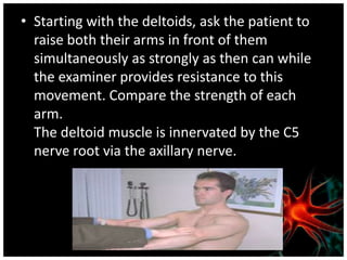
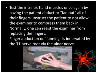
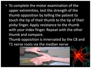
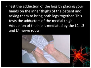
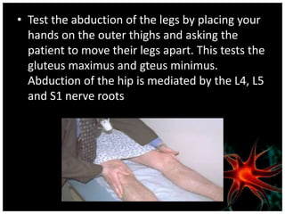
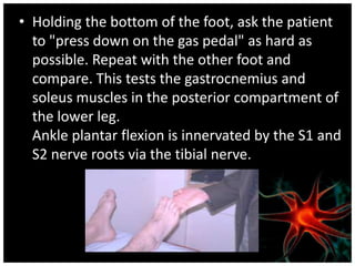
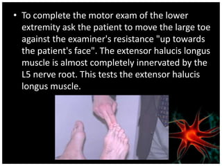
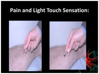
TOPIC INTRODUCTION
Neurological assessment helps to:
• Identify which component of the
  neurological system are affected
• If possible, determine the precise
  location of the problem.
• Screening for the presence of discrete
  abnormalities in patients at risk for the
  development of neurological disorders
: loss of ability to recognize
objects through a particular sensory
system; may be visual, auditory, or
tactile.
         inability to coordinate muscle
movements, resulting in difficulty in
walking, talking, and performing self-
care activities
abnormal movement marked by
alternating contraction and relaxation of
a muscle occurring in rapid succession.
               inability to tolerate light.
         increase in muscle tone at rest
characterized by increased resistance to
passive stretch.
            sustained increase in tension
of a muscle when it is passively
lengthened or stretched
Presenting complaint
• Ask about the symptoms:
• What are they?
• Which part of the body do they affect? Are they
  localised or more widespread?
• When did they start?
• How long do they last for?
• Were they sudden, rapid or gradual in onset? Is
  there a history of trauma?
Ask about any associated symptoms
(other features of neurological disease):
• Headache
• Numbness, pins and needles, cold or
  warmth
• Weakness, unsteadiness, stiffness or
  clumsiness
• Nausea or vomiting
• Visual disturbance
• Altered consciousness
• Some neurological problems can present years
  after a causative event.
• Enquire about other medical problems, past and
  present. These may give clues to the diagnosis. For
  example:
  – A person in atrial fibrillation may be producing multiple
    tiny emboli.
  – There may be vascular problems or recurrent
    miscarriage to suggest antiphospholipid syndrome.
  – There may be diabetes.
• Ask about pregnancy, delivery and neonatal
  health.
The systematic enquiry is very important
here. For example:
• Loss of weight and appetite may suggest
  malignancy and this may be a
  paraneoplastic syndrome.
• Gain in weight may have precipitated
  diabetes.
• Polyuria may suggest diabetes
• Consider if there may be a genetic basis or
  predisposition. For example:
• Huntingdon's chorea is unusual in that it is a
  familial disease that does not present until
  well into adult life.
• A family history of, e.g. type 2 diabetes,
  cerebral aneurysm, neuropathies, epilepsy,
  migraine or vascular disease may be
  important.
• Reflex Hammer
• 128 and 512 (or 1024) Hz Tuning Forks
• A Snellen Eye Chart or Pocket Vision
  Card
• Pen Light or Otoscope
• Wooden Handled Cotton Swabs
• Paper Clips
• A complete neurologic assessment consists of
  five steps:
• Mental status exam
• Cranial nerve assessment
• Reflex testing
• Motor system assessment
• Sensory system assessment
• Coordination
• Gait
• Intellect:
  (Memory, Orientation, Recognitio
  n, Calculations)
• New learning
• Judgement
• Perception
• Cranial Nerve I (Olfactory)
Cranial Nerve II (Optic)
Cranial Nerve III (Oculomotor)

• Assess pupil size and light reflex. A unilaterally
  dilated pupil with unilateral absent light reflex
  and/or if the eye will not turn upwards could
  indicate an internal carotid aneurysm or uncal
  herniation with increased intracranial
  pressure
Cranial Nerve IV (Trochlear) and
  Cranial Nerve VI (Abducens)
Cranial Nerve V (Trigeminal)
• Use a sharp implement.
• Ask the patient to close their eyes so that they
  receive no visual cues.
• Touch the sharp tip of the stick to the right and
  left side of the forehead, assessing the Ophthalmic
  branch.
• Touch the tip to the right and left side of the
  cheek area, assessing the Maxillary branch.
• Touch the tip to the right and left side of the jaw
  area, assessing the Mandibular branch.
• Place your hand on both Temporalis muscles,
  located on the lateral aspects of the forehead.
• Ask the patient to tightly close their jaw,
  causing the muscles beneath your fingers to
  become taught.
• Then place your hands on both Masseter
  muscles, located just in from of the Tempero-
  Mandibular joints.
Cranial Nerve VII (Facial)
Cranial Nerve VIII (Acoustic or
        Vestibulocochlear)
• Weber Test:
Rinne Test:
CN9 (Glosopharyngeal) and CN 10
           (Vagus):
Cranial Nerve XI (Spinal Accessory)
Cranial Nerve XII (Hypoglossal)
• Ask the patient to stick their
  tongue straight out of their
  mouth.
• If there is any suggestion of
  deviation to one
  side/weakness, direct them
  to push the tip of their
  tongue into either cheek
  while you provide counter
  pressure from the outside
Reflex Testing:
• Reflexes should be graded on a 0 to 4 "plus"
  scale:
              Grade                Description

 0                      Absent

 1+ or +                Hypoactive

 2+ or ++               "Normal"

 3+ or +++              Hyperactive without clonus

 4+ or ++++             Hyperactive with clonus
Achilles: (S1, S2)
Patellar: (L3, L4)
Biceps: (C5, C6)
Triceps: (C6, C7)
Brachioradialis (C5, C6) :
Babinski reflex:
The Motor System Examination
• The motor system evaluation is divided into
  the following: body positioning, involuntary
  movements, muscle tone and muscle
  strength.
• Note the appearance or muscularity of the
  muscle (wasted, highly developed, normal).
• Feel the tone of the muscle (flaccid, clonic,
  normal).
• Test the strength of the muscle group.
Muscle Strength
      Grade                     Description
0/5           No muscle movement
              Visible muscle movement, but no movement at
1/5
              the joint
2/5           Movement at the joint, but not against gravity
              Movement against gravity, but not against added
3/5
              resistance
              Movement against resistance, but less than
4/5
              normal
5/5           Normal strength
• Starting with the deltoids, ask the patient to
  raise both their arms in front of them
  simultaneously as strongly as then can while
  the examiner provides resistance to this
  movement. Compare the strength of each
  arm.
  The deltoid muscle is innervated by the C5
  nerve root via the axillary nerve.
• Ask the patient to extend and raise both arms
  in front of them as if they were carrying a
  pizza. Ask the patient to keep their arms in
  place while they close their eyes and count to
  10. Normally their arms will remain in place. If
  there is upper extremity weakness there will
  be a positive pronator drift, in which the
  affected arm will pronate and fall. This is one
  of the most sensitive tests for upper extremity
  weakness.
• Test the strength of lower arm flexion by
  holding the patient's wrist from above and
  instructing them to "flex their hand up to their
  shoulder". Provide resistance at the wrist.
  Repeat and compare to the opposite arm. This
  tests the biceps muscle.
  The biceps muscle is innervated by the C5 and
  C6 nerve roots via the musculocutaneous nerve
• Now have the patient extend their forearm
  against the examiner's resistance. Make certain
  that the patient begins their extension from a
  fully flexed position because this part of the
  movement is most sensitive to a loss in
  strength. This tests the triceps. Note any
  asymmetry in the other arm.

  The triceps muscle is innervated by the C6 and
  C7 nerve roots via the radial nerve.
• Test the intrinsic hand muscles once again by
  having the patient abduct or "fan out" all of
  their fingers. Instruct the patient to not allow
  the examiner to compress them back in.
  Normally, one can resist the examiner from
  replacing the fingers.
  Finger abduction or "fanning" is innervated by
  the T1 nerve root via the ulnar nerve.
• To complete the motor examination of the
  upper extremities, test the strength of the
  thumb opposition by telling the patient to
  touch the tip of their thumb to the tip of their
  pinky finger. Apply resistance to the thumb
  with your index finger. Repeat with the other
  thumb and compare.
  Thumb opposition is innervated by the C8 and
  T1 nerve roots via the median nerve
• Test the adduction of the legs by placing your
  hands on the inner thighs of the patient and
  asking them to bring both legs together. This
  tests the adductors of the medial thigh.
  Adduction of the hip is mediated by the L2, L3
  and L4 nerve roots.
• Test the abduction of the legs by placing your
  hands on the outer thighs and asking the
  patient to move their legs apart. This tests the
  gluteus maximus and gteus minimus.
  Abduction of the hip is mediated by the L4, L5
  and S1 nerve roots
• Holding the bottom of the foot, ask the patient
  to "press down on the gas pedal" as hard as
  possible. Repeat with the other foot and
  compare. This tests the gastrocnemius and
  soleus muscles in the posterior compartment of
  the lower leg.
  Ankle plantar flexion is innervated by the S1 and
  S2 nerve roots via the tibial nerve.
• To complete the motor exam of the lower
  extremity ask the patient to move the large toe
  against the examiner's resistance "up towards
  the patient's face". The extensor halucis longus
  muscle is almost completely innervated by the
  L5 nerve root. This tests the extensor halucis
  longus muscle.
The Sensory System Examination
The sensory exam includes testing for: pain
sensation (pin prick), light touch sensation
(brush), position sense, stereognosia,
graphesthesia, and extinction. Diabetes
mellitus, thiamine deficiency and neurotoxin
damage (e.g. insecticides) are the most
common causes of sensory disturbances
Pain and Light Touch Sensation:
Stereognosia

• Test stereognosis by asking the patient to
  close their eyes and identify the object
  you place in their hand. Place a coin or
  pen in their hand. Repeat this with the
  other hand using a different object
• Astereognosis refers to the inability to
  recognize objects placed in the hand.
  Without a corresponding dorsal column
  system lesion, these abnormalities suggest a
  lesion in the sensory cortex of the parietal
  lobe.
Graphesthesia

• Test graphesthesia by asking the patient to
  close their eyes and identify the number or
  letter you will write with the back of a pen on
  their palm. Repeat on the other hand with a
  different letter or number
• Apraxias are problems with executing
  movements despite intact strength,
  coordination, position sense and
  comprehension. This finding is a defect in
  higher intell-ectual functioning and
  associated with cortical damage
Coordination and Gait
Rapid Alternating Movements
• Ask the patient to strike one hand on the
  thigh, raise the hand, turn it over, and then
  strike it back down as fast as possible.
• Ask the patient to tap the distal thumb with
  the tip of the index finger as fast as possible.
• Ask the patient to tap your hand with the ball
  of each foot as fast as possible.
Point-to-Point Movements
• Ask the patient to touch your index finger and
  their nose alternately several times. Move your
  finger about as the patient performs this task.
• Hold your finger still so that the patient can
  touch it with one arm and finger outstretched.
  Ask the patient to move their arm and return
  to your finger with their eyes closed.
Romberg:
• Be prepared to catch the patient if they are
  unstable.
• Ask the patient to stand with the feet together
  and eyes closed for 5-10 seconds without
  support.
• The test is said to be positive if the patient
  becomes unstable (indicating a vestibular or
  proprioceptive problem).
Gait:
Ask the patient to:
• Walk across the room, turn and come back
• Walk heel-to-toe in a straight line
• Walk on their toes in a straight line
• Walk on their heels in a straight line
• Hop in place on each foot
• Do a shallow knee bend
• Rise from a sitting position
BIBLIOGRAPHY
Neurological assessment ppt

Neurological assessment ppt

  • 2.
    TOPIC INTRODUCTION Neurological assessmenthelps to: • Identify which component of the neurological system are affected • If possible, determine the precise location of the problem. • Screening for the presence of discrete abnormalities in patients at risk for the development of neurological disorders
  • 3.
    : loss ofability to recognize objects through a particular sensory system; may be visual, auditory, or tactile. inability to coordinate muscle movements, resulting in difficulty in walking, talking, and performing self- care activities
  • 4.
    abnormal movement markedby alternating contraction and relaxation of a muscle occurring in rapid succession. inability to tolerate light. increase in muscle tone at rest characterized by increased resistance to passive stretch. sustained increase in tension of a muscle when it is passively lengthened or stretched
  • 6.
    Presenting complaint • Askabout the symptoms: • What are they? • Which part of the body do they affect? Are they localised or more widespread? • When did they start? • How long do they last for? • Were they sudden, rapid or gradual in onset? Is there a history of trauma?
  • 7.
    Ask about anyassociated symptoms (other features of neurological disease): • Headache • Numbness, pins and needles, cold or warmth • Weakness, unsteadiness, stiffness or clumsiness • Nausea or vomiting • Visual disturbance • Altered consciousness
  • 8.
    • Some neurologicalproblems can present years after a causative event. • Enquire about other medical problems, past and present. These may give clues to the diagnosis. For example: – A person in atrial fibrillation may be producing multiple tiny emboli. – There may be vascular problems or recurrent miscarriage to suggest antiphospholipid syndrome. – There may be diabetes. • Ask about pregnancy, delivery and neonatal health.
  • 9.
    The systematic enquiryis very important here. For example: • Loss of weight and appetite may suggest malignancy and this may be a paraneoplastic syndrome. • Gain in weight may have precipitated diabetes. • Polyuria may suggest diabetes
  • 10.
    • Consider ifthere may be a genetic basis or predisposition. For example: • Huntingdon's chorea is unusual in that it is a familial disease that does not present until well into adult life. • A family history of, e.g. type 2 diabetes, cerebral aneurysm, neuropathies, epilepsy, migraine or vascular disease may be important.
  • 11.
    • Reflex Hammer •128 and 512 (or 1024) Hz Tuning Forks • A Snellen Eye Chart or Pocket Vision Card • Pen Light or Otoscope • Wooden Handled Cotton Swabs • Paper Clips
  • 12.
    • A completeneurologic assessment consists of five steps: • Mental status exam • Cranial nerve assessment • Reflex testing • Motor system assessment • Sensory system assessment • Coordination • Gait
  • 13.
    • Intellect: (Memory, Orientation, Recognitio n, Calculations) • New learning • Judgement • Perception
  • 14.
    • Cranial NerveI (Olfactory)
  • 15.
  • 16.
    Cranial Nerve III(Oculomotor) • Assess pupil size and light reflex. A unilaterally dilated pupil with unilateral absent light reflex and/or if the eye will not turn upwards could indicate an internal carotid aneurysm or uncal herniation with increased intracranial pressure
  • 17.
    Cranial Nerve IV(Trochlear) and Cranial Nerve VI (Abducens)
  • 18.
    Cranial Nerve V(Trigeminal) • Use a sharp implement. • Ask the patient to close their eyes so that they receive no visual cues. • Touch the sharp tip of the stick to the right and left side of the forehead, assessing the Ophthalmic branch. • Touch the tip to the right and left side of the cheek area, assessing the Maxillary branch. • Touch the tip to the right and left side of the jaw area, assessing the Mandibular branch.
  • 19.
    • Place yourhand on both Temporalis muscles, located on the lateral aspects of the forehead. • Ask the patient to tightly close their jaw, causing the muscles beneath your fingers to become taught. • Then place your hands on both Masseter muscles, located just in from of the Tempero- Mandibular joints.
  • 20.
  • 21.
    Cranial Nerve VIII(Acoustic or Vestibulocochlear) • Weber Test:
  • 22.
  • 23.
  • 24.
    Cranial Nerve XI(Spinal Accessory)
  • 25.
    Cranial Nerve XII(Hypoglossal) • Ask the patient to stick their tongue straight out of their mouth. • If there is any suggestion of deviation to one side/weakness, direct them to push the tip of their tongue into either cheek while you provide counter pressure from the outside
  • 26.
    Reflex Testing: • Reflexesshould be graded on a 0 to 4 "plus" scale: Grade Description 0 Absent 1+ or + Hypoactive 2+ or ++ "Normal" 3+ or +++ Hyperactive without clonus 4+ or ++++ Hyperactive with clonus
  • 27.
  • 28.
  • 29.
  • 30.
  • 31.
  • 32.
  • 33.
    The Motor SystemExamination • The motor system evaluation is divided into the following: body positioning, involuntary movements, muscle tone and muscle strength. • Note the appearance or muscularity of the muscle (wasted, highly developed, normal). • Feel the tone of the muscle (flaccid, clonic, normal). • Test the strength of the muscle group.
  • 34.
    Muscle Strength Grade Description 0/5 No muscle movement Visible muscle movement, but no movement at 1/5 the joint 2/5 Movement at the joint, but not against gravity Movement against gravity, but not against added 3/5 resistance Movement against resistance, but less than 4/5 normal 5/5 Normal strength
  • 35.
    • Starting withthe deltoids, ask the patient to raise both their arms in front of them simultaneously as strongly as then can while the examiner provides resistance to this movement. Compare the strength of each arm. The deltoid muscle is innervated by the C5 nerve root via the axillary nerve.
  • 36.
    • Ask thepatient to extend and raise both arms in front of them as if they were carrying a pizza. Ask the patient to keep their arms in place while they close their eyes and count to 10. Normally their arms will remain in place. If there is upper extremity weakness there will be a positive pronator drift, in which the affected arm will pronate and fall. This is one of the most sensitive tests for upper extremity weakness.
  • 37.
    • Test thestrength of lower arm flexion by holding the patient's wrist from above and instructing them to "flex their hand up to their shoulder". Provide resistance at the wrist. Repeat and compare to the opposite arm. This tests the biceps muscle. The biceps muscle is innervated by the C5 and C6 nerve roots via the musculocutaneous nerve
  • 38.
    • Now havethe patient extend their forearm against the examiner's resistance. Make certain that the patient begins their extension from a fully flexed position because this part of the movement is most sensitive to a loss in strength. This tests the triceps. Note any asymmetry in the other arm. The triceps muscle is innervated by the C6 and C7 nerve roots via the radial nerve.
  • 39.
    • Test theintrinsic hand muscles once again by having the patient abduct or "fan out" all of their fingers. Instruct the patient to not allow the examiner to compress them back in. Normally, one can resist the examiner from replacing the fingers. Finger abduction or "fanning" is innervated by the T1 nerve root via the ulnar nerve.
  • 40.
    • To completethe motor examination of the upper extremities, test the strength of the thumb opposition by telling the patient to touch the tip of their thumb to the tip of their pinky finger. Apply resistance to the thumb with your index finger. Repeat with the other thumb and compare. Thumb opposition is innervated by the C8 and T1 nerve roots via the median nerve
  • 41.
    • Test theadduction of the legs by placing your hands on the inner thighs of the patient and asking them to bring both legs together. This tests the adductors of the medial thigh. Adduction of the hip is mediated by the L2, L3 and L4 nerve roots.
  • 42.
    • Test theabduction of the legs by placing your hands on the outer thighs and asking the patient to move their legs apart. This tests the gluteus maximus and gteus minimus. Abduction of the hip is mediated by the L4, L5 and S1 nerve roots
  • 43.
    • Holding thebottom of the foot, ask the patient to "press down on the gas pedal" as hard as possible. Repeat with the other foot and compare. This tests the gastrocnemius and soleus muscles in the posterior compartment of the lower leg. Ankle plantar flexion is innervated by the S1 and S2 nerve roots via the tibial nerve.
  • 44.
    • To completethe motor exam of the lower extremity ask the patient to move the large toe against the examiner's resistance "up towards the patient's face". The extensor halucis longus muscle is almost completely innervated by the L5 nerve root. This tests the extensor halucis longus muscle.
  • 45.
    The Sensory SystemExamination The sensory exam includes testing for: pain sensation (pin prick), light touch sensation (brush), position sense, stereognosia, graphesthesia, and extinction. Diabetes mellitus, thiamine deficiency and neurotoxin damage (e.g. insecticides) are the most common causes of sensory disturbances
  • 46.
    Pain and LightTouch Sensation:
  • 47.
    Stereognosia • Test stereognosisby asking the patient to close their eyes and identify the object you place in their hand. Place a coin or pen in their hand. Repeat this with the other hand using a different object
  • 48.
    • Astereognosis refersto the inability to recognize objects placed in the hand. Without a corresponding dorsal column system lesion, these abnormalities suggest a lesion in the sensory cortex of the parietal lobe.
  • 49.
    Graphesthesia • Test graphesthesiaby asking the patient to close their eyes and identify the number or letter you will write with the back of a pen on their palm. Repeat on the other hand with a different letter or number
  • 50.
    • Apraxias areproblems with executing movements despite intact strength, coordination, position sense and comprehension. This finding is a defect in higher intell-ectual functioning and associated with cortical damage
  • 51.
    Coordination and Gait RapidAlternating Movements • Ask the patient to strike one hand on the thigh, raise the hand, turn it over, and then strike it back down as fast as possible. • Ask the patient to tap the distal thumb with the tip of the index finger as fast as possible. • Ask the patient to tap your hand with the ball of each foot as fast as possible.
  • 52.
    Point-to-Point Movements • Askthe patient to touch your index finger and their nose alternately several times. Move your finger about as the patient performs this task. • Hold your finger still so that the patient can touch it with one arm and finger outstretched. Ask the patient to move their arm and return to your finger with their eyes closed.
  • 53.
    Romberg: • Be preparedto catch the patient if they are unstable. • Ask the patient to stand with the feet together and eyes closed for 5-10 seconds without support. • The test is said to be positive if the patient becomes unstable (indicating a vestibular or proprioceptive problem).
  • 54.
    Gait: Ask the patientto: • Walk across the room, turn and come back • Walk heel-to-toe in a straight line • Walk on their toes in a straight line • Walk on their heels in a straight line • Hop in place on each foot • Do a shallow knee bend • Rise from a sitting position
  • 57.