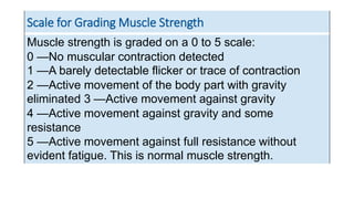
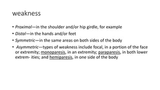
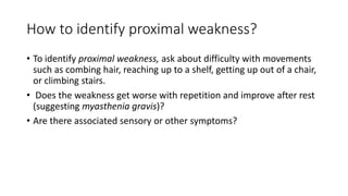
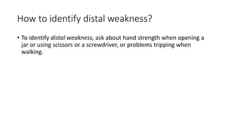
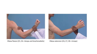
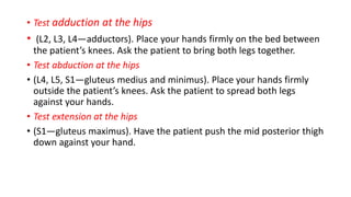
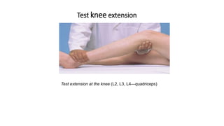
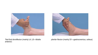
Physical exam
Fatemeh torabi
• Muscle Strength. Normal strength varies widely, so your standard of
normal should allow for factors like age, sex, and muscular training.
The patient’s dominant side is usually slightly stronger than the
nondominant side, though differences can be hard to detect. Keep
this difference in mind as you compare sides.
• Impaired strength or weakness is called paresis.
• Absent strength is paralysis, or plegia.
• Hemiparesis refers to weakness of one half of the body.
• hemiplegia refers to paralysis of one half of the body.
• Paraplegia means paralysis of the legs.
• quadriplegia means paralysis of all four limbs.
Weakness
• It is important to clarify what the patient means—fatigue, apathy,
drowsiness, or actual loss of strength.
• True motor weakness can arise from the CNS, a peripheral nerve, the
neuromuscular junction, or a muscle.
• Time course and location are especially relevant. Is the onset sudden,
gradual or subacute, or chronic, over a long period of time?
Muscle strength test
• Test muscle strength by asking the patient to actively resist your
movement.
• Remember that a muscle is strongest when shortest, and weakest when
longest. Give the patient the advantage as you try to overcome the
resistance and judge true the muscle’s true strength. Some patients give
way during tests of muscle strength due to pain, misunderstanding of the
test, an effort to help the examiner, conversion disorder, or malingering.
• If the muscles are too weak to overcome resistance, test them against
gravity alone or with gravity eliminated.
• . Finally, if the patient fails to move the body part, observe or palpate for
weak muscular contraction.
Scale for Grading Muscle Strength
Muscle strength is graded on a 0 to 5 scale:
0 —No muscular contraction detected
1 —A barely detectable flicker or trace of contraction
2 —Active movement of the body part with gravity
eliminated 3 —Active movement against gravity
4 —Active movement against gravity and some
resistance
5 —Active movement against full resistance without
evident fatigue. This is normal muscle strength.
weakness
• Proximal—in the shoulder and/or hip girdle, for example
• Distal—in the hands and/or feet
• Symmetric—in the same areas on both sides of the body
• Asymmetric—types of weakness include focal, in a portion of the face
or extremity; monoparesis, in an extremity; paraparesis, in both lower
extrem- ities; and hemiparesis, in one side of the body
How to identify proximal weakness?
• To identify proximal weakness, ask about difficulty with movements
such as combing hair, reaching up to a shelf, getting up out of a chair,
or climbing stairs.
• Does the weakness get worse with repetition and improve after rest
(suggesting myasthenia gravis)?
• Are there associated sensory or other symptoms?
How to identify distal weakness?
• To identify distal weakness, ask about hand strength when opening a
jar or using scissors or a screwdriver, or problems tripping when
walking.
‫عضالنی‬ ‫نیروی‬ ‫بندی‬ ‫درجه‬
‫توصیف‬ ‫درجه‬
complete paralysis ‫عضالنی‬ ‫انقباض‬ ‫بدون‬ ۰
Flicker of contraction possible ‫تشخیص‬ ‫قابل‬ ‫انقباض‬ ‫میزان‬ ‫حداقل‬ ۱
Movement possible if gravity
eliminated
‫جاذبه‬ ‫نیروی‬ ‫حذف‬ ‫با‬ ‫فعال‬ ‫حرکات‬ ۲
Movement against gravity but not
resistance
‫جاذبه‬ ‫نیروی‬ ‫خالف‬ ‫بر‬ ‫فعال‬ ‫حرکات‬ ۳
Movement possible against some
resistance
‫جاذبه‬ ‫نیروی‬ ‫خالف‬ ‫بر‬ ‫فعال‬ ‫حرکات‬
‫اندک‬ ‫مقاومت‬ ‫و‬
۴
Power normal ‫و‬ ‫جاذبه‬ ‫نیروی‬ ‫خالف‬ ‫بر‬ ‫فعال‬ ‫حرکات‬
‫کامل‬ ‫مقاومت‬
۵
. Many clinicians make further distinctions by adding plus or minus signs toward the
stronger end of this scale.
Thus, 4+ indicates good but not full strength, while 5− means a trace of weakness.
•Methods for testing individual major muscle groups
Elbow flexion (C5, C6—biceps and brachioradialis) Elbow extension (C6, C7, C8—triceps)
Extension of wrist
Test extension at the wrist
(C6, C7, C8, radial nerve) by asking the patient to make a fist and resist as you press down .
Extensor weakness is seen in peripheral radial nerve damage, and in the hemiplegia of
CNS disease seen in stroke or multiple sclerosis.
Test of grip
Test the grip (C7, C8, T1)
A weak grip is seen in cervical radiculopathy, median or
ulnar peripheral nerve disease, and pain from de Quervain
tenosynovitis, carpal tunnel syndrome, arthritis, and
epicondylitis.
Test finger abduction.
Test finger abduction (C8, T1, ulnar nerve)
Weak finger abduction occurs in ulnar nerve disorders.
Test opposition of the thumb
Test opposition of the thumb (C8, T1, median nerve)
Inspect for weak opposition of the thumb in median nerve
disorders such as carpal tunnel syndrome
Test hip flexion.
1.Test flexion at the hip (L2, L3, L4—iliopsoas)
• Test adduction at the hips
• (L2, L3, L4—adductors). Place your hands firmly on the bed between
the patient’s knees. Ask the patient to bring both legs together.
• Test abduction at the hips
• (L4, L5, S1—gluteus medius and minimus). Place your hands firmly
outside the patient’s knees. Ask the patient to spread both legs
against your hands.
• Test extension at the hips
• (S1—gluteus maximus). Have the patient push the mid posterior thigh
down against your hand.
Test knee extension
Test extension at the knee (L2, L3, L4—quadriceps)
Test knee flexion.
Test flexion at the knee (L4, L5, S1, S2—hamstrings)
Test foot dorsiflexion (mainly L4, L5—tibialis
anterior)
plantar flexion (mainly S1—gastrocnemius, soleus)
THE END

Presentation1.pptx

  • 1.
  • 2.
    • Muscle Strength.Normal strength varies widely, so your standard of normal should allow for factors like age, sex, and muscular training. The patient’s dominant side is usually slightly stronger than the nondominant side, though differences can be hard to detect. Keep this difference in mind as you compare sides.
  • 3.
    • Impaired strengthor weakness is called paresis. • Absent strength is paralysis, or plegia. • Hemiparesis refers to weakness of one half of the body. • hemiplegia refers to paralysis of one half of the body. • Paraplegia means paralysis of the legs. • quadriplegia means paralysis of all four limbs.
  • 4.
    Weakness • It isimportant to clarify what the patient means—fatigue, apathy, drowsiness, or actual loss of strength. • True motor weakness can arise from the CNS, a peripheral nerve, the neuromuscular junction, or a muscle. • Time course and location are especially relevant. Is the onset sudden, gradual or subacute, or chronic, over a long period of time?
  • 5.
    Muscle strength test •Test muscle strength by asking the patient to actively resist your movement. • Remember that a muscle is strongest when shortest, and weakest when longest. Give the patient the advantage as you try to overcome the resistance and judge true the muscle’s true strength. Some patients give way during tests of muscle strength due to pain, misunderstanding of the test, an effort to help the examiner, conversion disorder, or malingering. • If the muscles are too weak to overcome resistance, test them against gravity alone or with gravity eliminated. • . Finally, if the patient fails to move the body part, observe or palpate for weak muscular contraction.
  • 6.
    Scale for GradingMuscle Strength Muscle strength is graded on a 0 to 5 scale: 0 —No muscular contraction detected 1 —A barely detectable flicker or trace of contraction 2 —Active movement of the body part with gravity eliminated 3 —Active movement against gravity 4 —Active movement against gravity and some resistance 5 —Active movement against full resistance without evident fatigue. This is normal muscle strength.
  • 7.
    weakness • Proximal—in theshoulder and/or hip girdle, for example • Distal—in the hands and/or feet • Symmetric—in the same areas on both sides of the body • Asymmetric—types of weakness include focal, in a portion of the face or extremity; monoparesis, in an extremity; paraparesis, in both lower extrem- ities; and hemiparesis, in one side of the body
  • 8.
    How to identifyproximal weakness? • To identify proximal weakness, ask about difficulty with movements such as combing hair, reaching up to a shelf, getting up out of a chair, or climbing stairs. • Does the weakness get worse with repetition and improve after rest (suggesting myasthenia gravis)? • Are there associated sensory or other symptoms?
  • 9.
    How to identifydistal weakness? • To identify distal weakness, ask about hand strength when opening a jar or using scissors or a screwdriver, or problems tripping when walking.
  • 10.
    ‫عضالنی‬ ‫نیروی‬ ‫بندی‬‫درجه‬ ‫توصیف‬ ‫درجه‬ complete paralysis ‫عضالنی‬ ‫انقباض‬ ‫بدون‬ ۰ Flicker of contraction possible ‫تشخیص‬ ‫قابل‬ ‫انقباض‬ ‫میزان‬ ‫حداقل‬ ۱ Movement possible if gravity eliminated ‫جاذبه‬ ‫نیروی‬ ‫حذف‬ ‫با‬ ‫فعال‬ ‫حرکات‬ ۲ Movement against gravity but not resistance ‫جاذبه‬ ‫نیروی‬ ‫خالف‬ ‫بر‬ ‫فعال‬ ‫حرکات‬ ۳ Movement possible against some resistance ‫جاذبه‬ ‫نیروی‬ ‫خالف‬ ‫بر‬ ‫فعال‬ ‫حرکات‬ ‫اندک‬ ‫مقاومت‬ ‫و‬ ۴ Power normal ‫و‬ ‫جاذبه‬ ‫نیروی‬ ‫خالف‬ ‫بر‬ ‫فعال‬ ‫حرکات‬ ‫کامل‬ ‫مقاومت‬ ۵ . Many clinicians make further distinctions by adding plus or minus signs toward the stronger end of this scale. Thus, 4+ indicates good but not full strength, while 5− means a trace of weakness.
  • 11.
    •Methods for testingindividual major muscle groups
  • 12.
    Elbow flexion (C5,C6—biceps and brachioradialis) Elbow extension (C6, C7, C8—triceps)
  • 13.
    Extension of wrist Testextension at the wrist (C6, C7, C8, radial nerve) by asking the patient to make a fist and resist as you press down . Extensor weakness is seen in peripheral radial nerve damage, and in the hemiplegia of CNS disease seen in stroke or multiple sclerosis.
  • 14.
    Test of grip Testthe grip (C7, C8, T1) A weak grip is seen in cervical radiculopathy, median or ulnar peripheral nerve disease, and pain from de Quervain tenosynovitis, carpal tunnel syndrome, arthritis, and epicondylitis.
  • 15.
    Test finger abduction. Testfinger abduction (C8, T1, ulnar nerve) Weak finger abduction occurs in ulnar nerve disorders.
  • 16.
    Test opposition ofthe thumb Test opposition of the thumb (C8, T1, median nerve) Inspect for weak opposition of the thumb in median nerve disorders such as carpal tunnel syndrome
  • 17.
    Test hip flexion. 1.Testflexion at the hip (L2, L3, L4—iliopsoas)
  • 18.
    • Test adductionat the hips • (L2, L3, L4—adductors). Place your hands firmly on the bed between the patient’s knees. Ask the patient to bring both legs together. • Test abduction at the hips • (L4, L5, S1—gluteus medius and minimus). Place your hands firmly outside the patient’s knees. Ask the patient to spread both legs against your hands. • Test extension at the hips • (S1—gluteus maximus). Have the patient push the mid posterior thigh down against your hand.
  • 19.
    Test knee extension Testextension at the knee (L2, L3, L4—quadriceps)
  • 20.
    Test knee flexion. Testflexion at the knee (L4, L5, S1, S2—hamstrings)
  • 21.
    Test foot dorsiflexion(mainly L4, L5—tibialis anterior) plantar flexion (mainly S1—gastrocnemius, soleus)
  • 22.