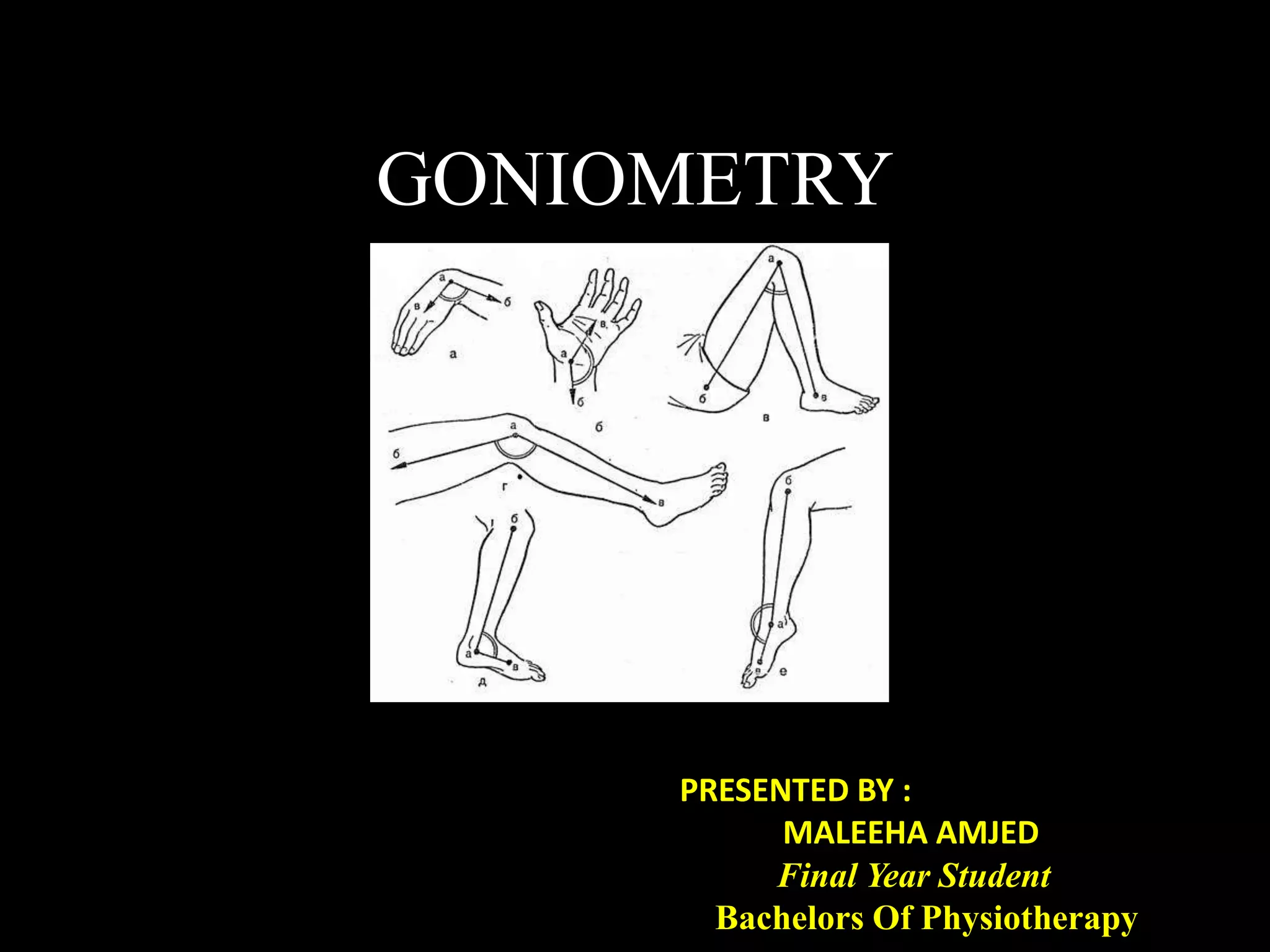
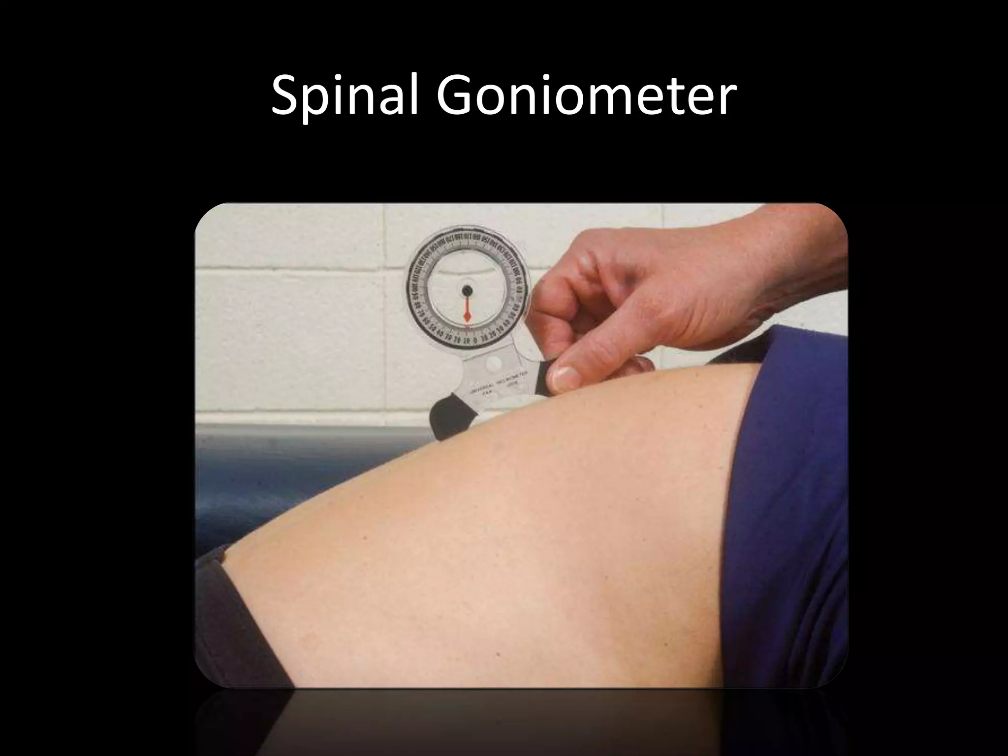
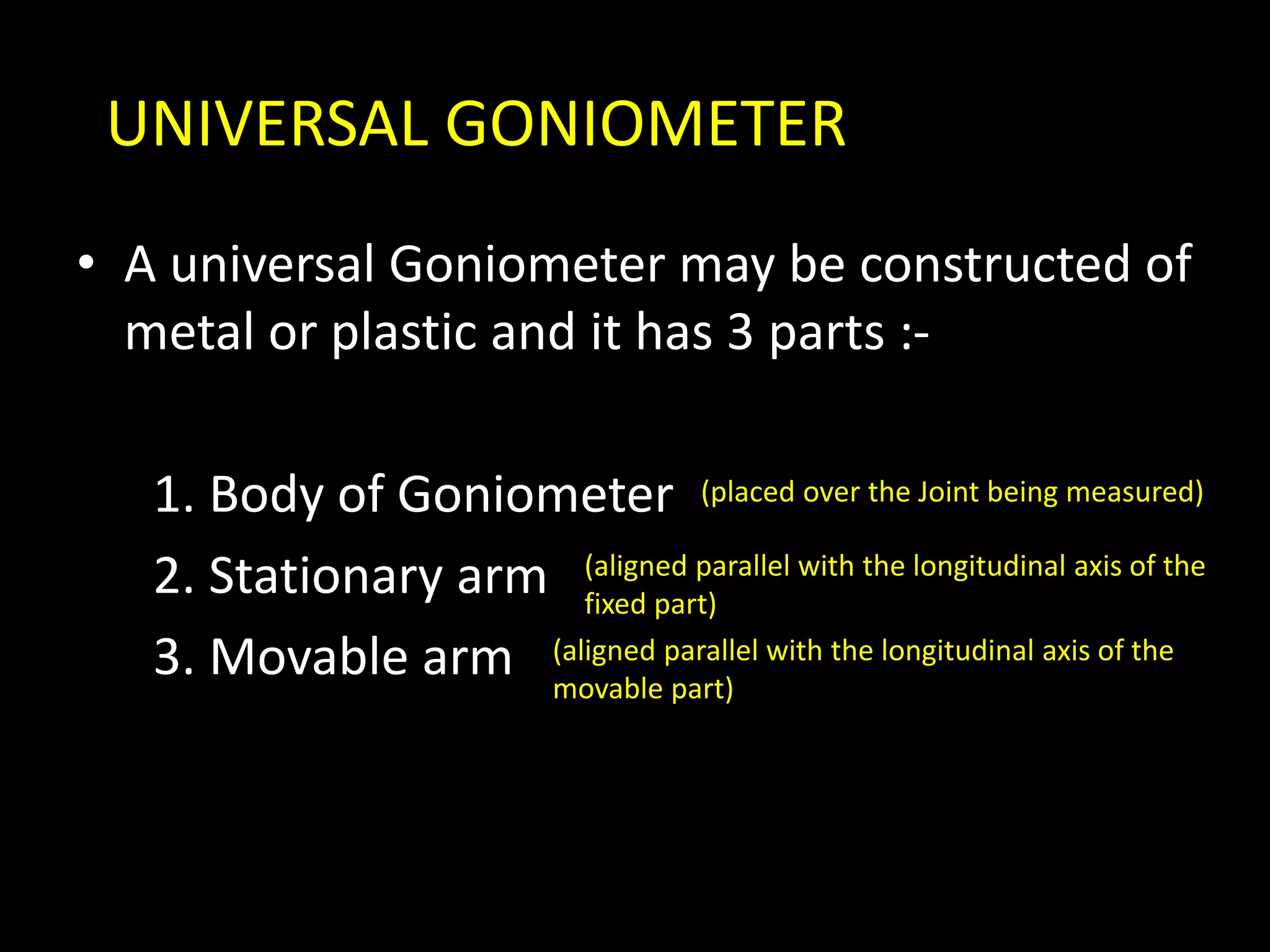
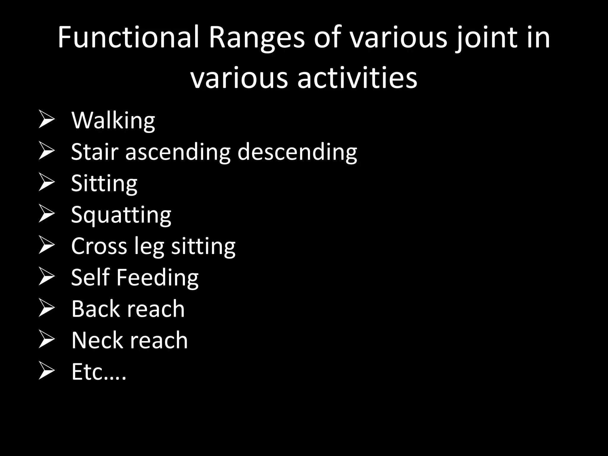
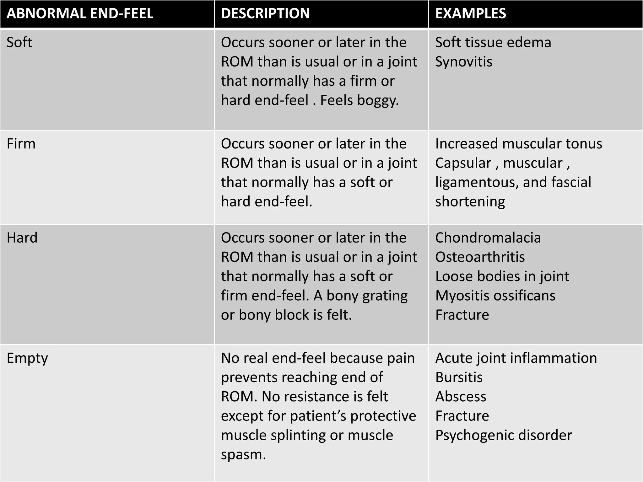
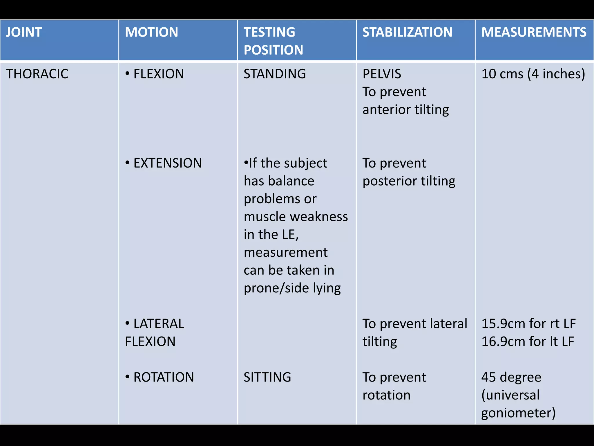
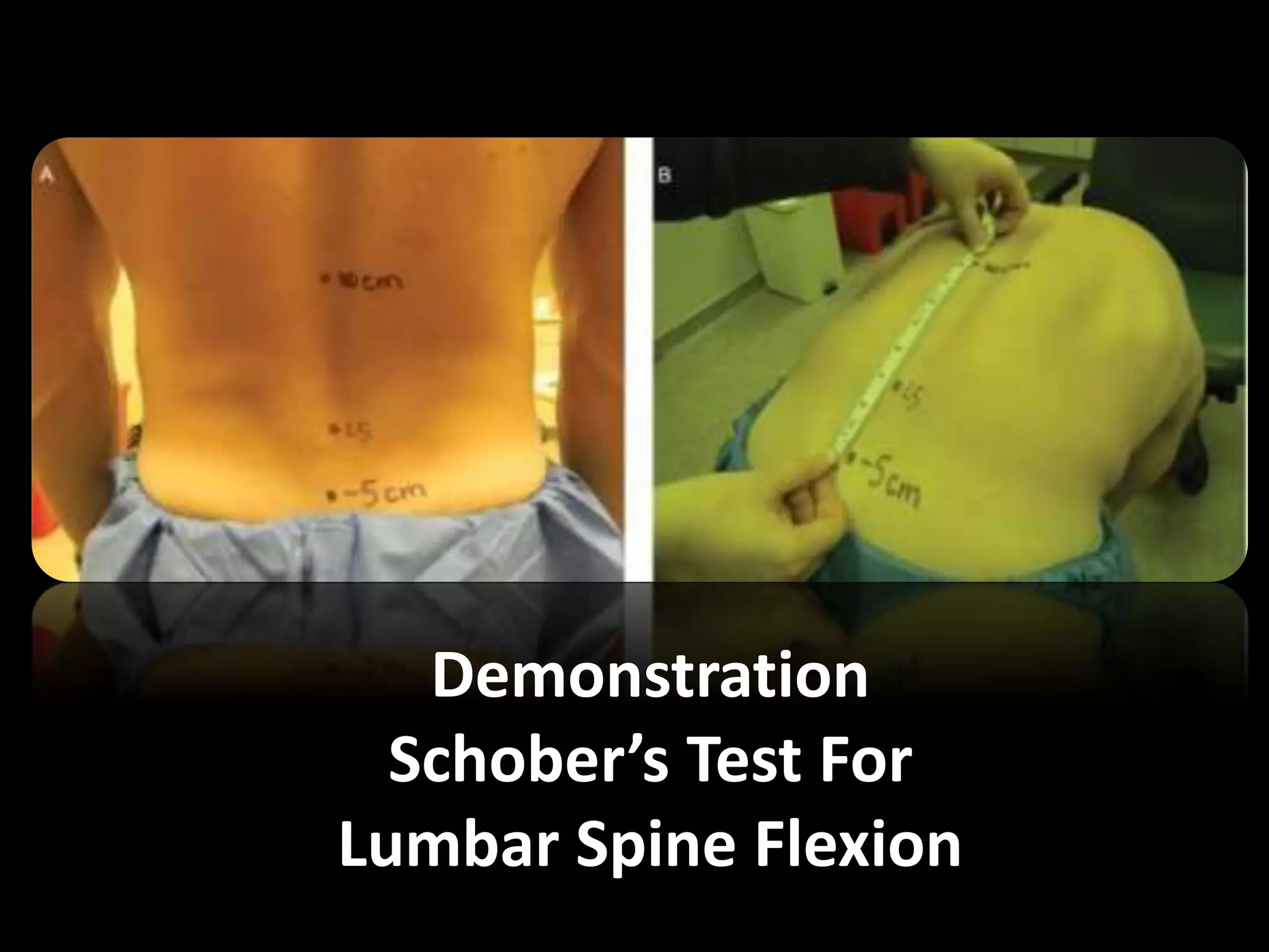
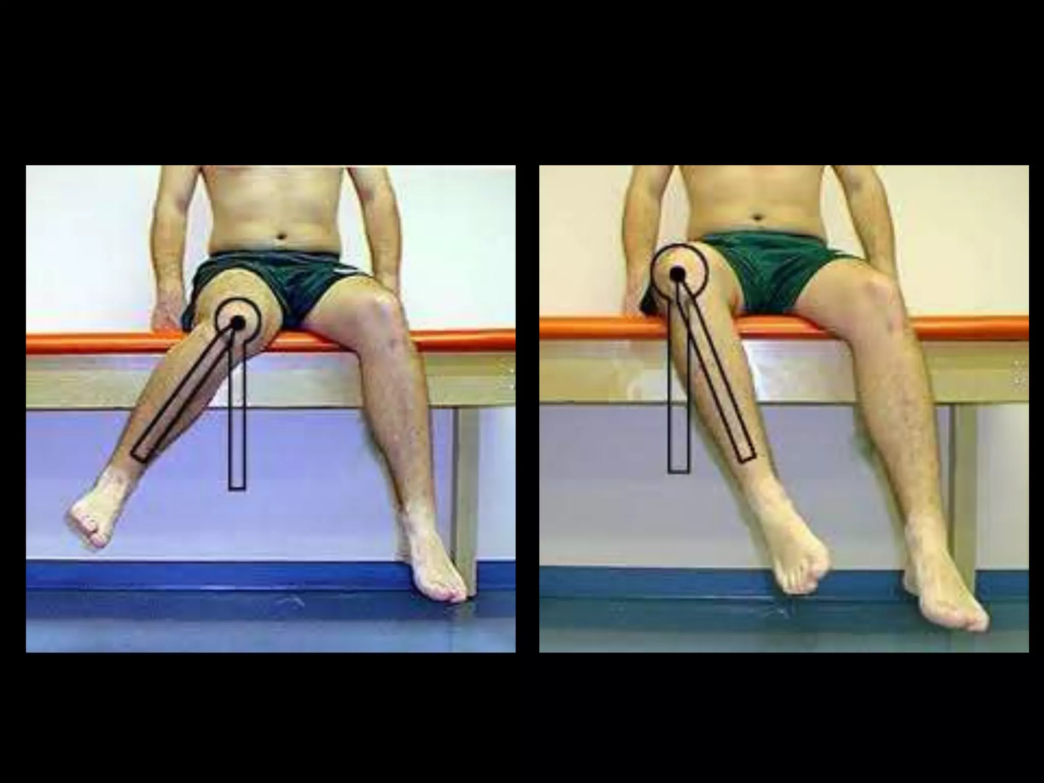
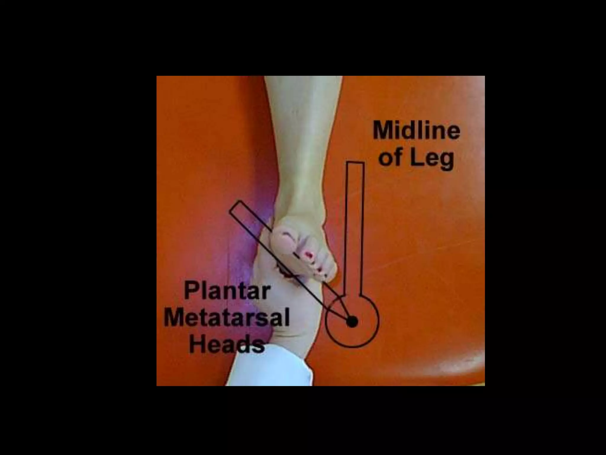
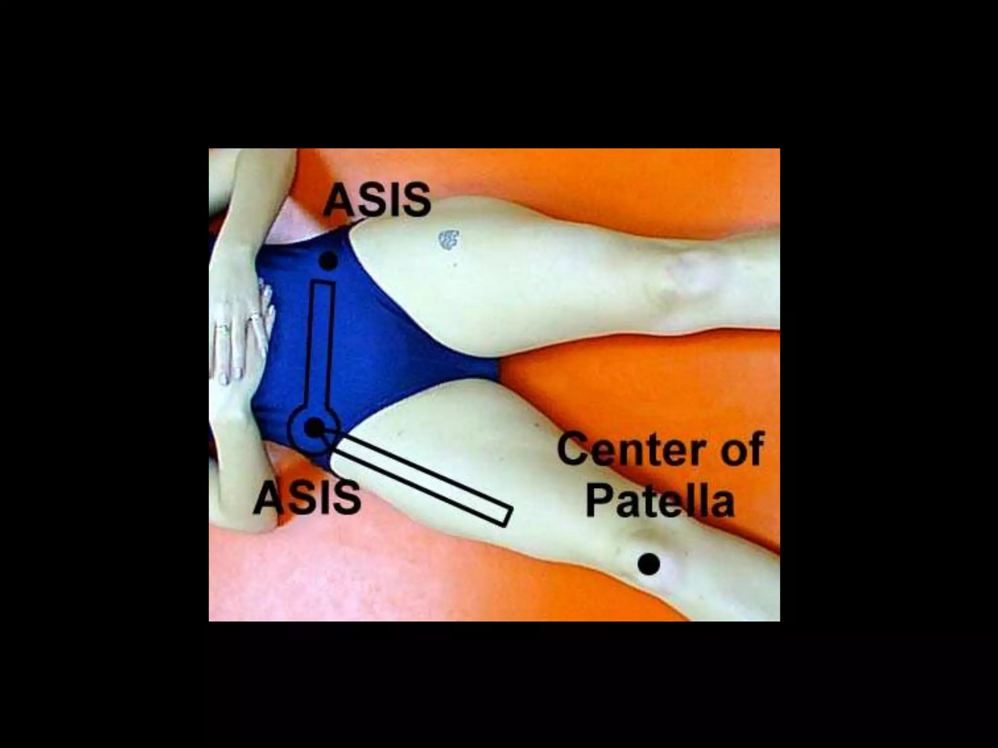
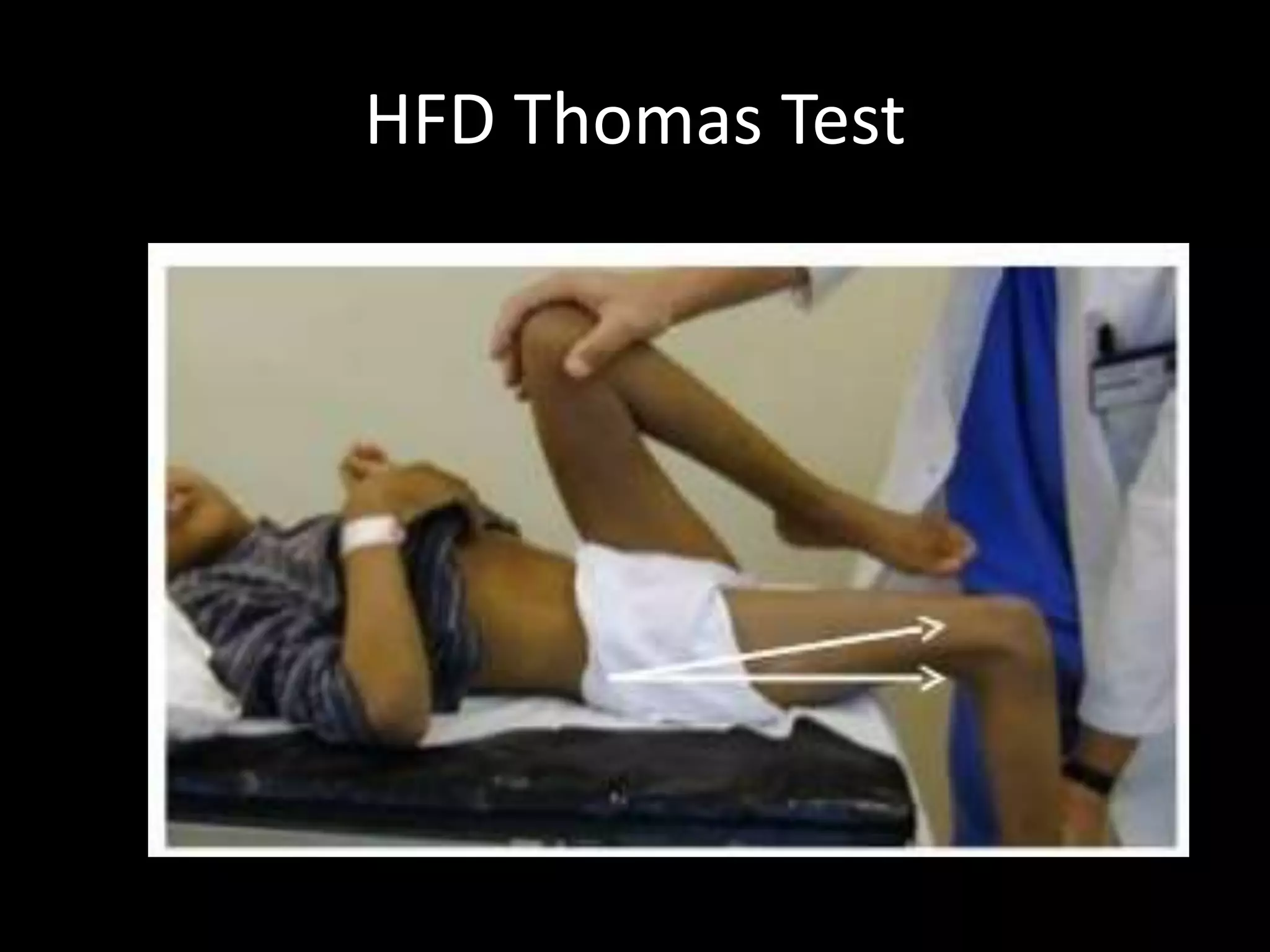
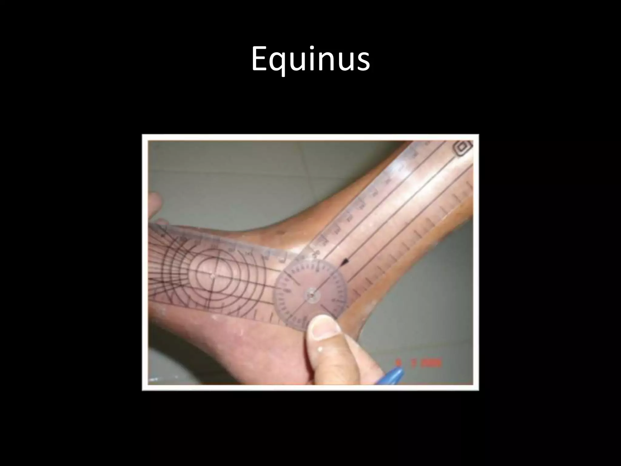
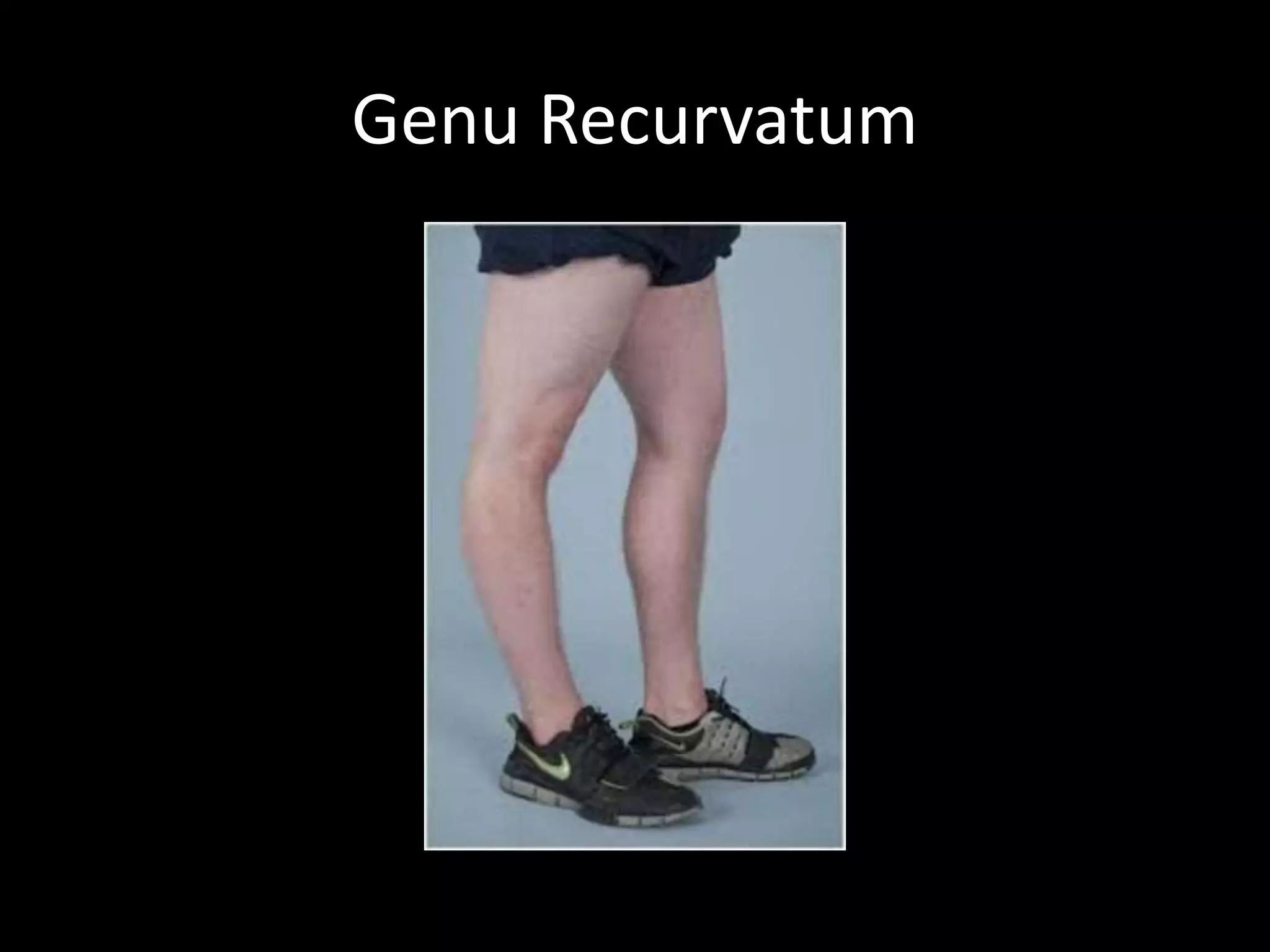
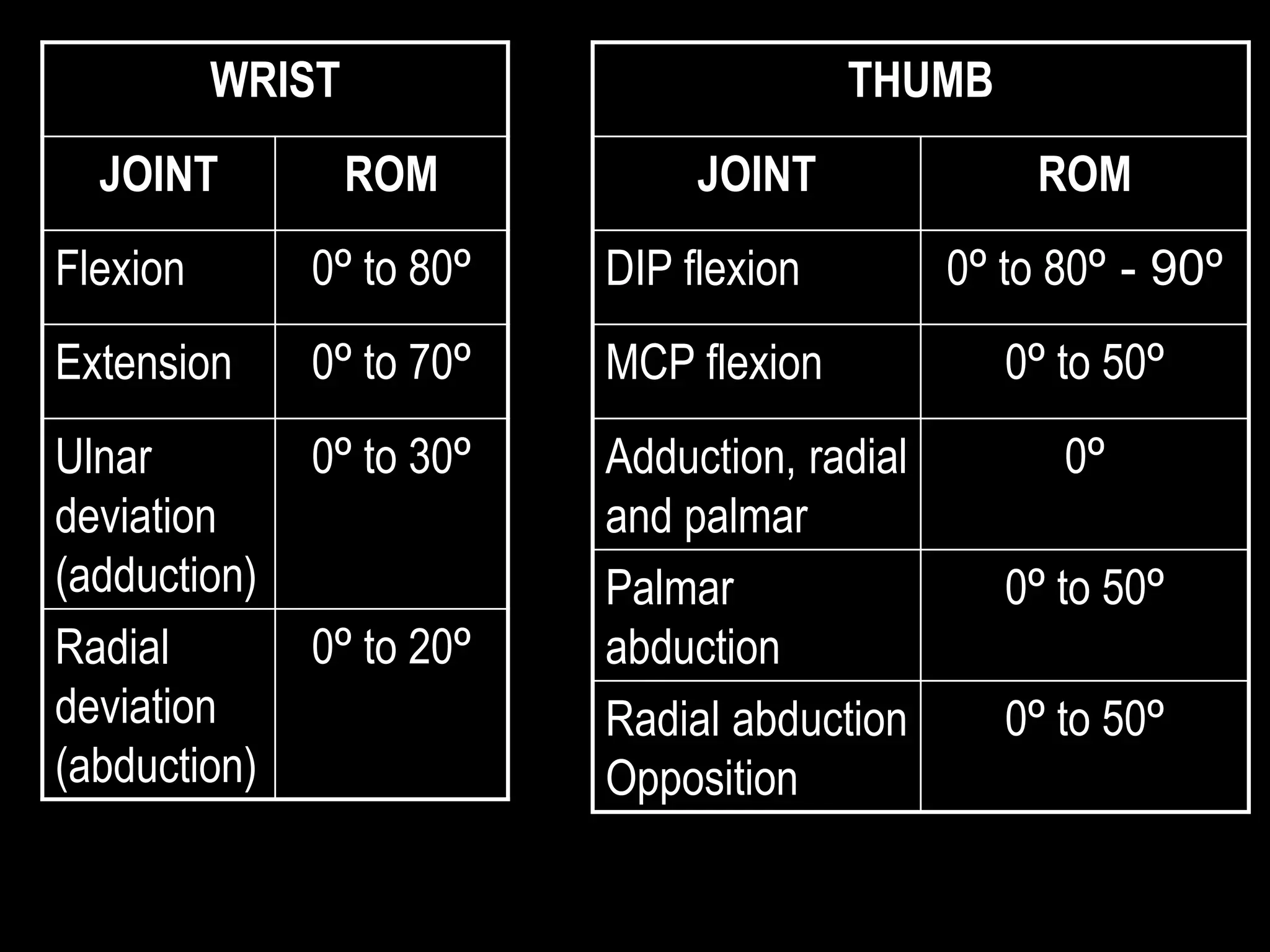
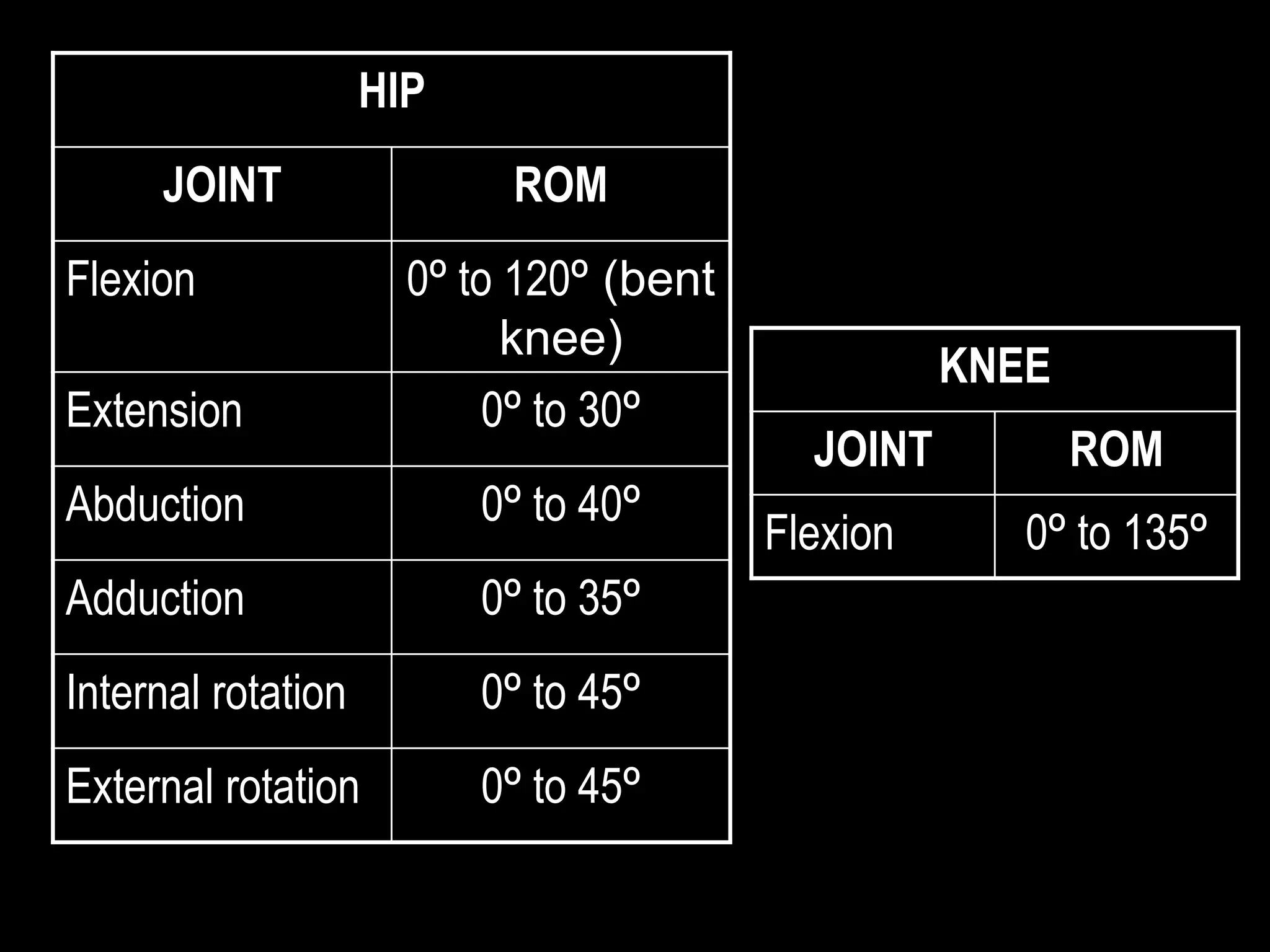
Goniometry refers to the measurement of joint angles in the human body. It is an important part of a physical examination to determine range of motion, evaluate progress, and modify treatment. There are different types of goniometers used to measure motion in various planes at joints like the shoulder, elbow, wrist, fingers, hip, and spine. Factors like a person's age, joint health, surrounding soft tissues, and pathological conditions can impact the normal range of motion values. Proper positioning, stabilization, and identification of bony landmarks is required to accurately measure and document a joint's range of motion.
Co-ordination Exercise,Definitions,Nervous control,Motor pathway,Cerebral cortex,Kinesthetic sensation,Causes of Incoordination,Flaccidity
Spasticity ,Cerebellar ataxia,Loss of kinesthetic sensation,Types of coordination,Posterior column,Test for Incoordination.
Co-ordination Exercise,Definitions,Nervous control,Motor pathway,Cerebral cortex,Kinesthetic sensation,Causes of Incoordination,Flaccidity
Spasticity ,Cerebellar ataxia,Loss of kinesthetic sensation,Types of coordination,Posterior column,Test for Incoordination.
Goniometry is the measuring of angles created by the bones of the body at the joints.1, 2, 3
The term goniometry is derived from two Greek words, gonia meaning angle and metron, meaning measure. 1, 2, 3, 4, 5,
System to measure the joint ranges in each plane of the joint is termed goniometry. 4
These measurements are done with instrument such as goniometer, a tape measure, inclinometers or by visual estimate.
Brian Mulligan described novel concept of the simultaneous application of therapist applied accessory mobilizations and patient generated active movements
Posture - a perquisite for functional abilities in daily life. Posture is a combination of anatomy and physiology with inherent application of bio-mechanics and kinematics. Sitting, standing, walking are all functional activities depending on the ability of the body to support that posture to carry out each activity. Injuries and pathologies either postural or structural can massively change the bio-mechanics of posture and thus affect functional abilities.
A chronicle on muscle strengthening:
MMT is a procedure for the evaluation of strength of individual
muscle or muscles group, based upon the effective performance of a movement in relation to the forces of gravity or manual resistance through the available ROM.
Some slides are taken from different textbooks of medicine like Davidson, Kumar and Clark and Oxford, and some from other presentations made by respected tutors. I'm barely responsible for compilation of various resources per my interest. These resources are free for use, and I do not claim any copyright. Hoping knowledge remains free for all, forever.
The Gram stain is a fundamental technique in microbiology used to classify bacteria based on their cell wall structure. It provides a quick and simple method to distinguish between Gram-positive and Gram-negative bacteria, which have different susceptibilities to antibiotics
263778731218 Abortion Clinic /Pills In Harare ,sisternakatoto
263778731218 Abortion Clinic /Pills In Harare ,ABORTION WOMEN’S CLINIC +27730423979 IN women clinic we believe that every woman should be able to make choices in her pregnancy. Our job is to provide compassionate care, safety,affordable and confidential services. That’s why we have won the trust from all generations of women all over the world. we use non surgical method(Abortion pills) to terminate…Dr.LISA +27730423979women Clinic is committed to providing the highest quality of obstetrical and gynecological care to women of all ages. Our dedicated staff aim to treat each patient and her health concerns with compassion and respect.Our dedicated group ABORTION WOMEN’S CLINIC +27730423979 IN women clinic we believe that every woman should be able to make choices in her pregnancy. Our job is to provide compassionate care, safety,affordable and confidential services. That’s why we have won the trust from all generations of women all over the world. we use non surgical method(Abortion pills) to terminate…Dr.LISA +27730423979women Clinic is committed to providing the highest quality of obstetrical and gynecological care to women of all ages. Our dedicated staff aim to treat each patient and her health concerns with compassion and respect.Our dedicated group of receptionists, nurses, and physicians have worked together as a teamof receptionists, nurses, and physicians have worked together as a team wwww.lisywomensclinic.co.za/
NVBDCP.pptx Nation vector borne disease control programSapna Thakur
NVBDCP was launched in 2003-2004 . Vector-Borne Disease: Disease that results from an infection transmitted to humans and other animals by blood-feeding arthropods, such as mosquitoes, ticks, and fleas. Examples of vector-borne diseases include Dengue fever, West Nile Virus, Lyme disease, and malaria.
These lecture slides, by Dr Sidra Arshad, offer a quick overview of the physiological basis of a normal electrocardiogram.
Learning objectives:
1. Define an electrocardiogram (ECG) and electrocardiography
2. Describe how dipoles generated by the heart produce the waveforms of the ECG
3. Describe the components of a normal electrocardiogram of a typical bipolar lead (limb II)
4. Differentiate between intervals and segments
5. Enlist some common indications for obtaining an ECG
6. Describe the flow of current around the heart during the cardiac cycle
7. Discuss the placement and polarity of the leads of electrocardiograph
8. Describe the normal electrocardiograms recorded from the limb leads and explain the physiological basis of the different records that are obtained
9. Define mean electrical vector (axis) of the heart and give the normal range
10. Define the mean QRS vector
11. Describe the axes of leads (hexagonal reference system)
12. Comprehend the vectorial analysis of the normal ECG
13. Determine the mean electrical axis of the ventricular QRS and appreciate the mean axis deviation
14. Explain the concepts of current of injury, J point, and their significance
Study Resources:
1. Chapter 11, Guyton and Hall Textbook of Medical Physiology, 14th edition
2. Chapter 9, Human Physiology - From Cells to Systems, Lauralee Sherwood, 9th edition
3. Chapter 29, Ganong’s Review of Medical Physiology, 26th edition
4. Electrocardiogram, StatPearls - https://www.ncbi.nlm.nih.gov/books/NBK549803/
5. ECG in Medical Practice by ABM Abdullah, 4th edition
6. Chapter 3, Cardiology Explained, https://www.ncbi.nlm.nih.gov/books/NBK2214/
7. ECG Basics, http://www.nataliescasebook.com/tag/e-c-g-basics
Basavarajeeyam is an important text for ayurvedic physician belonging to andhra pradehs. It is a popular compendium in various parts of our country as well as in andhra pradesh. The content of the text was presented in sanskrit and telugu language (Bilingual). One of the most famous book in ayurvedic pharmaceutics and therapeutics. This book contains 25 chapters called as prakaranas. Many rasaoushadis were explained, pioneer of dhatu druti, nadi pareeksha, mutra pareeksha etc. Belongs to the period of 15-16 century. New diseases like upadamsha, phiranga rogas are explained.
Recomendações da OMS sobre cuidados maternos e neonatais para uma experiência pós-natal positiva.
Em consonância com os ODS – Objetivos do Desenvolvimento Sustentável e a Estratégia Global para a Saúde das Mulheres, Crianças e Adolescentes, e aplicando uma abordagem baseada nos direitos humanos, os esforços de cuidados pós-natais devem expandir-se para além da cobertura e da simples sobrevivência, de modo a incluir cuidados de qualidade.
Estas diretrizes visam melhorar a qualidade dos cuidados pós-natais essenciais e de rotina prestados às mulheres e aos recém-nascidos, com o objetivo final de melhorar a saúde e o bem-estar materno e neonatal.
Uma “experiência pós-natal positiva” é um resultado importante para todas as mulheres que dão à luz e para os seus recém-nascidos, estabelecendo as bases para a melhoria da saúde e do bem-estar a curto e longo prazo. Uma experiência pós-natal positiva é definida como aquela em que as mulheres, pessoas que gestam, os recém-nascidos, os casais, os pais, os cuidadores e as famílias recebem informação consistente, garantia e apoio de profissionais de saúde motivados; e onde um sistema de saúde flexível e com recursos reconheça as necessidades das mulheres e dos bebês e respeite o seu contexto cultural.
Estas diretrizes consolidadas apresentam algumas recomendações novas e já bem fundamentadas sobre cuidados pós-natais de rotina para mulheres e neonatos que recebem cuidados no pós-parto em unidades de saúde ou na comunidade, independentemente dos recursos disponíveis.
É fornecido um conjunto abrangente de recomendações para cuidados durante o período puerperal, com ênfase nos cuidados essenciais que todas as mulheres e recém-nascidos devem receber, e com a devida atenção à qualidade dos cuidados; isto é, a entrega e a experiência do cuidado recebido. Estas diretrizes atualizam e ampliam as recomendações da OMS de 2014 sobre cuidados pós-natais da mãe e do recém-nascido e complementam as atuais diretrizes da OMS sobre a gestão de complicações pós-natais.
O estabelecimento da amamentação e o manejo das principais intercorrências é contemplada.
Recomendamos muito.
Vamos discutir essas recomendações no nosso curso de pós-graduação em Aleitamento no Instituto Ciclos.
Esta publicação só está disponível em inglês até o momento.
Prof. Marcus Renato de Carvalho
www.agostodourado.com
Adv. biopharm. APPLICATION OF PHARMACOKINETICS : TARGETED DRUG DELIVERY SYSTEMSAkankshaAshtankar
MIP 201T & MPH 202T
ADVANCED BIOPHARMACEUTICS & PHARMACOKINETICS : UNIT 5
APPLICATION OF PHARMACOKINETICS : TARGETED DRUG DELIVERY SYSTEMS By - AKANKSHA ASHTANKAR
Knee anatomy and clinical tests 2024.pdfvimalpl1234
This includes all relevant anatomy and clinical tests compiled from standard textbooks, Campbell,netter etc..It is comprehensive and best suited for orthopaedicians and orthopaedic residents.
Title: Sense of Taste
Presenter: Dr. Faiza, Assistant Professor of Physiology
Qualifications:
MBBS (Best Graduate, AIMC Lahore)
FCPS Physiology
ICMT, CHPE, DHPE (STMU)
MPH (GC University, Faisalabad)
MBA (Virtual University of Pakistan)
Learning Objectives:
Describe the structure and function of taste buds.
Describe the relationship between the taste threshold and taste index of common substances.
Explain the chemical basis and signal transduction of taste perception for each type of primary taste sensation.
Recognize different abnormalities of taste perception and their causes.
Key Topics:
Significance of Taste Sensation:
Differentiation between pleasant and harmful food
Influence on behavior
Selection of food based on metabolic needs
Receptors of Taste:
Taste buds on the tongue
Influence of sense of smell, texture of food, and pain stimulation (e.g., by pepper)
Primary and Secondary Taste Sensations:
Primary taste sensations: Sweet, Sour, Salty, Bitter, Umami
Chemical basis and signal transduction mechanisms for each taste
Taste Threshold and Index:
Taste threshold values for Sweet (sucrose), Salty (NaCl), Sour (HCl), and Bitter (Quinine)
Taste index relationship: Inversely proportional to taste threshold
Taste Blindness:
Inability to taste certain substances, particularly thiourea compounds
Example: Phenylthiocarbamide
Structure and Function of Taste Buds:
Composition: Epithelial cells, Sustentacular/Supporting cells, Taste cells, Basal cells
Features: Taste pores, Taste hairs/microvilli, and Taste nerve fibers
Location of Taste Buds:
Found in papillae of the tongue (Fungiform, Circumvallate, Foliate)
Also present on the palate, tonsillar pillars, epiglottis, and proximal esophagus
Mechanism of Taste Stimulation:
Interaction of taste substances with receptors on microvilli
Signal transduction pathways for Umami, Sweet, Bitter, Sour, and Salty tastes
Taste Sensitivity and Adaptation:
Decrease in sensitivity with age
Rapid adaptation of taste sensation
Role of Saliva in Taste:
Dissolution of tastants to reach receptors
Washing away the stimulus
Taste Preferences and Aversions:
Mechanisms behind taste preference and aversion
Influence of receptors and neural pathways
Impact of Sensory Nerve Damage:
Degeneration of taste buds if the sensory nerve fiber is cut
Abnormalities of Taste Detection:
Conditions: Ageusia, Hypogeusia, Dysgeusia (parageusia)
Causes: Nerve damage, neurological disorders, infections, poor oral hygiene, adverse drug effects, deficiencies, aging, tobacco use, altered neurotransmitter levels
Neurotransmitters and Taste Threshold:
Effects of serotonin (5-HT) and norepinephrine (NE) on taste sensitivity
Supertasters:
25% of the population with heightened sensitivity to taste, especially bitterness
Increased number of fungiform papillae
Title: Sense of Smell
Presenter: Dr. Faiza, Assistant Professor of Physiology
Qualifications:
MBBS (Best Graduate, AIMC Lahore)
FCPS Physiology
ICMT, CHPE, DHPE (STMU)
MPH (GC University, Faisalabad)
MBA (Virtual University of Pakistan)
Learning Objectives:
Describe the primary categories of smells and the concept of odor blindness.
Explain the structure and location of the olfactory membrane and mucosa, including the types and roles of cells involved in olfaction.
Describe the pathway and mechanisms of olfactory signal transmission from the olfactory receptors to the brain.
Illustrate the biochemical cascade triggered by odorant binding to olfactory receptors, including the role of G-proteins and second messengers in generating an action potential.
Identify different types of olfactory disorders such as anosmia, hyposmia, hyperosmia, and dysosmia, including their potential causes.
Key Topics:
Olfactory Genes:
3% of the human genome accounts for olfactory genes.
400 genes for odorant receptors.
Olfactory Membrane:
Located in the superior part of the nasal cavity.
Medially: Folds downward along the superior septum.
Laterally: Folds over the superior turbinate and upper surface of the middle turbinate.
Total surface area: 5-10 square centimeters.
Olfactory Mucosa:
Olfactory Cells: Bipolar nerve cells derived from the CNS (100 million), with 4-25 olfactory cilia per cell.
Sustentacular Cells: Produce mucus and maintain ionic and molecular environment.
Basal Cells: Replace worn-out olfactory cells with an average lifespan of 1-2 months.
Bowman’s Gland: Secretes mucus.
Stimulation of Olfactory Cells:
Odorant dissolves in mucus and attaches to receptors on olfactory cilia.
Involves a cascade effect through G-proteins and second messengers, leading to depolarization and action potential generation in the olfactory nerve.
Quality of a Good Odorant:
Small (3-20 Carbon atoms), volatile, water-soluble, and lipid-soluble.
Facilitated by odorant-binding proteins in mucus.
Membrane Potential and Action Potential:
Resting membrane potential: -55mV.
Action potential frequency in the olfactory nerve increases with odorant strength.
Adaptation Towards the Sense of Smell:
Rapid adaptation within the first second, with further slow adaptation.
Psychological adaptation greater than receptor adaptation, involving feedback inhibition from the central nervous system.
Primary Sensations of Smell:
Camphoraceous, Musky, Floral, Pepperminty, Ethereal, Pungent, Putrid.
Odor Detection Threshold:
Examples: Hydrogen sulfide (0.0005 ppm), Methyl-mercaptan (0.002 ppm).
Some toxic substances are odorless at lethal concentrations.
Characteristics of Smell:
Odor blindness for single substances due to lack of appropriate receptor protein.
Behavioral and emotional influences of smell.
Transmission of Olfactory Signals:
From olfactory cells to glomeruli in the olfactory bulb, involving lateral inhibition.
Primitive, less old, and new olfactory systems with different path
3. What is Goniometry?
• The term goniometry is derived from two Greek words :
Gonia-metron
• Therefore, goniometry refers to the measurement of angles, in
particular the measurement of angles created at human joints
by the bones.
ANGLE MEASURE
4. PARTS OF MOTOR EXAMINATION
1. Nutrition Of Muscle
2. Muscle Tone
3. Reflexes
4. Range Of Motion and TCD’s
5. Manual Muscle Testing
5. Why Is It Performed ?
• Determining the presence of joint impairment
• Developing treatment goals.
• Evaluating progress or lack of progress.
• Modifying treatment.
• Motivating the subject.
• Research
7. PLANES AND AXIS
• Osteo-kinematic motions are described to be taking place in 3
cardinal planes and axis
8.
9. Synovial joint
• Most evolved & hence most mobile type of
joints
• The ends of bony components are free to
move in relation to one another
• Bony components are indirectly connected to
one another by means of a joint capsule that
encloses the joint
10. Joint Ranges
Active ROM Passive ROM
• Active motion is the unassisted voluntary
movement of a joint. (Quality of ROM)
• Passive motion is attained by the examiner
without the patient’s assistance. (Quantity of ROM
)
• ** Normally, PROM is slightly greater than AROM
because joints have a small amount of motion at
the end range that is not under voluntary control.
12. physiologic motion is limited
by a physiologic barrier
tension develops within the
surrounding tissues
(joint capsule, ligaments
and connective tissue)
13. additional amount of passive
range of motion can be performed the anatomic
barrier cannot be
exceeded without
disrupting the
joints integrity
16. ACTIVE INSUFFICIENCY?
Flex your wrist
completely
Attempt to
tighten your
fist
• A muscle
cantcontractmaximally
across both joints
together
Much force
than in
slightly
extended
position
• The multi joint long
finger flexors enter
active insufficiency
when wrist flexes
• Shortest possible
length of muscle
17. PASSIVE INSUFFICIENCY?
FLEXION OF THE FINGERS
IS A RESULT OF
INSUFFICIENT
EXTENSIBILTY OF THE
FINGER FLEXORS
STRETCHED OVER
EXTENDED WRIST
EXTENSION OF THE
FINGERS IS A RESULT OF
INSUFFICIENT LENGTH OF
THE FINGER EXTENSORS
STRETCHED OVER FLEXED
WRIST
• LONGEST POSSIBLE LENGTH OF MUSCLE
• Muscle cant stretch maximally at both joints together
18. Other Examples of AI PI In Body and its
clinical relevance with Goniometry
• BICEPS : At the top of curl, (when biceps begin to smash against
forearm), when elbows are lifted
**Shortens biceps over both the shoulder & elbow blade
• Simultaneously lengthening the TRICEPS
• HAMS : When reaching to touch toes
**Lengthening felt as a stretch
• RECTUS FEMORIS : Hip flexion with knee extension(70 degree) is
less than hip flexion with knees bent (120 degree)
• GASTROCNEMIUS : Seated calf / heel raise places the
gastrocnemius into active insufficiency since the knee flexes too
much & ankle performs plantarflexion
19. MEASURING
JOINT RANGE OF MOTION
• Range Of Motion (ROM) is the arc of motion that
occurs at a joint or a series of joints.
• Three notation systems have been used to
define ROM :
1. The 0 to 180 degree system
2. The 180 to 0 degree system
3. The 360 degree system
Most commonly used is the 0 to 180 degree
notation system
20. Prerequisite Knowledge For Measuring ROM
a) Normal ROM’s (Range)
b) Joint Structure And Function
c) Recommended positioning for self and patient
d) Bony landmarks related to each joint
e) Alignment of Goniometer
f) Normal end-feel
g) Factors that can alter normal ROM
21. FACTORS DETERMINING AMOUNT OF ROM
Integrity Of
Joint
SurfaceRELIABILITY
Amount Of
Scarring
Present
AGE
GENDER
Shape Of
Articulating
Surface
Health
Of
Joint
Various
diseases/
pathological
conditions
Health Of
Surrounding
Tissues
Mobilty &
Pliabilty Of Soft
Tissue
22. Common pathological causes of ROM
Restriction
• Skin/soft tissue contracture
• Arthritis
• Fracture
• Burns
• Muscle weakness/paralysis
• Pain
• Edema
• Spasticity
• Presence of foreign body in the joint
23. Prerequisite Skills For Measuring ROM
• The therapist should be skilled in
Correct positioning (Pt/ Pt Jt/ PT And GM)
Stabilization for measurement
Palpation
Alignment
Recording measurements accurately
Documentation
24. • Visual observation of the joint and its adjacent
area is important to look for :
a) Compensatory motions
b) Posture
c) Muscle contour
d) Skin creases
e) Facial expressions
25. Testing Procedure
PLACE THE SUBJECT IN TESTING
POSITION
STABILIZE THE PROXIMAL JOINT SEGMENT
MOVE THE DISTAL JOINT SEGMENT TO ZERO STARTING POSITION. SLOWLY MOVE
THE DISTAL JOINT SEGMENT TO THE END OF PASSIVE ROM AND DETERMINE END FEEL
MAKE VISUAL ESTIMATE OF THE ROM
RETURN THE DISTAL JOINT SEGMENT TO THE STARTING POSITION
PALPATE THE BONY ANATOMICAL LANDMARKS
ALIGN THE GONIOMETER
26. READ & RECORD THE STARTING POSITION.
REMOVE THE GONIOMETER
STABILIZE THE PROXIMAL JOINT SEGMENT
MOVE THE DISTAL SEGMENT
THROUGH FULL ROM
REPLACE & REALIGN THE GONIOMETER. PALPATE THE ANATOMICAL LAND
MARKS AGAIN IF NECESSARY
READ & RECORD THE ROM
27. Joint Mobility Scale
Hyper Mobility
(Mild, Moderate,
Severe)
Exercise, Bracing
surgery
Normal mobility Normal function
Hypo Mobility
(Mild, Moderate,
Severe)
Exercise, Mobilization,
surgery
N
28. Documentation
• Hypo Mobility : A motion that does not start with 0
degree or ends prematurely indicates joint
hypomobility
Example : if knee joint has 30 degree of hypomobility in
flexion, it would be recorded as 30 – 135 deg
• Hyper Mobility : Joint hypermobility at the beginning
of the range is noted by inclusion of a zero between the
starting & ending measurements
Example : if the elbow joint has 5 degree of
hypermobility in extension and 140 degree of flexion ,
it would be recorded as 5 – 0 – 140 deg
29. Types of Goniometer
• Full Circle Manual Universal Goniometer (360)
• Half circle manual Goniometer (180)
• Gravity Goniometer :-
• a) Double Inclinometer (used for spine goniometry)
• b) Pendulum Inclinometer
• c) Bubble Goniometer
• Electrogoniometer
• Digital Goniometer
• Tape Measurements
• Smartphone Devices
• Use of malleable wires/sheets (in cases of deformities)
33. UNIVERSAL GONIOMETER
• A universal Goniometer may be constructed of
metal or plastic and it has 3 parts :-
1. Body of Goniometer
2. Stationary arm
3. Movable arm
(placed over the Joint being measured)
(aligned parallel with the longitudinal axis of the
fixed part)
(aligned parallel with the longitudinal axis of the
movable part)
36. Precautions !!!
1. Joint irritability status
2. Presence of Pain
3. Instability
4. Recent trauma
5. Is it really important to assess accurate ROM ??
37. Functional Ranges of various joint in
various activities
Walking
Stair ascending descending
Sitting
Squatting
Cross leg sitting
Self Feeding
Back reach
Neck reach
Etc….
38. ROM Required In ADL’s
ASCENDING STAIRS REQUIRES
BETWEEN
47 - 66 DEGREE OF HIP FLEXION
DEPENDING ON STAIR
DIMENSION
DESCENDING STAIRS REQUIRES AN
AVERAGE OF
21 - 36 DEGREE OF DORSIFLEXION,
86.9 - 107 DEGREE OF KNEE FLEXION
DEPENDING ON STAIR DIMENSIONS
39. Rising from a chair requires a mean range of
knee flexion of 90.1 - 95.0 degree and
full dorsiflexion ROM depending on height
of seat
Sitting in a chair with an
average seat height requires
112 degrees of hip flexion
40. Drinking from a cup requires about
130 degree of elbow flexion
36 to 52 degrees of shoulder flexion
Reaching objects on a high shelf
require
148 degrees of shoulder flexion
41. Using a telephone requires
approx 40 degrees of wrist
extension
Approximately
50 degrees of pronation
occur while reading a newspaper
Reaching behind the head
requires about
112 degrees of abduction
of the shoulder
42. END-FEEL
• The end of each motion at each joint is limited
from further movement by particular
anatomical structures.
• The type of structure that limits a joint motion
has a characteristic feel, which may be detected
by the therapist performing the passive ROM.
• This feeling, which is experienced by the
therapist as resistance or a barrier to further
motion, is called the end-feel.
43. NORMAL END-FEEL DESCRIPTION EXAMPLE
Soft Soft Tissue Approximation Knee flexion (contact
between soft tissue of
posterior leg and posterior
thigh)
Firm Muscular stretch
Capsular stretch
Ligamentous stretch
Hip flexion with knee
straight (passive elastic
tension of hamstring
muscles)
Extension of
metacarpophalangeal joints
of fingers
Forearm supination (tension
in the palmar radioulnar
ligament of the inferior
radioulnar joint)
Hard Bone contacting bone Elbow extension (olecranon
process of the ulna and
olecranon fossa of humerus)
44. ABNORMAL END-FEEL DESCRIPTION EXAMPLES
Soft Occurs sooner or later in the
ROM than is usual or in a joint
that normally has a firm or
hard end-feel . Feels boggy.
Soft tissue edema
Synovitis
Firm Occurs sooner or later in the
ROM than is usual or in a joint
that normally has a soft or
hard end-feel.
Increased muscular tonus
Capsular , muscular ,
ligamentous, and fascial
shortening
Hard Occurs sooner or later in the
ROM than is usual or in a joint
that normally has a soft or
firm end-feel. A bony grating
or bony block is felt.
Chondromalacia
Osteoarthritis
Loose bodies in joint
Myositis ossificans
Fracture
Empty No real end-feel because pain
prevents reaching end of
ROM. No resistance is felt
except for patient’s protective
muscle splinting or muscle
spasm.
Acute joint inflammation
Bursitis
Abscess
Fracture
Psychogenic disorder
45. JOINT MOTION TESTING
POSITION
STABILIZATION MEASUREMENTS
CERVICAL • FLEXION
• EXTENSION
• SIDE FLEXION
• ROTATION
Sitting Shoulder & chest
Shoulder & chest
to prevent
extension of
thoracic &
lumbar spine
To prevent side
flexion of
thoracic &
lumbar spine
To prevent
rotation of
thoracic &
lumbar spine
1 cm– 4.3 cm
18.5 cm–22.4cm
10.7cm-12.9cm
11cm-13.2cm
TAPE MEASUREMENTS OF THE SPINE
46. JOINT MOTION TESTING
POSITION
STABILIZATION MEASUREMENTS
THORACIC • FLEXION
• EXTENSION
• LATERAL
FLEXION
• ROTATION
STANDING
•If the subject
has balance
problems or
muscle weakness
in the LE,
measurement
can be taken in
prone/side lying
SITTING
PELVIS
To prevent
anterior tilting
To prevent
posterior tilting
To prevent lateral
tilting
To prevent
rotation
10 cms (4 inches)
15.9cm for rt LF
16.9cm for lt LF
45 degree
(universal
goniometer)
47. JOINT MOTION TESTING
POSITION
STABILIZATION MEASUREMENTS
LUMBAR • FLEXION
•EXTENSION
•LATERAL
FLEXION
STANDING PELVIS
To prevent
anterior tilting
To prevent
posterior tilting
To prevent
lateral tilting
6.7cm in males
5.8cm in females
Average
6.3cm-6.9cm
(Modified
Schober test)
1.6cm (Modified
Schober Test)
25 – 30 degree
by AMA (double
inclinometer)
49. Capsular & Non-capsular Pattern Of
Movement Restriction
• Cyriax proposed that pathological conditions
involving the entire joint capsule cause a
particular pattern of restriction involving most
of the passive motions of the joint. This pattern
is called as capsular pattern
• Restriction caused by condition involving
structures other than the entire joint capsule is
called as non-capsular pattern
• Example – Adhesive Capsulitis Shoulder
64. CERVICAL SPINE
JOINT ROM
Flexion 0º to 45º
Extension 0º to 45º
Lateral flexion 0º to 45º
Rotation 0º to 60º
THORACIC AND LUMBAR
SPINE
JOINT ROM
Flexion 0º to 80º
Extension 0º to 30º
Lateral flexion 0º to 40º
Rotation 0º to 45º
65. SHOULDER
JOINT ROM
Flexion 0º to 180º
Extension 0º to 60º
Abduction 0º to 180º
Adduction 0º
Horizontal abduction 0º to 40º
Horizontal Adduction 0º to 130º
Internal rotation
Arm in Abduction 0º to 70º
Arm in Adduction 0º to 60º
External rotation
Arm in Abduction 0º to 90º
Arm in Adduction 0º to 80º
66. ELBOW
JOINT ROM
Flexion 0º to 135º - 150º
Extension 0º
FOREARM
JOINT ROM
Pronation 0º to 80º - 90º
Supination 0º to 80º - 90º
67. WRIST
JOINT ROM
Flexion 0º to 80º
Extension 0º to 70º
Ulnar
deviation
(adduction)
0º to 30º
Radial
deviation
(abduction)
0º to 20º
THUMB
JOINT ROM
DIP flexion 0º to 80º - 90º
MCP flexion 0º to 50º
Adduction, radial
and palmar
0º
Palmar
abduction
0º to 50º
Radial abduction
Opposition
0º to 50º
68. FINGERS
JOINT ROM
MCP flexion 0º to 90º
MCP hyperextension 0º to 15º - 45º
PIP flexion 0º to 110º
DIP flexion 0º to 80º
abduction 0º to 25º
69. HIP
JOINT ROM
Flexion 0º to 120º (bent
knee)
Extension 0º to 30º
Abduction 0º to 40º
Adduction 0º to 35º
Internal rotation 0º to 45º
External rotation 0º to 45º
KNEE
JOINT ROM
Flexion 0º to 135º
70. ANKLE AND FOOT
JOINT ROM
Plantar flexion 0º to 50º
Dorsiflexion 0º to 15º
Inversion 0º to 35º
Eversion 0º to 20º
71. SOURCES
• Measurement of Joint Motion : A Guide
to Goniometry, 4th Edition, by Cynthia C. Norkin
• Physical Rehabilitation 6th Edition SuSan B.
O’Sullivan
• Magee (2002). Orthopedic physical Assessment (4th
ed.). Phil: Saunders.
• Kisner C, & Colby LA (2002). Therapeutic
exercise: Foundations and techniques (4th ed.). PA:
FA Davis.
• The Principles of Exercise Therapy (Fourth
Edition): M. Dena Gardiner.