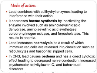
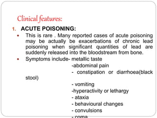
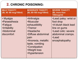
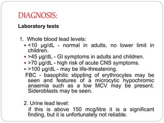
CLINICAL TOXICOLOGY
PREPARED BY,
NIBY,
IV-PHARM-D
ARSENIC AND LEAD POISONING
ARSENIC POISONING
arsenic:
 Arsenic is thought to occur through out the
universe.
 It is the twentieth most common element in the
earth’s crust, having a concentration of 1.8
ppm.
 Arsenic is today the commonest source of
acute heavy metal poisoning , and is second
only to lead in the incidence of chronic toxicity.
 It exists in compounds that may be organic or
inorganic.
 Elemental arsenic is the least toxic.
Physical appearance:
 Arsenic is a metalloid i.e. it is an element
which resembles a metal in some respects,
and is by itself not very toxic.
 Arsenic is a silver-grey or tin white, shiny,
brittle, crystalline, and metallic looking
element.
 It is rarely found in its isolated, elemental
form.
 More commonly it is present in mineral
species in alloys or as an oxide or other
compound form.
Inorganic and organic arsenicals
CHEMICAL NAME USES
1 ELEMENTAL ARSENIC In alloys
2 ARSINE Lead plating,soldering,galvanising,
and in electronic components
3 TRIMETHYL ARSINE Present in sewage
4 ARSENIC TRIOXIDE Manufacture of glass ,insecticide,
rodenticide,previously used in medicine for
fever(Flower’s solution)
5 ARSENIC TRICHLORIDE Pottery
6 SODIUM ARSENITE Wood preservative,insecticide, veterinary use
7 ARSENIC PENTOXIDE Manufacture of coloured glass,insecticide ,wood
preservative
8 LEAD ARSENATE Insecticide
9 SULPHIDES OF ARSENIC Depilatory
10 COPPER ARSENATE Colouring agent for toys, wallpaper,etc.
11 ORGANIC
ARSENICALS(carbarsone
Previously used in therapeutics
USUAL FATAL DOSE:
 200-300 mg for arsenic trioxide.
 In general,the pentavalent form of arsenic
(arsenate) is less toxic than the trivalent
form(arsenite) because it is less water
soluble.
 The most toxic form is arsine gas(25-30 ppm
can be lethal in 30 minutes)
Toxicokinetics and mode of action:
 Arsenic is absorbed through all portals of entry
including oral, inhalational, and cutaneous routes.
 After absorption it is redistributed to the liver ,
lungs, intestinal wall, spleen, where it binds to the
sulfhydryl groups of tissue proteins.
 Arsenic replaces phosphorus in the bone where it
may remain for years. It gets deposited also in
hair.
 While arsenic does not crosses the blood brain
barrier easily, it crosses the placenta readily and
can give rise to intrauterine death of the feotus.
 In less severe intoxications it can cause respiratory
distress of the newborn due to pulmonary
haemorrhage and hyaline membrane formation.
Clinical features:
SYSTEM ACUTE CHRONIC
DERMAL Hair loss,transverse bands of
opacity in nails.
melanosis(neck,yelids,nipples)
bowen’s disease,facial
oedema,hyperkeratosis,hyperpig
mentation,skin cancer.
OCULAR Conjunctivitis, lacrimation Dimness of vision
GI Abdominal pain, metallic taste,
dysphagia, vomiting, bloody or
rice-water diarrhoea. Garlicky
odour in breath
Anorexia, nausea, vomiting,
diarrhoea,weight loss
AIRWAYS Irritation of upper airways Perforation of nasal septum,
chronic laryngitis, bronchitis
LIVER Fatty degeneration Hepatomegaly , jaundice,
cirrhosis
KIDNEY Oliguria , uraemia Nephritic changes
NEUROLOGICA
L
Hyperpyrexia, convulsions,coma Encephalopathy, polyneuritiss,
tremor, ataxia, limb tenderness,
difficulty in walking
CARDIAC Tachycardia, hypotension, Hypotensin, myocarditis
Diagnosis:
1. Urine level: if the 24 hr excretion of arsenic
exceeds 100mcg, it is indicative of toxicity.
2. Blood level: less reliable than urine level because
of short half life of arsenic in blood.A level of
arsenic less than 7mcg/100ml-normal range.
3. Hair level: possibility of scalp hair being
contaminated with arsenic from the environment.
4. Radiography: arsenic is radio-opaque,abdominal
x-ray reveals its presence in GIT in acute
poisoning.
5. Additional investigation:
 Monitor CBC, serum electrolytes, urinalysis, liver
and renal functional tests.
 Obtain ECG- Cardiac monitoring.
 Chest radiography with severe poisoning or
Treatment:
1. Supportive measures: gastric lavage,
intravenous fluids, cardiac monitoring, etc.
2. Chelation therapy: This can be done with –
 BAL(British Anti Lewesite or dimercaprol)-
3to5mg/kg IM every 4 hrs until the urinary
excretion dips below 50mcg/24hrs . Usual
duration of therapy is 7to10 days.
 Penicillamine – can be given orally at a dose of
100mg/kg/day, 6th hourly for 5 days.
 DMSA(Dimercapto succinic acid), or
DMPS(Dimercapto propane sulfonic acid).-
superior to BAL and penicillamine.
Principles of chelation :
 Begin chelation therapy in symptomatic patients. The
urine arsenic level which should prompt chelation in
an asymptomatic patient has been recommended as
200mcg/litre.
 Repeat courses of chelation therapy should be
prescribed in severe poisonings until the 24-hr urine
arsenic level falls below 50 mcg/litre.
 Chelation therapy is not very effective for chronic
poisoning and is totally ineffective in arsine poisoning.
The later should be treated with emphasis on
respiratory stabilisation and haemodialysis.
3. Haemodialysis or exchange transfusion.
LEAD POISONING
LEAD:
 Lead is the commonest metal involved in
chronic poisoning.
 It was one of the first metals known to man
and has been widely used during the last two
thousand years of domestic, industrial, and
therapeutic purposes.
 Lead is abundant in soil, being distributed
throughout the earth’s crust.
Physical appearance:
 Elemental lead exists as a highly lustrous,
heavy, silvery-grey metal with a cubic crystal
structure that assumes a bluish tint as it
tarnishes in air.
 It is quite soft and malleable.
 Several of its salts occur as variously coloured
powders or liquids and are widely in industry
and at home producing cumulative toxicity on
chronic exposure.
Uses:
 Lead acetate(sugar of lead) has been used in
therapeutics.
 Lead carbonate(white lead) is still used in paints.
 Lead oxide(litharge) is essential for glazing of
pottery and enamel ware.
 Tetraethyl lead is mixed with petrol as an antiknock
to prevent detonation in internal combustion
engines.
 Lead tetroxide is the most common compound in
vermilion(“sindoor”).
 Lead sulfide is used as a collyrium(“surma”).
SOURCES OF LEAD EXPOSURE:
ENVIRONMENTA
L
DOMESTIC OCCUPATIONAL NON-
OCCUPATIONAL
•Automobile
exhaust
•Drug abuse
•Soil
•water
•Ceramic ware
•Coloured picture
books
•Contaminated
flour
•Cosmetics
•“health “foods
•House paint
•Indigenous
medicines
•Pencils
•Toys
•Auto repair works
•Battery making
•Glass
manufacture
•Mining
•Plastics
manufacture
•Plumbing
•Pottery
•Printing
•Rubber industry
•Ship building
•Smelting&
refining
•Soldering(electro
nics
•Candle with
lead-containing
wicks
•Ayurvedic
medicines
•Paint
•Retained bullets
•ink
•Automobile
storage battery
casing
•Ceramic glazes
•Lead pipes
•Silvery jewellery
workers
•Renovation
Usual fatal dose:
 This is not relevant to lead since acute poisoning is very
rare.
 The average lethal dose is said to be 10gm/70kg for most
lead salts, while it is 100mg/kg for tetraethyl lead.
TOXICOKINETICS:
 Lead is absorbed through all portals of entry.
-Occupational exposure results mainly from inhalation,
while in most other situations the mode of intake is
ingestion
-Tetraethyl lead can be absorbed rapidly through intact
skin.
 Following absorption it is stored in the bones as
phosphate and carbonate. In children about 70% of total
Mode of action:
 Lead combines with sulfhydryl enzymes leading to
interference with their action.
 It decreases haeme synthesis by inactivating the
enzyme involved such as aminolaevulinic acid
dehydrase, aminolaevulinic acid synthetase,
corporphyrinogen oxidase, and ferrochelatase. This
results in anemia.
 Lead increases haemolysis as a result of which
immature red cells are released into circulation such as
reticulocytes and basophilic stipped cells.
 In CNS, lead causes oedema and has a direct cytotoxic
effect leading to decreased nerve conduction, increased
psychomotor activity,lower IQ, and behavioural
disorders.
Clinical features:
1. ACUTE POISONING:
 This is rare . Many reported cases of acute poisoning
may be actually be exacerbations of chronic lead
poisoning when significant quantities of lead are
suddenly released into the bloodstream from bone.
 Symptoms include- metallic taste
-abdominal pain
- constipation or diarrhoea(black
stool)
- vomiting
-hyperactivity or lethargy
- ataxia
- behavioural changes
- convulsions
2. CHRONIC POISONING:
MILD TOXICITY
(BL 40 -60 mcg/100ml)
MODERATE TOXICITY
(BL 60-100 mcg/100ml)
SEVERE TOXICITY
(BL more than
100mcg/ml)
• Myalgia
•Paraesthesia
•Fatigue
•Irritability
•Abdominal
discomfort
•Arthralgia
•Muscular
exhaustibility
•Tremor
•Headache
•Diffuse abdominal
pain
•Anorexia, metallic
taste, vomiting
•Constipation
•Weight loss
•Hypertension
•Lead palsy: wrist or
foot drop
•A bluish black lead
line on gums
•Lead colic: severe
abdominal cramps.
•Lead
encephalopathy
DIAGNOSIS:
Laboratory tests
1. Whole blood lead levels:
 <10 μg/dL - normal in adults, no lower limit in
children.
 >45 μg/dL - GI symptoms in adults and children.
 >70 μg/dL - high risk of acute CNS symptoms.
 >100 μg/dL - may be life-threatening.
FBC - basophilic stippling of erythrocytes may be
seen and features of a microcytic hypochromic
anaemia such as a low MCV may be present.
Sideroblasts may be seen.
2. Urine lead level:
if this is above 150 mcg/litre it is a significant
finding, but it is unfortunately not reliable.
 Renal function tests to detect renal complications and
uric acid levels to detect gout may also be advisable.
 Nerve conduction tests should be considered if
neuropathy is suspected.
 Psychometric testing should be considered if clinically
indicated.
Radio-imaging
 Plain X-ray may show transverse lines in tubular bones.
These are actually areas of arrested bone growth and
may persist for a long time after exposure ends. They
are not seen in the early phase of exposure.
 Plain abdominal X-rays may show radio-opaque flecks
in cases of suspected lead foreign body ingestion (eg,
pica in children).
 X-ray fluorescence works by detecting specific
emissions from tissues when bombarded with X-rays. It
is a sensitive method of detecting low levels of lead in
the body.
TREATMENT:
1.SEVERE ACUTE POISONING WITH ENCEPHALOPATHY:
a. BAL -4mg/kg immediately (in children)
b. Cranial CT scan: to rule out cerebral oedema
Cerebral oedema managed by:
o Controlled hyperventillation, maintaining an arterial
CO2 tension of 25-30 mmHg can reduce intracranial
pressure.
o DIURETICS- Mannitol 20%- 1-1.5 gm/kg by
infusion over 10-20 minutes(adult) ,0.5-1gm/kg by IV
infusion(child)
Glycerol- 0.3-1gm/kg orally.
o CORTICOSTEROIDS- Dexamethasone- 16mg/day
in divided doses(low dose) & 1-2mg/day in divided
c. For seizures: IV Diazepam (Adult:up to 10mg
slowly, repeat if necessary; Child: 0.-0.3mg/kg
slowly).
d. CaNa2 EDTA 75mg/kg/day IV Infusion.
e. After the initial dose of BAL, reapeat the same dose at 4
hourly itervals until blood level falls below 40
mcg/100ml.Then reduce BAL to 12mg/kg/day in 3
divided doses.
f. Reduce CaNa2 EDTA to 50 mg/kg/day as condition
improves.
- continue the above regimen until patient is
2. SEVERE ACUTE POISONING WITH
ENCEPHALOPATHY:
(BL more than 70mcg/100ml)
a. BAL - 12 mg/kg/day.
b. EDTA -50mg/kg/day.
c. Discontinue BAL when the BL falls below
40mcg/100ml,but continue EDTA for 5 more days.
d. Change to oral chelation subsequently which may
have to be continued until the BL falls below
15mcg/100ml, or 3 months have been completed.
3.MODERATE POISONING:
(BL level between 45 and 70 mcg/100ml)
a. EDTA 50mg/kg/day.
b. When blood lead falls below 40 mcg/100ml, begin
oral chelation.
4.MILD POISONING:
BL level between20 and 35 mcg/100ml
a. D-penicillamine 30 mg/kg/day in 3 divided doses.
Start with ¼ th of the calculated
dose. Double this after 1 week. Double again after
1 week. Continue this until the BL level falls to less
than 15 mcg/100ml .
5. SUPPORTIVE MEASURES:
a. Thiamine 10-50 mg/kg to improve neurological
manifestation.
b. Lead colic usually responds to Ivcalcium
gluconate.
c. Correct iron defficiency present.
d. IV fluids
e. Finally the sine qua non treatment of heavy metal
REFERENCE:
TEXTBOOK-
MODERN MEDICAL TOXICOLOGY- VV PILLAI.
THANK YOU…..

ARSENIC AND LEAD POISONING

  • 1.
  • 2.
  • 3.
    arsenic:  Arsenic isthought to occur through out the universe.  It is the twentieth most common element in the earth’s crust, having a concentration of 1.8 ppm.  Arsenic is today the commonest source of acute heavy metal poisoning , and is second only to lead in the incidence of chronic toxicity.  It exists in compounds that may be organic or inorganic.  Elemental arsenic is the least toxic.
  • 4.
    Physical appearance:  Arsenicis a metalloid i.e. it is an element which resembles a metal in some respects, and is by itself not very toxic.  Arsenic is a silver-grey or tin white, shiny, brittle, crystalline, and metallic looking element.  It is rarely found in its isolated, elemental form.  More commonly it is present in mineral species in alloys or as an oxide or other compound form.
  • 5.
    Inorganic and organicarsenicals CHEMICAL NAME USES 1 ELEMENTAL ARSENIC In alloys 2 ARSINE Lead plating,soldering,galvanising, and in electronic components 3 TRIMETHYL ARSINE Present in sewage 4 ARSENIC TRIOXIDE Manufacture of glass ,insecticide, rodenticide,previously used in medicine for fever(Flower’s solution) 5 ARSENIC TRICHLORIDE Pottery 6 SODIUM ARSENITE Wood preservative,insecticide, veterinary use 7 ARSENIC PENTOXIDE Manufacture of coloured glass,insecticide ,wood preservative 8 LEAD ARSENATE Insecticide 9 SULPHIDES OF ARSENIC Depilatory 10 COPPER ARSENATE Colouring agent for toys, wallpaper,etc. 11 ORGANIC ARSENICALS(carbarsone Previously used in therapeutics
  • 6.
    USUAL FATAL DOSE: 200-300 mg for arsenic trioxide.  In general,the pentavalent form of arsenic (arsenate) is less toxic than the trivalent form(arsenite) because it is less water soluble.  The most toxic form is arsine gas(25-30 ppm can be lethal in 30 minutes)
  • 7.
    Toxicokinetics and modeof action:  Arsenic is absorbed through all portals of entry including oral, inhalational, and cutaneous routes.  After absorption it is redistributed to the liver , lungs, intestinal wall, spleen, where it binds to the sulfhydryl groups of tissue proteins.  Arsenic replaces phosphorus in the bone where it may remain for years. It gets deposited also in hair.  While arsenic does not crosses the blood brain barrier easily, it crosses the placenta readily and can give rise to intrauterine death of the feotus.  In less severe intoxications it can cause respiratory distress of the newborn due to pulmonary haemorrhage and hyaline membrane formation.
  • 8.
    Clinical features: SYSTEM ACUTECHRONIC DERMAL Hair loss,transverse bands of opacity in nails. melanosis(neck,yelids,nipples) bowen’s disease,facial oedema,hyperkeratosis,hyperpig mentation,skin cancer. OCULAR Conjunctivitis, lacrimation Dimness of vision GI Abdominal pain, metallic taste, dysphagia, vomiting, bloody or rice-water diarrhoea. Garlicky odour in breath Anorexia, nausea, vomiting, diarrhoea,weight loss AIRWAYS Irritation of upper airways Perforation of nasal septum, chronic laryngitis, bronchitis LIVER Fatty degeneration Hepatomegaly , jaundice, cirrhosis KIDNEY Oliguria , uraemia Nephritic changes NEUROLOGICA L Hyperpyrexia, convulsions,coma Encephalopathy, polyneuritiss, tremor, ataxia, limb tenderness, difficulty in walking CARDIAC Tachycardia, hypotension, Hypotensin, myocarditis
  • 9.
    Diagnosis: 1. Urine level:if the 24 hr excretion of arsenic exceeds 100mcg, it is indicative of toxicity. 2. Blood level: less reliable than urine level because of short half life of arsenic in blood.A level of arsenic less than 7mcg/100ml-normal range. 3. Hair level: possibility of scalp hair being contaminated with arsenic from the environment. 4. Radiography: arsenic is radio-opaque,abdominal x-ray reveals its presence in GIT in acute poisoning. 5. Additional investigation:  Monitor CBC, serum electrolytes, urinalysis, liver and renal functional tests.  Obtain ECG- Cardiac monitoring.  Chest radiography with severe poisoning or
  • 10.
    Treatment: 1. Supportive measures:gastric lavage, intravenous fluids, cardiac monitoring, etc. 2. Chelation therapy: This can be done with –  BAL(British Anti Lewesite or dimercaprol)- 3to5mg/kg IM every 4 hrs until the urinary excretion dips below 50mcg/24hrs . Usual duration of therapy is 7to10 days.  Penicillamine – can be given orally at a dose of 100mg/kg/day, 6th hourly for 5 days.  DMSA(Dimercapto succinic acid), or DMPS(Dimercapto propane sulfonic acid).- superior to BAL and penicillamine.
  • 11.
    Principles of chelation:  Begin chelation therapy in symptomatic patients. The urine arsenic level which should prompt chelation in an asymptomatic patient has been recommended as 200mcg/litre.  Repeat courses of chelation therapy should be prescribed in severe poisonings until the 24-hr urine arsenic level falls below 50 mcg/litre.  Chelation therapy is not very effective for chronic poisoning and is totally ineffective in arsine poisoning. The later should be treated with emphasis on respiratory stabilisation and haemodialysis. 3. Haemodialysis or exchange transfusion.
  • 12.
  • 13.
    LEAD:  Lead isthe commonest metal involved in chronic poisoning.  It was one of the first metals known to man and has been widely used during the last two thousand years of domestic, industrial, and therapeutic purposes.  Lead is abundant in soil, being distributed throughout the earth’s crust.
  • 14.
    Physical appearance:  Elementallead exists as a highly lustrous, heavy, silvery-grey metal with a cubic crystal structure that assumes a bluish tint as it tarnishes in air.  It is quite soft and malleable.  Several of its salts occur as variously coloured powders or liquids and are widely in industry and at home producing cumulative toxicity on chronic exposure.
  • 15.
    Uses:  Lead acetate(sugarof lead) has been used in therapeutics.  Lead carbonate(white lead) is still used in paints.  Lead oxide(litharge) is essential for glazing of pottery and enamel ware.  Tetraethyl lead is mixed with petrol as an antiknock to prevent detonation in internal combustion engines.  Lead tetroxide is the most common compound in vermilion(“sindoor”).  Lead sulfide is used as a collyrium(“surma”).
  • 16.
    SOURCES OF LEADEXPOSURE: ENVIRONMENTA L DOMESTIC OCCUPATIONAL NON- OCCUPATIONAL •Automobile exhaust •Drug abuse •Soil •water •Ceramic ware •Coloured picture books •Contaminated flour •Cosmetics •“health “foods •House paint •Indigenous medicines •Pencils •Toys •Auto repair works •Battery making •Glass manufacture •Mining •Plastics manufacture •Plumbing •Pottery •Printing •Rubber industry •Ship building •Smelting& refining •Soldering(electro nics •Candle with lead-containing wicks •Ayurvedic medicines •Paint •Retained bullets •ink •Automobile storage battery casing •Ceramic glazes •Lead pipes •Silvery jewellery workers •Renovation
  • 17.
    Usual fatal dose: This is not relevant to lead since acute poisoning is very rare.  The average lethal dose is said to be 10gm/70kg for most lead salts, while it is 100mg/kg for tetraethyl lead. TOXICOKINETICS:  Lead is absorbed through all portals of entry. -Occupational exposure results mainly from inhalation, while in most other situations the mode of intake is ingestion -Tetraethyl lead can be absorbed rapidly through intact skin.  Following absorption it is stored in the bones as phosphate and carbonate. In children about 70% of total
  • 18.
    Mode of action: Lead combines with sulfhydryl enzymes leading to interference with their action.  It decreases haeme synthesis by inactivating the enzyme involved such as aminolaevulinic acid dehydrase, aminolaevulinic acid synthetase, corporphyrinogen oxidase, and ferrochelatase. This results in anemia.  Lead increases haemolysis as a result of which immature red cells are released into circulation such as reticulocytes and basophilic stipped cells.  In CNS, lead causes oedema and has a direct cytotoxic effect leading to decreased nerve conduction, increased psychomotor activity,lower IQ, and behavioural disorders.
  • 19.
    Clinical features: 1. ACUTEPOISONING:  This is rare . Many reported cases of acute poisoning may be actually be exacerbations of chronic lead poisoning when significant quantities of lead are suddenly released into the bloodstream from bone.  Symptoms include- metallic taste -abdominal pain - constipation or diarrhoea(black stool) - vomiting -hyperactivity or lethargy - ataxia - behavioural changes - convulsions
  • 20.
    2. CHRONIC POISONING: MILDTOXICITY (BL 40 -60 mcg/100ml) MODERATE TOXICITY (BL 60-100 mcg/100ml) SEVERE TOXICITY (BL more than 100mcg/ml) • Myalgia •Paraesthesia •Fatigue •Irritability •Abdominal discomfort •Arthralgia •Muscular exhaustibility •Tremor •Headache •Diffuse abdominal pain •Anorexia, metallic taste, vomiting •Constipation •Weight loss •Hypertension •Lead palsy: wrist or foot drop •A bluish black lead line on gums •Lead colic: severe abdominal cramps. •Lead encephalopathy
  • 21.
    DIAGNOSIS: Laboratory tests 1. Wholeblood lead levels:  <10 μg/dL - normal in adults, no lower limit in children.  >45 μg/dL - GI symptoms in adults and children.  >70 μg/dL - high risk of acute CNS symptoms.  >100 μg/dL - may be life-threatening. FBC - basophilic stippling of erythrocytes may be seen and features of a microcytic hypochromic anaemia such as a low MCV may be present. Sideroblasts may be seen. 2. Urine lead level: if this is above 150 mcg/litre it is a significant finding, but it is unfortunately not reliable.
  • 22.
     Renal functiontests to detect renal complications and uric acid levels to detect gout may also be advisable.  Nerve conduction tests should be considered if neuropathy is suspected.  Psychometric testing should be considered if clinically indicated. Radio-imaging  Plain X-ray may show transverse lines in tubular bones. These are actually areas of arrested bone growth and may persist for a long time after exposure ends. They are not seen in the early phase of exposure.  Plain abdominal X-rays may show radio-opaque flecks in cases of suspected lead foreign body ingestion (eg, pica in children).  X-ray fluorescence works by detecting specific emissions from tissues when bombarded with X-rays. It is a sensitive method of detecting low levels of lead in the body.
  • 23.
    TREATMENT: 1.SEVERE ACUTE POISONINGWITH ENCEPHALOPATHY: a. BAL -4mg/kg immediately (in children) b. Cranial CT scan: to rule out cerebral oedema Cerebral oedema managed by: o Controlled hyperventillation, maintaining an arterial CO2 tension of 25-30 mmHg can reduce intracranial pressure. o DIURETICS- Mannitol 20%- 1-1.5 gm/kg by infusion over 10-20 minutes(adult) ,0.5-1gm/kg by IV infusion(child) Glycerol- 0.3-1gm/kg orally. o CORTICOSTEROIDS- Dexamethasone- 16mg/day in divided doses(low dose) & 1-2mg/day in divided
  • 24.
    c. For seizures:IV Diazepam (Adult:up to 10mg slowly, repeat if necessary; Child: 0.-0.3mg/kg slowly). d. CaNa2 EDTA 75mg/kg/day IV Infusion. e. After the initial dose of BAL, reapeat the same dose at 4 hourly itervals until blood level falls below 40 mcg/100ml.Then reduce BAL to 12mg/kg/day in 3 divided doses. f. Reduce CaNa2 EDTA to 50 mg/kg/day as condition improves. - continue the above regimen until patient is
  • 25.
    2. SEVERE ACUTEPOISONING WITH ENCEPHALOPATHY: (BL more than 70mcg/100ml) a. BAL - 12 mg/kg/day. b. EDTA -50mg/kg/day. c. Discontinue BAL when the BL falls below 40mcg/100ml,but continue EDTA for 5 more days. d. Change to oral chelation subsequently which may have to be continued until the BL falls below 15mcg/100ml, or 3 months have been completed. 3.MODERATE POISONING: (BL level between 45 and 70 mcg/100ml) a. EDTA 50mg/kg/day. b. When blood lead falls below 40 mcg/100ml, begin oral chelation.
  • 26.
    4.MILD POISONING: BL levelbetween20 and 35 mcg/100ml a. D-penicillamine 30 mg/kg/day in 3 divided doses. Start with ¼ th of the calculated dose. Double this after 1 week. Double again after 1 week. Continue this until the BL level falls to less than 15 mcg/100ml . 5. SUPPORTIVE MEASURES: a. Thiamine 10-50 mg/kg to improve neurological manifestation. b. Lead colic usually responds to Ivcalcium gluconate. c. Correct iron defficiency present. d. IV fluids e. Finally the sine qua non treatment of heavy metal
  • 27.