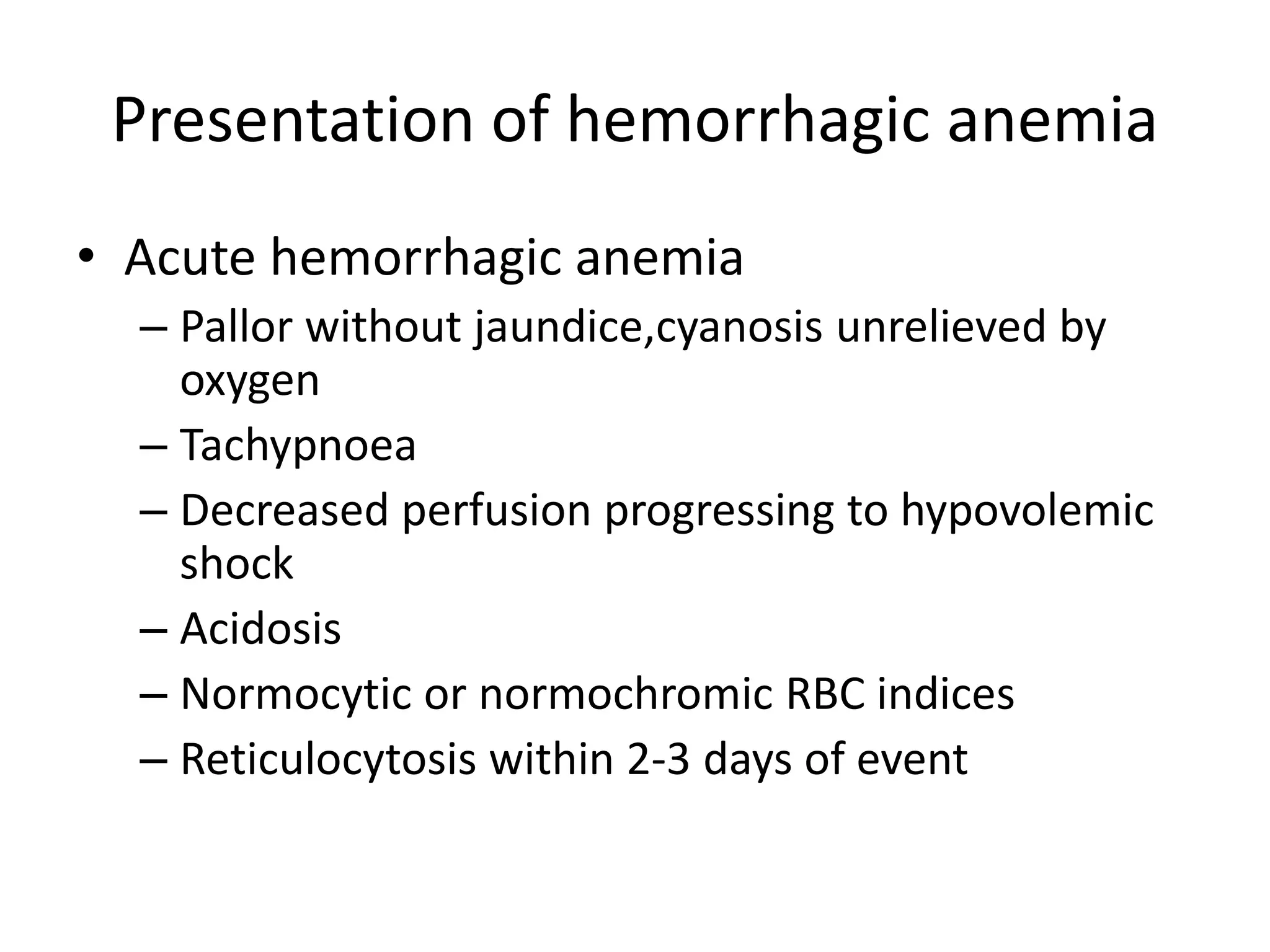
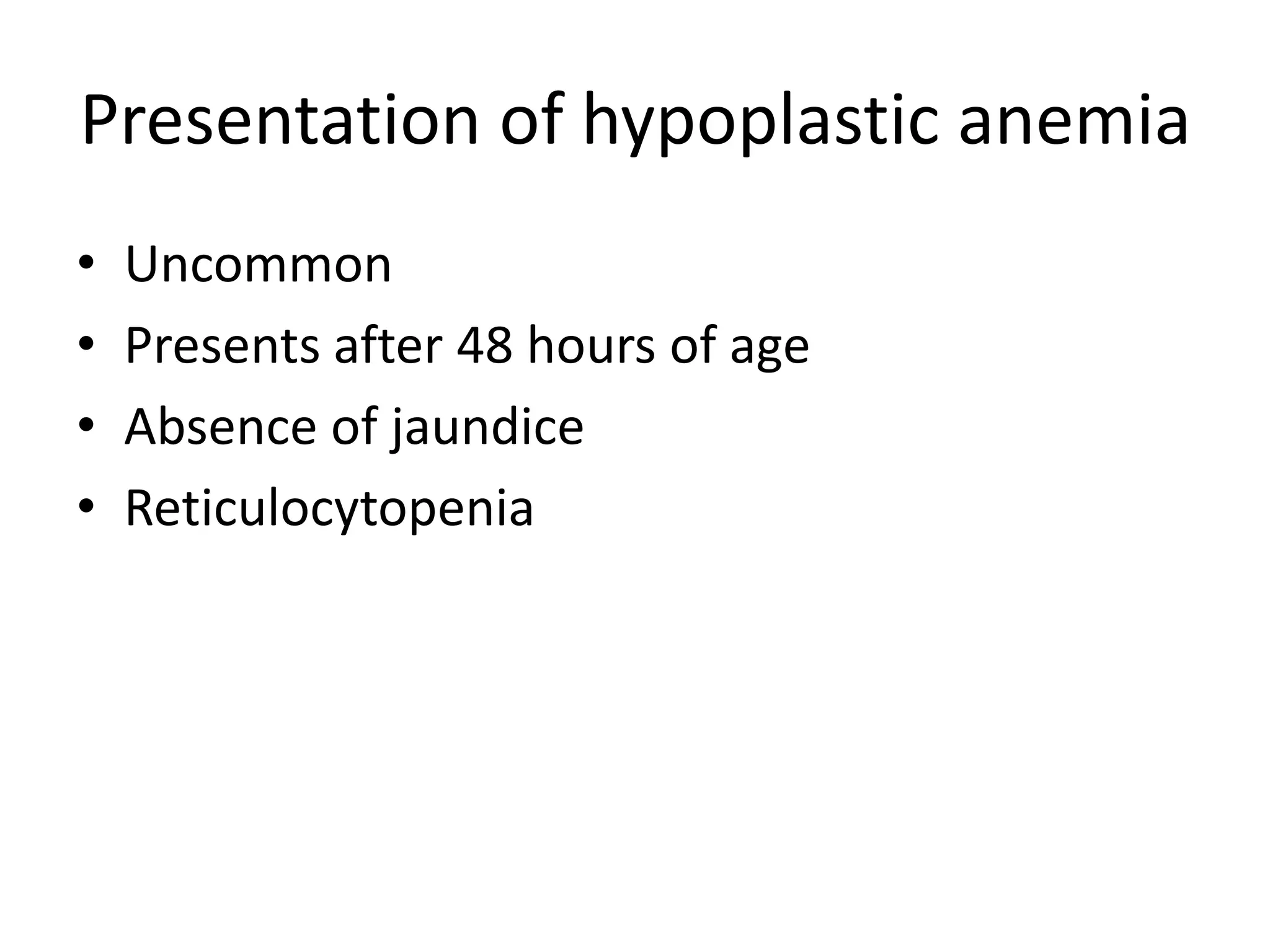
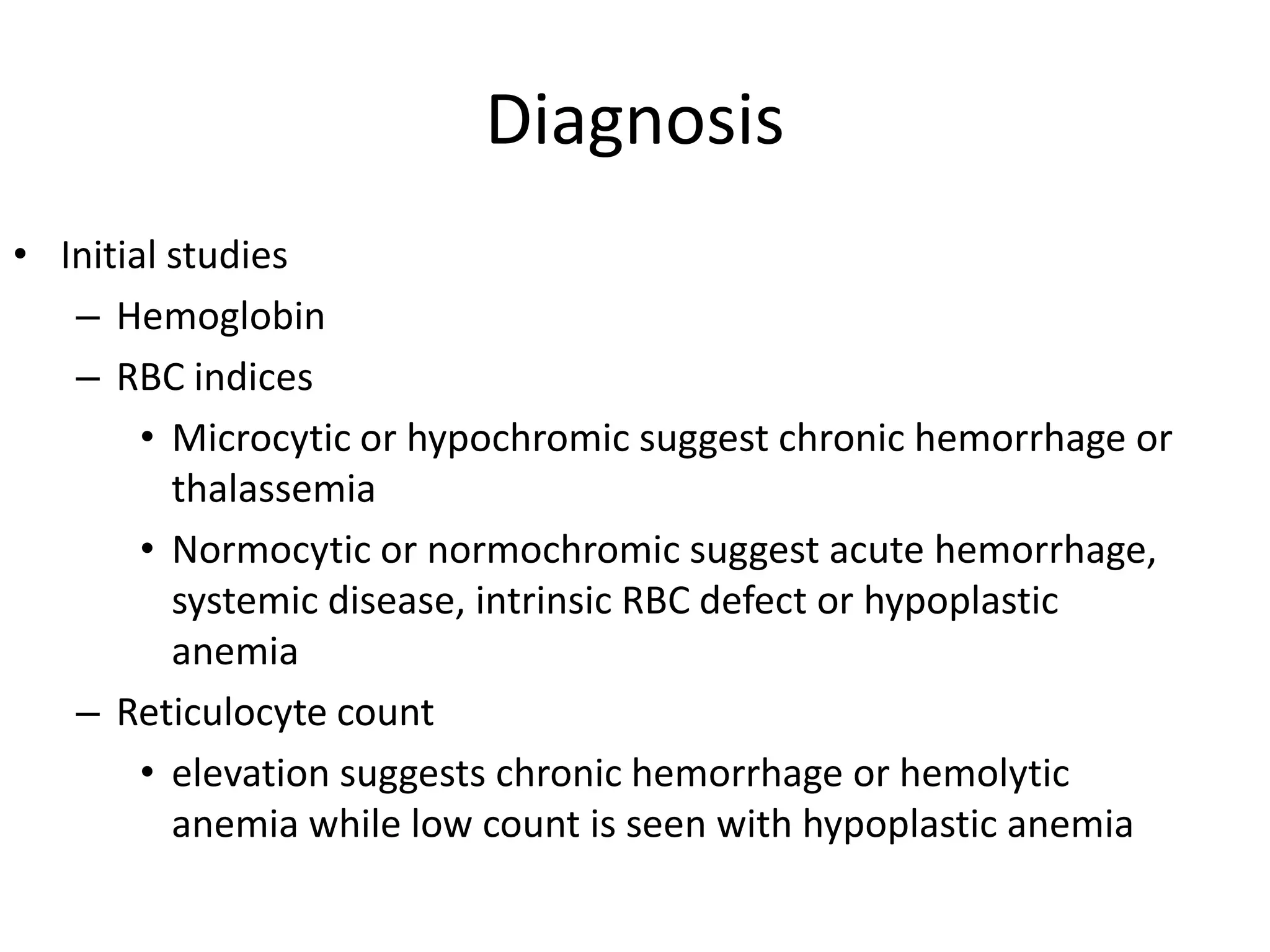
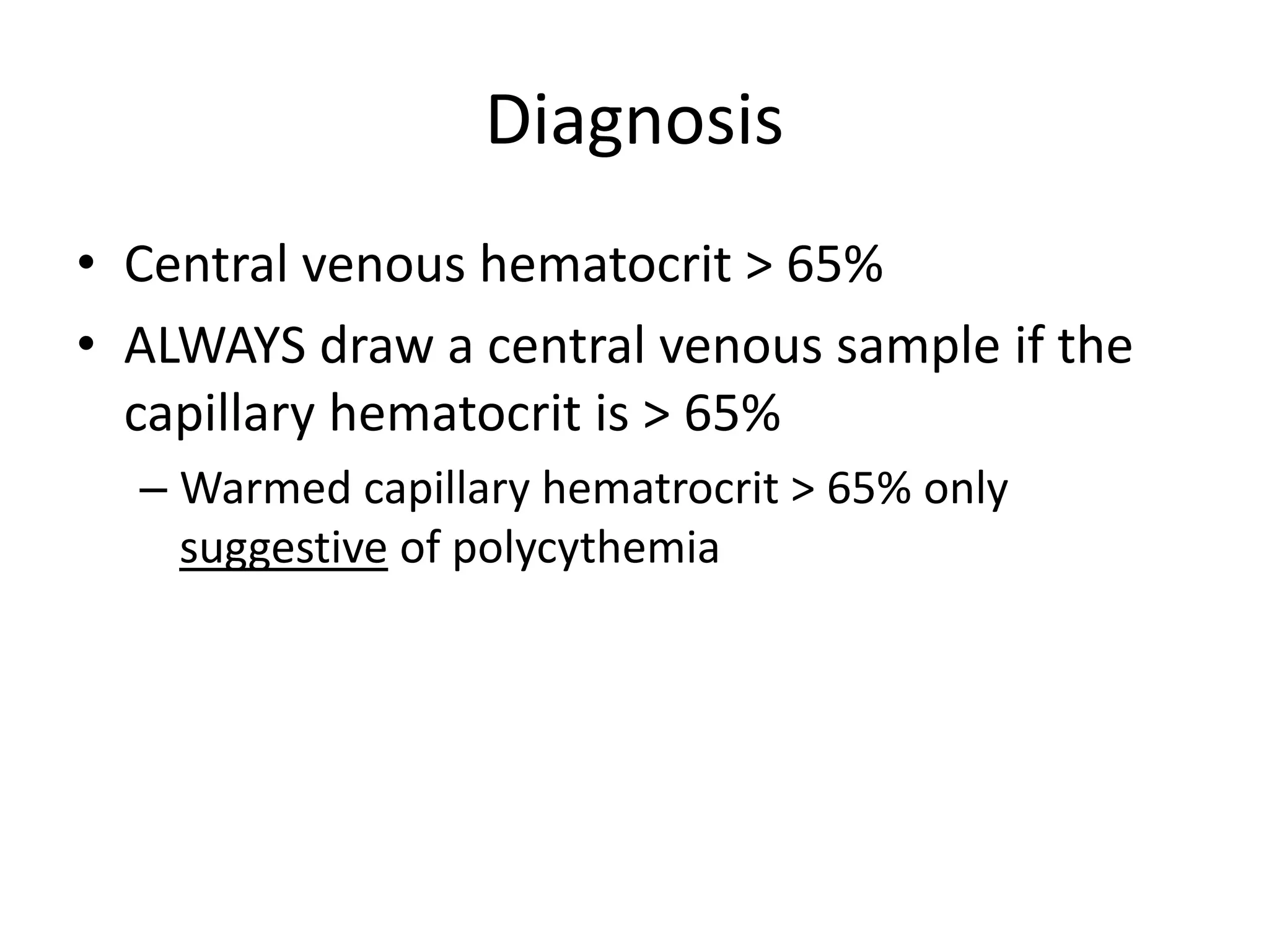
ANEMIA,POLYCYTHEMIA IN A
NEWBORN
-Dr.Apoorva
Pediatrics pg
ANEMIA IN NEONATES
Physiologic Anemia Of Infancy
• In utero,due to high oxygen saturation(45%) in fetal
aorta,erythropoietin levels are high &hence,RBC
production is rapid.
• At one week postnatally, all RBC indices begin
declining to a minimum value reached at about 8-12
weeks of age (11g/dl)
– decreased RBC production
– plasma dilution associated with increasing blood volume
– shorter life span of neonatal RBCs (50-70 days)
– more fragile RBCs
• Switch from HbF to HbA (switch to HbA provides for
greater unloading of oxygen to tissues d/t lower
oxygen affinity of HbA relative to HbF.)
• Seldom produces symptoms.
• As the hemoglobin levels reach nadir,oxygen delivery
to tissues is impaired,erythropoietin stimulated,RBC
production increases.
• Iron stores rapidly utilized for this process.
Hence,iron has to be supplied.
Anemia of Prematurity
• Occurs in low birth weight infants.
• The nadir is lower and is reached sooner.
• Average nadir is 7-9 g/dL and is reached at 4-8 weeks of
age.
• Due to a combination of :
decreased RBC mass at birth,
increased iatrogenic losses from lab draws,
shorter RBC life span,
inadequate erythropoietin production,
low iron stores,
rapid rate of growth,
Vitamin E deficiency.
• Signs and Symptoms :
apnea
poor weight gain
pallor
decreased activity
Tachycardia.
• Iron administration does not alter nadir reached or its
rate of reduction.
Pathophysiology
• Anemia in the newborn results from three
processes
– Loss of RBCs: hemorrhagic anemia
• Most common cause
– Increased destruction: hemolytic anemia
– Underproduction of RBCs: hypoplastic anemia
Hemorrhagic anemia
• Antepartum period
– Loss of placental integrity
• Abruption, previa, traumatic amniocentesis.
– Anomalies of the umbilical cord or placental vessels
• Velamentous insertion of the cord , communicating vessels, cord
hematoma, entanglement of the cord,vasa previa.
– Twin-twin transfusion syndrome
• Only in monozygotic multiple births
• 13-33% of twin pregnancies have TTTS
• Difference in hemoglobin usually > 5 g/dL
• Congestive heart disease common in anemic twin and hyperviscosity
common in plethoric twin
Hemorrhagic anemia
• Intrapartum period
– Fetomaternal hemorrhage
• Increased risk with ECV,ICV,breech delivery,placental
malformations
– Traumatic rupture of the cord
– Failure of placental transfusion due to cord occlusion
(nuchal or prolapsed cord)
– Obstetric trauma causing occult visceral or intracranial
hemorrhage
Hemorrhagic anemia
• Neonatal period
– Enclosed hemorrhage: suggests obstetric trauma or severe
perinatal hypoxia
• Hemorrhagic caput succedaneum, cephalhematoma, intracranial
hemorrhage, visceral hemorrhage
– Defects in hemostasis
• Congenital coagulation factor deficiency
• Consumption coagulopathy: DIC, sepsis
• Vitamin K dependent factor deficiency
• Thrombocytopenia: immune, or congenital with absent radii
– Iatrogenic blood loss due to blood draws
Hemolytic anemia
• Immune hemolysis:
Rh/ ABO /minor blood group incompatibility or
autoimmune hemolysis
• Nonimmune:
sepsis, TORCH infection
• Congenital erythrocyte defect
– G6PD, thalassemia, membrane defects (hereditary
spherocytosis,elliptocytosis)
• Systemic diseases: galactosemia, osteopetrosis
• Nutritional deficiency: vitamin E
Hypoplastic anemia
• Congenital
– Diamond-Blackfan syndrome, congenital leukemia,
sideroblastic anemia
• Acquired
– Infection: Rubella and parvovirus are the most
common
– Drug induced
Clinical presentation
• Determine the following factors :
– Age at presentation
– Associated clinical features
– Hemodynamic status of the infant
– Presence or absence of compensatory
reticulocytosis
– Family history,obstetric history
Presentation of hemorrhagic anemia
• Acute hemorrhagic anemia
– Pallor without jaundice,cyanosis unrelieved by
oxygen
– Tachypnoea
– Decreased perfusion progressing to hypovolemic
shock
– Acidosis
– Normocytic or normochromic RBC indices
– Reticulocytosis within 2-3 days of event
• Chronic
– Pallor
– Minimal signs of respiratory distress
– Microcytic or hypochromic RBC indices
– Compensatory reticulocytosis
– Enlarged liver d/t extramedullary erythropoiesis
Presentation of hemolytic anemia
• Jaundice is usually the first symptom
• Compensatory reticulocytosis
• Pallor
• Hepatosplenomegaly
Presentation of hypoplastic anemia
• Uncommon
• Presents after 48 hours of age
• Absence of jaundice
• Reticulocytopenia
Presentation of other forms
• Twin-twin transfusion
– Growth failure in the anemic twin
• Occult internal hemorrhage
– Intracranial: bulging anterior fontanelle and neurologic
signs (altered mental status, apnea, seizures)
– Visceral hemorrhage: most often liver is damaged and
leads to abdominal mass
– Pulmonary hemorrhage: radiographic opacification of a
hemithorax with bloody tracheal secretions
Diagnosis
• Initial studies
– Hemoglobin
– RBC indices
• Microcytic or hypochromic suggest chronic hemorrhage or
thalassemia
• Normocytic or normochromic suggest acute hemorrhage,
systemic disease, intrinsic RBC defect or hypoplastic
anemia
– Reticulocyte count
• elevation suggests chronic hemorrhage or hemolytic
anemia while low count is seen with hypoplastic anemia
Diagnosis
– Blood smear to look for
• spherocytes (hereditary spherocytosis,immune
hemolysis)
• elliptocytes (hereditary elliptocytosis)
• pyknocytes ,bite cells,heinz bodies(G6PD)
• Schistocytes,fragmented RBC’s (consumption
coagulopathy)
– Direct Coombs test: positive in isoimmune or
autoimmune hemolysis
Other diagnostic studies
• Blood type and Rh in isoimmune hemolysis
• Kleihauer-Betke test on maternal blood to look for
fetomaternal hemorrhage
• CXR for pulmonary hemorrhage
• Bone marrow aspiration for congenital hypoplastic or
aplastic anemia
• TORCH: IgM levels, urine for CMV
• DIC panel, platelets looking for consumption
• Occult hemorrhage: cranial or abdominal ultrasound
• Intrinsic RBC defects: enzyme studies, globin chain ratios,
membrane studies
Management
• Simple replacement transfusion
– Indications:
• acute hemorrhage
– Use 15-20 ml/kg O, RH- packed RBCs or blood cross-
matched to mother and adjust hct to 50%
– Give via UVC
– Draw diagnostic studies before transfusion
• ongoing deficit replacement
• maintenance of effective oxygen-carrying capacity
– Hct >35% in severe cardiopulmonary disease
– Hct >40% in mild-moderate cardiopulmonary disease,
apnea, symptomatic anemia, need for surgery
Management
• Exchange transfusion
– Indications
• Chronic hemolytic anemia
• Severe isoimmune hemolytic anemia
• Consumption coagulopathy
• Nutritional replacement: iron, folate, vitamin E
• Erythropoietin
– Increased erythropoiesis without significant side
effects
POLYCYTHEMIA
IN NEONATES
• Polycythemia is increased total RBC mass
– Central venous hematocrit > 65%
• Polycythemic hyperviscosity is increased
viscosity of the blood resulting from increased
numbers of RBCs
– Not all polycythemic infants have symptoms of
hyperviscosity
Incidence
• Polycythemia occurs in 2-4% of newborns
– Half of these are symptomatic
• Hyperviscosity occurs in 25% of infants with
hematocrit 60-64%
Pathophysiology
• Clinical signs result from regional effects of
hyperviscosity and from the formation of
microthrombi
– Tissue hypoxia
– Acidosis
– Hypoglycemia in the substrate
• Organs affected: CNS, kidneys, adrenals,
cardiopulmonary system, GI tract
What affects hyperviscosity?
• Hematocrit
– Increased hct is the most important single factor
– Results from increase in circulating RBCs or decreased
plasma volume (dehydration)
• Plasma viscosity
– Higher plasma proteins = increased viscosity
• Especially fibrinogen (typically low in neonates)
– Not usually an issue in neonates
• RBC aggregation
– Occurs in areas of low blood flow = venous
microcirculation
– Not a large factor in neonates
• Deformability of RBC membrane: usually normal
Conditions that alter incidence
• Altitude: increased RBC mass
• Neonatal age
– Physiologic increase in hematocrit due to fluid
shifts away from intravascular compartment with
maximum at 2-4 hours of age
• Obstetric factors: delayed cord clamping or
“stripping” of the umbilical cord
• High-risk delivery, especially if precipitous
Perinatal processes
• Enhanced fetal erythropoiesis usually related
to fetal hypoxia
– Placental insufficiency
• Maternal hypertension, abruption, post-dates, IUGR,
maternal smoking
– Endocrine disorders: due to increased oxygen
consumption
• IDM (>40% incidence), congenital thyrotoxicosis, CAH,
Beckwith-Wiedemann syndrome (hyperinsulinism)
DUE TO : Hypertransfusion
• Delayed cord clamping
• Should be done within 1 minute
• Gravity: positioning below the placenta will
increase placental transfusion
• Meds: oxytocin can increase contractions and
thus transfusion
• Decreased in c-section ( no contractions )
• Twin-twin transfusion
• Intrapartum asphyxia
• Enhances net umbilical flow toward the infant, while acidosis
increases capillary leak leading to reduced plasma volume
Clinical presentation
• Symptoms are non-specific!
• CNS: lethargy, hyperirritability, proximal muscle
hypotonia, vasomotor instability, vomiting,
seizures, cerebral infarction (rare)
• Cardiopulmonary: respiratory distress,
tachycardia, CHF, pulmonary hypertension
• GI: feeding intolerance, sometimes NEC
• GU: oliguria, ARF, renal vein thrombosis, priapism
• Metabolic: hypo-glycemia/-calcemia/-
magnesemia
• Heme: hyperbili, thrombocytopenia
• Skin: ruddiness
Diagnosis
• Central venous hematocrit > 65%
• ALWAYS draw a central venous sample if the
capillary hematocrit is > 65%
– Warmed capillary hematrocrit > 65% only
suggestive of polycythemia
Management
• Asymptomatic infants
– Expectant observation unless central venous hematocrit
>75% (consider partial exchange transfusion)
– Can do a trial of rehydration over 6-8 hr if dehydrated
• Give 130-150 ml/kg/d
– Check central hematocrit q6 hourly
Management
• Symptomatic infants with central hct > 65%
– Partial exchange transfusion is advisable but debatable
– For exchange can use normal saline, 5% albumin, or FFP
– Volume exchanged =
• (Weight (kg) x blood volume) x (hct - desired hct) / hct
Other investigations
• Serum glucose
– Hypoglycemia is common with polycythemia
• Serum bilirubin
– Increased bilirubin due to increased RBC turnover
• Serum sodium, BUN, urine specific gravity
– Usually high if baby is deyhdrated
• Blood gas to rule-out inadequate oxygenation as
cause of symptoms
• Platelets, as thyrombocytopenia can be present
• Serum calcium-hypocalcemia can be seen
Prognosis
• Increased risk of GI disorders and NEC with partial
exchange transfusion (PET)
• Older trials show decreased neurologic complications
from hyperviscosity with PET, but newer trials show
no real benefit
– PET is controversial!
• Infants with asymptomatic polycythemia have an
increased risk for neurologic sequelae
– Normocythemic controls with the same perinatal history
have a similarly increased risk
THANK YOU!

Anemia & polycythemia in neonates

  • 1.
  • 2.
  • 3.
    Physiologic Anemia OfInfancy • In utero,due to high oxygen saturation(45%) in fetal aorta,erythropoietin levels are high &hence,RBC production is rapid. • At one week postnatally, all RBC indices begin declining to a minimum value reached at about 8-12 weeks of age (11g/dl) – decreased RBC production – plasma dilution associated with increasing blood volume – shorter life span of neonatal RBCs (50-70 days) – more fragile RBCs
  • 4.
    • Switch fromHbF to HbA (switch to HbA provides for greater unloading of oxygen to tissues d/t lower oxygen affinity of HbA relative to HbF.) • Seldom produces symptoms. • As the hemoglobin levels reach nadir,oxygen delivery to tissues is impaired,erythropoietin stimulated,RBC production increases. • Iron stores rapidly utilized for this process. Hence,iron has to be supplied.
  • 5.
    Anemia of Prematurity •Occurs in low birth weight infants. • The nadir is lower and is reached sooner. • Average nadir is 7-9 g/dL and is reached at 4-8 weeks of age. • Due to a combination of : decreased RBC mass at birth, increased iatrogenic losses from lab draws, shorter RBC life span, inadequate erythropoietin production, low iron stores, rapid rate of growth, Vitamin E deficiency.
  • 6.
    • Signs andSymptoms : apnea poor weight gain pallor decreased activity Tachycardia. • Iron administration does not alter nadir reached or its rate of reduction.
  • 7.
    Pathophysiology • Anemia inthe newborn results from three processes – Loss of RBCs: hemorrhagic anemia • Most common cause – Increased destruction: hemolytic anemia – Underproduction of RBCs: hypoplastic anemia
  • 8.
    Hemorrhagic anemia • Antepartumperiod – Loss of placental integrity • Abruption, previa, traumatic amniocentesis. – Anomalies of the umbilical cord or placental vessels • Velamentous insertion of the cord , communicating vessels, cord hematoma, entanglement of the cord,vasa previa. – Twin-twin transfusion syndrome • Only in monozygotic multiple births • 13-33% of twin pregnancies have TTTS • Difference in hemoglobin usually > 5 g/dL • Congestive heart disease common in anemic twin and hyperviscosity common in plethoric twin
  • 9.
    Hemorrhagic anemia • Intrapartumperiod – Fetomaternal hemorrhage • Increased risk with ECV,ICV,breech delivery,placental malformations – Traumatic rupture of the cord – Failure of placental transfusion due to cord occlusion (nuchal or prolapsed cord) – Obstetric trauma causing occult visceral or intracranial hemorrhage
  • 10.
    Hemorrhagic anemia • Neonatalperiod – Enclosed hemorrhage: suggests obstetric trauma or severe perinatal hypoxia • Hemorrhagic caput succedaneum, cephalhematoma, intracranial hemorrhage, visceral hemorrhage – Defects in hemostasis • Congenital coagulation factor deficiency • Consumption coagulopathy: DIC, sepsis • Vitamin K dependent factor deficiency • Thrombocytopenia: immune, or congenital with absent radii – Iatrogenic blood loss due to blood draws
  • 11.
    Hemolytic anemia • Immunehemolysis: Rh/ ABO /minor blood group incompatibility or autoimmune hemolysis • Nonimmune: sepsis, TORCH infection • Congenital erythrocyte defect – G6PD, thalassemia, membrane defects (hereditary spherocytosis,elliptocytosis) • Systemic diseases: galactosemia, osteopetrosis • Nutritional deficiency: vitamin E
  • 12.
    Hypoplastic anemia • Congenital –Diamond-Blackfan syndrome, congenital leukemia, sideroblastic anemia • Acquired – Infection: Rubella and parvovirus are the most common – Drug induced
  • 13.
    Clinical presentation • Determinethe following factors : – Age at presentation – Associated clinical features – Hemodynamic status of the infant – Presence or absence of compensatory reticulocytosis – Family history,obstetric history
  • 14.
    Presentation of hemorrhagicanemia • Acute hemorrhagic anemia – Pallor without jaundice,cyanosis unrelieved by oxygen – Tachypnoea – Decreased perfusion progressing to hypovolemic shock – Acidosis – Normocytic or normochromic RBC indices – Reticulocytosis within 2-3 days of event
  • 15.
    • Chronic – Pallor –Minimal signs of respiratory distress – Microcytic or hypochromic RBC indices – Compensatory reticulocytosis – Enlarged liver d/t extramedullary erythropoiesis
  • 16.
    Presentation of hemolyticanemia • Jaundice is usually the first symptom • Compensatory reticulocytosis • Pallor • Hepatosplenomegaly
  • 17.
    Presentation of hypoplasticanemia • Uncommon • Presents after 48 hours of age • Absence of jaundice • Reticulocytopenia
  • 18.
    Presentation of otherforms • Twin-twin transfusion – Growth failure in the anemic twin • Occult internal hemorrhage – Intracranial: bulging anterior fontanelle and neurologic signs (altered mental status, apnea, seizures) – Visceral hemorrhage: most often liver is damaged and leads to abdominal mass – Pulmonary hemorrhage: radiographic opacification of a hemithorax with bloody tracheal secretions
  • 19.
    Diagnosis • Initial studies –Hemoglobin – RBC indices • Microcytic or hypochromic suggest chronic hemorrhage or thalassemia • Normocytic or normochromic suggest acute hemorrhage, systemic disease, intrinsic RBC defect or hypoplastic anemia – Reticulocyte count • elevation suggests chronic hemorrhage or hemolytic anemia while low count is seen with hypoplastic anemia
  • 20.
    Diagnosis – Blood smearto look for • spherocytes (hereditary spherocytosis,immune hemolysis) • elliptocytes (hereditary elliptocytosis) • pyknocytes ,bite cells,heinz bodies(G6PD) • Schistocytes,fragmented RBC’s (consumption coagulopathy) – Direct Coombs test: positive in isoimmune or autoimmune hemolysis
  • 21.
    Other diagnostic studies •Blood type and Rh in isoimmune hemolysis • Kleihauer-Betke test on maternal blood to look for fetomaternal hemorrhage • CXR for pulmonary hemorrhage • Bone marrow aspiration for congenital hypoplastic or aplastic anemia • TORCH: IgM levels, urine for CMV • DIC panel, platelets looking for consumption • Occult hemorrhage: cranial or abdominal ultrasound • Intrinsic RBC defects: enzyme studies, globin chain ratios, membrane studies
  • 22.
    Management • Simple replacementtransfusion – Indications: • acute hemorrhage – Use 15-20 ml/kg O, RH- packed RBCs or blood cross- matched to mother and adjust hct to 50% – Give via UVC – Draw diagnostic studies before transfusion • ongoing deficit replacement • maintenance of effective oxygen-carrying capacity – Hct >35% in severe cardiopulmonary disease – Hct >40% in mild-moderate cardiopulmonary disease, apnea, symptomatic anemia, need for surgery
  • 23.
    Management • Exchange transfusion –Indications • Chronic hemolytic anemia • Severe isoimmune hemolytic anemia • Consumption coagulopathy • Nutritional replacement: iron, folate, vitamin E
  • 24.
    • Erythropoietin – Increasederythropoiesis without significant side effects
  • 25.
  • 26.
    • Polycythemia isincreased total RBC mass – Central venous hematocrit > 65% • Polycythemic hyperviscosity is increased viscosity of the blood resulting from increased numbers of RBCs – Not all polycythemic infants have symptoms of hyperviscosity
  • 27.
    Incidence • Polycythemia occursin 2-4% of newborns – Half of these are symptomatic • Hyperviscosity occurs in 25% of infants with hematocrit 60-64%
  • 28.
    Pathophysiology • Clinical signsresult from regional effects of hyperviscosity and from the formation of microthrombi – Tissue hypoxia – Acidosis – Hypoglycemia in the substrate • Organs affected: CNS, kidneys, adrenals, cardiopulmonary system, GI tract
  • 29.
    What affects hyperviscosity? •Hematocrit – Increased hct is the most important single factor – Results from increase in circulating RBCs or decreased plasma volume (dehydration) • Plasma viscosity – Higher plasma proteins = increased viscosity • Especially fibrinogen (typically low in neonates) – Not usually an issue in neonates • RBC aggregation – Occurs in areas of low blood flow = venous microcirculation – Not a large factor in neonates • Deformability of RBC membrane: usually normal
  • 30.
    Conditions that alterincidence • Altitude: increased RBC mass • Neonatal age – Physiologic increase in hematocrit due to fluid shifts away from intravascular compartment with maximum at 2-4 hours of age • Obstetric factors: delayed cord clamping or “stripping” of the umbilical cord • High-risk delivery, especially if precipitous
  • 31.
    Perinatal processes • Enhancedfetal erythropoiesis usually related to fetal hypoxia – Placental insufficiency • Maternal hypertension, abruption, post-dates, IUGR, maternal smoking – Endocrine disorders: due to increased oxygen consumption • IDM (>40% incidence), congenital thyrotoxicosis, CAH, Beckwith-Wiedemann syndrome (hyperinsulinism)
  • 32.
    DUE TO :Hypertransfusion • Delayed cord clamping • Should be done within 1 minute • Gravity: positioning below the placenta will increase placental transfusion • Meds: oxytocin can increase contractions and thus transfusion • Decreased in c-section ( no contractions ) • Twin-twin transfusion • Intrapartum asphyxia • Enhances net umbilical flow toward the infant, while acidosis increases capillary leak leading to reduced plasma volume
  • 33.
    Clinical presentation • Symptomsare non-specific! • CNS: lethargy, hyperirritability, proximal muscle hypotonia, vasomotor instability, vomiting, seizures, cerebral infarction (rare) • Cardiopulmonary: respiratory distress, tachycardia, CHF, pulmonary hypertension • GI: feeding intolerance, sometimes NEC • GU: oliguria, ARF, renal vein thrombosis, priapism • Metabolic: hypo-glycemia/-calcemia/- magnesemia • Heme: hyperbili, thrombocytopenia • Skin: ruddiness
  • 34.
    Diagnosis • Central venoushematocrit > 65% • ALWAYS draw a central venous sample if the capillary hematocrit is > 65% – Warmed capillary hematrocrit > 65% only suggestive of polycythemia
  • 35.
    Management • Asymptomatic infants –Expectant observation unless central venous hematocrit >75% (consider partial exchange transfusion) – Can do a trial of rehydration over 6-8 hr if dehydrated • Give 130-150 ml/kg/d – Check central hematocrit q6 hourly
  • 36.
    Management • Symptomatic infantswith central hct > 65% – Partial exchange transfusion is advisable but debatable – For exchange can use normal saline, 5% albumin, or FFP – Volume exchanged = • (Weight (kg) x blood volume) x (hct - desired hct) / hct
  • 37.
    Other investigations • Serumglucose – Hypoglycemia is common with polycythemia • Serum bilirubin – Increased bilirubin due to increased RBC turnover • Serum sodium, BUN, urine specific gravity – Usually high if baby is deyhdrated • Blood gas to rule-out inadequate oxygenation as cause of symptoms • Platelets, as thyrombocytopenia can be present • Serum calcium-hypocalcemia can be seen
  • 38.
    Prognosis • Increased riskof GI disorders and NEC with partial exchange transfusion (PET) • Older trials show decreased neurologic complications from hyperviscosity with PET, but newer trials show no real benefit – PET is controversial! • Infants with asymptomatic polycythemia have an increased risk for neurologic sequelae – Normocythemic controls with the same perinatal history have a similarly increased risk
  • 39.

Editor's Notes

  • #4 Liver-erythropoietin….due to low oxygen sat95% low eryhtropoietin
  • #6 Exaggeration of normal physiologic anemia
  • #9 Due to blood loss
  • #10 Chorangioma choriocarcinoma
  • #12 Maternal lupus RA