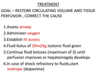
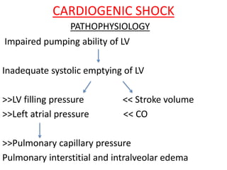
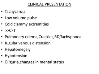
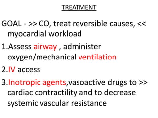
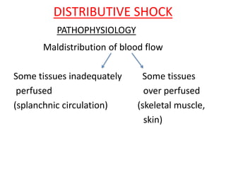
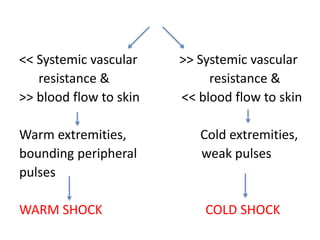
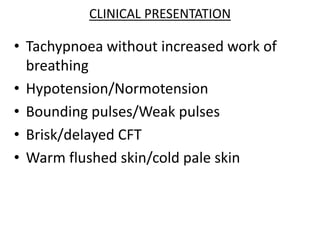
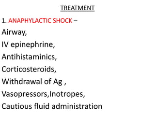
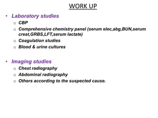
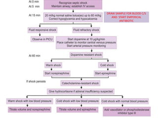
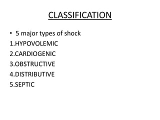
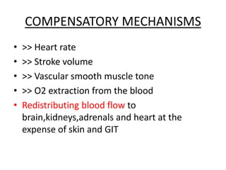
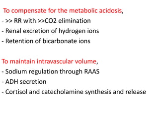
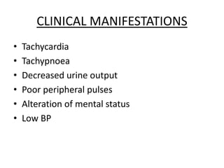
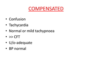
SHOCK IN CHILDREN
-Dr.Apoorva.E
PG,DCMS
• “ ACUTE CIRCULATORY FAILURE “
with inadequate or inappropriately
distributed tissue perfusion
resulting in generalized cellular
hypoxia
• Body’s inability to deliver adequate oxygen to
meet the metabolic demands of the tissues.
• Initially compensated
• Continued presence of an inciting trigger +
body’s exaggerated response
lead to progression of shock
if untreated,irreversible tissue injury
irreversible shock
CLASSIFICATION
• 5 major types of shock
1.HYPOVOLEMIC
2.CARDIOGENIC
3.OBSTRUCTIVE
4.DISTRIBUTIVE
5.SEPTIC
HYPOVOLEMIC SHOCK
• Characterized by fluid loss ( internal / external )
• Decreased preload
Water/electrolyte plasma blood
loss loss loss
CARDIOGENIC SHOCK
• Poor myocardial contractility leading to cardiac
pump failure
• Due to :
CHD
Myocarditis
Cardiomyopathies
Arrhythmias
OBSTRUCTIVE SHOCK
• Decreased cardiac output secondary to restriction
of all cardiac chambers
• Due to :
Tension pneumothorax
Pericardial tamponade
Pulmonary embolism
Anterior mediastinal masses
Coarctation of aorta
DISTRIBUTIVE SHOCK
• Caused by inadequate vasomotor tone
Capillary leak
Maldistribution of fluid into interstitium
• Post-spinal cord or brainstem injury
Anaphylaxis
Poisonings
SEPTIC SHOCK
• Complex interaction of
distributive,cardiogenic and hypovolemic
shock
• Bacterial/Viral/Fungal
Hypovolemic shock – blood VOLUME
problem
Cardiogenic shock - blood PUMP
problem
Distributive shock – blood VESSEL
problem
PATHOPHYSIOLOGY
COMPENSATORY MECHANISMS
• >> Heart rate
• >> Stroke volume
• >> Vascular smooth muscle tone
• >> O2 extraction from the blood
• Redistributing blood flow to
brain,kidneys,adrenals and heart at the
expense of skin and GIT
To compensate for the metabolic acidosis,
- >> RR with >>CO2 elimination
- Renal excretion of hydrogen ions
- Retention of bicarbonate ions
To maintain intravascular volume,
- Sodium regulation through RAAS
- ADH secretion
- Cortisol and catecholamine synthesis and release
CLINICAL MANIFESTATIONS
• Tachycardia
• Tachypnoea
• Decreased urine output
• Poor peripheral pulses
• Alteration of mental status
• Low BP
COMPENSATED
• Confusion
• Tachycardia
• Normal or mild tachypnoea
• >> CFT
• U/o-adequate
• BP normal
UNCOMPENSATED
• Drowsiness
• Marked tachycardia
• Tachypnoea and acidosis
• Very slow CFT
• Oliguria/Anuria
• Hypotension
HYPOTENSION FORMULA
Ages – 1 to 10 years
Hypotension is defined as SBP
< 70mmHg + [age in years X 2] mmHg
IRREVERSIBLE
• Child is unresponsive
• Bradycardia
• Apnoea
• Cold cyanotic skin
• Anuria
• Unrecordable BP
DIAGNOSIS
• Thorough history and physical exam
• Lab findings : thrombocytopenia
prolonged PT and apTT
reduced serum fibrinogen level
>> fibrin split products
anemia
neutrophilia
left shift of leukocytes
electrolyte disturbances
HYPOVOLEMIC SHOCK
<< intravascular volume
<< venous return and preload
<< decreased ventricular filling
<< decreased stroke volume
<< CO
<< tissue perfusion
PATHOPHYSIOLOGY
ASSESSMENT OF FLUID LOSS
TREATMENT
1.Assess airway
2.Administer oxygen
3.Establish IV access
4.Fluid bolus of 20ml/kg isotonic fluid given
5.Continue fluid boluses (maximum of 3) until
perfusion improves or hepatomegaly develops
6.In case of shock refractory to fluids,start
inotrope (dopamine)
GOAL – RESTORE CIRCULATING VOLUME AND TISSUE
PERFUSION , CORRECT THE CAUSE
CARDIOGENIC SHOCK
Impaired pumping ability of LV
Inadequate systolic emptying of LV
>>LV filling pressure << Stroke volume
>>Left atrial pressure << CO
>>Pulmonary capillary pressure
Pulmonary interstitial and intralveolar edema
PATHOPHYSIOLOGY
CLINICAL PRESENTATION
• Tachycardia
• Low volume pulse
• Cold clammy extremities
• >>CFT
• Pulmonary edema,Crackles,RD,Tachypnoea
• Jugular venous distension
• Hepatomegaly
• Hypotension
• Oliguria,changes in mental status
TREATMENT
GOAL - >> CO, treat reversible causes, <<
myocardial workload
1.Assess airway , administer
oxygen/mechanical ventilation
2.IV access
3.Inotropic agents,vasoactive drugs to >>
cardiac contractility and to decrease
systemic vascular resistance
4.Cautious administration of fluids
(5-10ml/kg boluses over longer time)
5. Morphine to decrease preload and
anxiety
6.Vasodilators for afterload reduction
7.Short acting beta blockers for refractory
tachycardia
OBSTRUCTIVE SHOCK
PATHOPHYSIOLOGY
Physical obstruction to blood flow
<< CO
<< Tissue perfusion
Compensatory >> in systemic vascular resistance
CLINICAL PRESENTATION
• Muffled heart sounds
• Distended neck veins
• Pulsus paradoxus ( << in SBP by
more than 10mmHg on inspiration )
• Signs of right heart failure plus
cyanosis,tachycardia,hypotension
PERICARDIAL
EFFUSION
PULMONARY
EMBOLISM
TREATMENT
• Pericardial drainage in case of
pericardial effusion
• Immediate needle decompression then
thoracostomy for chest tube in case of
tension pneumothorax
• Anticoagulants or embolectomy for
pulmonary embolism
DISTRIBUTIVE SHOCK
Maldistribution of blood flow
Some tissues inadequately Some tissues
perfused over perfused
(splanchnic circulation) (skeletal muscle,
skin)
PATHOPHYSIOLOGY
<< Systemic vascular >> Systemic vascular
resistance & resistance &
>> blood flow to skin << blood flow to skin
Warm extremities, Cold extremities,
bounding peripheral weak pulses
pulses
WARM SHOCK COLD SHOCK
CLINICAL PRESENTATION
• Tachypnoea without increased work of
breathing
• Hypotension/Normotension
• Bounding pulses/Weak pulses
• Brisk/delayed CFT
• Warm flushed skin/cold pale skin
TREATMENT
1. ANAPHYLACTIC SHOCK –
Airway,
IV epinephrine,
Antihistaminics,
Corticosteroids,
Withdrawal of Ag ,
Vasopressors,Inotropes,
Cautious fluid administration
2. NEUROGENIC SHOCK –
Cautious fluid administration,
Vasopressors,Inotropes,
Correct hypothermia,
Treat bradycardia with atropine,
Observe and prevent DVT (due to
peripheral pooling of blood)
SEPTIC SHOCK
PATHOPHYSIOLOGY
DEFINITIONS
SIRS
• Requires 2 of the following 4 features to be
present:
o Temperature >38.5° or <36.0° C
o Tachypnea >2SD ABOVE NORMAL FOR AGE
o Tachycardia  >2SD ABOVE NORMAL FOR AGE
o WBC ELEVATED OR DEPRESSED FOR AGE/>10%
IMMATURE NEUTROPHILS
INFECTION
• Suspected or proven infection or a clinical
syndrome associated with high probability
of infection
SEPSIS
• SIRS plus a suspected or proven infection
SEVERE SEPSIS
• Sepsis plus organ dysfunction,hypoperfusion
or hypotension
(including but not limited to lactic
acidosis,oliguria,acute mental status changes)
Identifying Acute Organ Dysfunction as a
Marker of Severe Sepsis
Tachycardia
Hypotension
 CVP
 PAOP
Jaundice
 Enzymes
 Albumin
 PT
Altered
Consciousness
Confusion
Psychosis
Tachypnea
PaO2 <70 mm Hg
SaO2 <90%
PaO2/FiO2 300
Oliguria
Anuria
 Creatinine
 Platelets
 PT/APTT
 Protein C
 D-dimer
MODS
• Presence of altered organ function such that
homeostasis cannot be maintained without
medical intervention
WORK UP
• Laboratory studies
o CBP
o Comprehensive chemistry panel (serum elec,abg,BUN,serum
creat,GRBS,LFT,serum lactate)
o Coagulation studies
o Blood & urine cultures
• Imaging studies
o Chest radiography
o Abdominal radiography
o Others according to the suspected cause.
DRAW SAMPLE FOR BLOOD C/S
AND START EMPERICAL
ANTIBIOTIC
•Antibiotics should be administered within the first hour of
recognition of septic shock
•Selection of antibiotic agents is empirically based on
an assessment of patient's immunity
the potential source of infection
the most likely responsible organisms.
•Antibiotic choice must be broad spectrum, covering gram-positive,
gram-negative, and anaerobic bacteria when the source is unknown
•Regimen for septic shock of unknown cause is
oGentamicin
o3rd generation cephalosporin
o if pseudomonas is suspected,ceftazidime
• Vancomycin must be added if resistant
staphylococci or enterococci are suspected.
• If there is an abdominal source, a drug effective
against anaerobes should be included
“metronidazole”
• Antibiotics are continued for at least 5 days after
shock resolves and evidence of infection subsides
• Abscesses must be drained and necrotic tissues
(eg, infarcted bowel) surgically excised.
DRUGS USED IN
SHOCK
1.Dopamine
-ionotrope at low dose
- peripheral vasoconstriction at
>10mcg/kg/min
- 3-20mcg/kg/min
-arrythmia at higher doses
2. Epinephrine
- >> HR and >> cardiac contractility
- Potent vasoconstrictor
- 0.05 – 3 mcg/kg/min
- << renal perfusion at higher doses,arrythmia at
higher doses
3. Norepinephrine
- Potent vasoconstrictor
- No significant effect on cardiac contractility
- 0.05 to 1.5 mcg/kg/min
4. Dobutamine
- >> cardiac contractility
- Peripheral vasodilator
- 1-10 mcg/kg/min
5.Phenylephrine
-potent vasoconstrictor
-0.5 to 2 mcg/kg/min
->> O2 consumption
-can cause sudden hypertension
Drug Indication Dose MOA Principal actions
Dopamine Renal perfusion 2-5 mcg/kg/min Dopaminergic Renal a. dilation
hypotension 5-10 mcg/kg/min 1 &
dopaminergic
+ inotrope
Hypotension >10 mcg/kg/min 1 vasoconstriction
Dobutamine Cardiogenic shock 2.5-25 mcg/kg/min Selective 1 + inotrope
Norepinephrine Hypotension 2-4 mcg/min 1 & 1 Vasoconstriction
Phenylephrine Hypotension 40-180 mcg/min Selective 1 Vasoconstriction
THANK YOU

Shock in children

  • 1.
  • 2.
    • “ ACUTECIRCULATORY FAILURE “ with inadequate or inappropriately distributed tissue perfusion resulting in generalized cellular hypoxia
  • 3.
    • Body’s inabilityto deliver adequate oxygen to meet the metabolic demands of the tissues. • Initially compensated • Continued presence of an inciting trigger + body’s exaggerated response lead to progression of shock if untreated,irreversible tissue injury irreversible shock
  • 5.
    CLASSIFICATION • 5 majortypes of shock 1.HYPOVOLEMIC 2.CARDIOGENIC 3.OBSTRUCTIVE 4.DISTRIBUTIVE 5.SEPTIC
  • 6.
    HYPOVOLEMIC SHOCK • Characterizedby fluid loss ( internal / external ) • Decreased preload Water/electrolyte plasma blood loss loss loss
  • 7.
    CARDIOGENIC SHOCK • Poormyocardial contractility leading to cardiac pump failure • Due to : CHD Myocarditis Cardiomyopathies Arrhythmias
  • 8.
    OBSTRUCTIVE SHOCK • Decreasedcardiac output secondary to restriction of all cardiac chambers • Due to : Tension pneumothorax Pericardial tamponade Pulmonary embolism Anterior mediastinal masses Coarctation of aorta
  • 9.
    DISTRIBUTIVE SHOCK • Causedby inadequate vasomotor tone Capillary leak Maldistribution of fluid into interstitium • Post-spinal cord or brainstem injury Anaphylaxis Poisonings
  • 10.
    SEPTIC SHOCK • Complexinteraction of distributive,cardiogenic and hypovolemic shock • Bacterial/Viral/Fungal
  • 11.
    Hypovolemic shock –blood VOLUME problem Cardiogenic shock - blood PUMP problem Distributive shock – blood VESSEL problem
  • 12.
  • 13.
    COMPENSATORY MECHANISMS • >>Heart rate • >> Stroke volume • >> Vascular smooth muscle tone • >> O2 extraction from the blood • Redistributing blood flow to brain,kidneys,adrenals and heart at the expense of skin and GIT
  • 14.
    To compensate forthe metabolic acidosis, - >> RR with >>CO2 elimination - Renal excretion of hydrogen ions - Retention of bicarbonate ions To maintain intravascular volume, - Sodium regulation through RAAS - ADH secretion - Cortisol and catecholamine synthesis and release
  • 15.
    CLINICAL MANIFESTATIONS • Tachycardia •Tachypnoea • Decreased urine output • Poor peripheral pulses • Alteration of mental status • Low BP
  • 16.
    COMPENSATED • Confusion • Tachycardia •Normal or mild tachypnoea • >> CFT • U/o-adequate • BP normal
  • 17.
    UNCOMPENSATED • Drowsiness • Markedtachycardia • Tachypnoea and acidosis • Very slow CFT • Oliguria/Anuria • Hypotension
  • 18.
    HYPOTENSION FORMULA Ages –1 to 10 years Hypotension is defined as SBP < 70mmHg + [age in years X 2] mmHg
  • 19.
    IRREVERSIBLE • Child isunresponsive • Bradycardia • Apnoea • Cold cyanotic skin • Anuria • Unrecordable BP
  • 20.
    DIAGNOSIS • Thorough historyand physical exam • Lab findings : thrombocytopenia prolonged PT and apTT reduced serum fibrinogen level >> fibrin split products anemia neutrophilia left shift of leukocytes electrolyte disturbances
  • 21.
    HYPOVOLEMIC SHOCK << intravascularvolume << venous return and preload << decreased ventricular filling << decreased stroke volume << CO << tissue perfusion PATHOPHYSIOLOGY
  • 22.
  • 23.
    TREATMENT 1.Assess airway 2.Administer oxygen 3.EstablishIV access 4.Fluid bolus of 20ml/kg isotonic fluid given 5.Continue fluid boluses (maximum of 3) until perfusion improves or hepatomegaly develops 6.In case of shock refractory to fluids,start inotrope (dopamine) GOAL – RESTORE CIRCULATING VOLUME AND TISSUE PERFUSION , CORRECT THE CAUSE
  • 24.
    CARDIOGENIC SHOCK Impaired pumpingability of LV Inadequate systolic emptying of LV >>LV filling pressure << Stroke volume >>Left atrial pressure << CO >>Pulmonary capillary pressure Pulmonary interstitial and intralveolar edema PATHOPHYSIOLOGY
  • 25.
    CLINICAL PRESENTATION • Tachycardia •Low volume pulse • Cold clammy extremities • >>CFT • Pulmonary edema,Crackles,RD,Tachypnoea • Jugular venous distension • Hepatomegaly • Hypotension • Oliguria,changes in mental status
  • 26.
    TREATMENT GOAL - >>CO, treat reversible causes, << myocardial workload 1.Assess airway , administer oxygen/mechanical ventilation 2.IV access 3.Inotropic agents,vasoactive drugs to >> cardiac contractility and to decrease systemic vascular resistance
  • 27.
    4.Cautious administration offluids (5-10ml/kg boluses over longer time) 5. Morphine to decrease preload and anxiety 6.Vasodilators for afterload reduction 7.Short acting beta blockers for refractory tachycardia
  • 28.
    OBSTRUCTIVE SHOCK PATHOPHYSIOLOGY Physical obstructionto blood flow << CO << Tissue perfusion Compensatory >> in systemic vascular resistance
  • 29.
    CLINICAL PRESENTATION • Muffledheart sounds • Distended neck veins • Pulsus paradoxus ( << in SBP by more than 10mmHg on inspiration ) • Signs of right heart failure plus cyanosis,tachycardia,hypotension PERICARDIAL EFFUSION PULMONARY EMBOLISM
  • 30.
    TREATMENT • Pericardial drainagein case of pericardial effusion • Immediate needle decompression then thoracostomy for chest tube in case of tension pneumothorax • Anticoagulants or embolectomy for pulmonary embolism
  • 31.
    DISTRIBUTIVE SHOCK Maldistribution ofblood flow Some tissues inadequately Some tissues perfused over perfused (splanchnic circulation) (skeletal muscle, skin) PATHOPHYSIOLOGY
  • 32.
    << Systemic vascular>> Systemic vascular resistance & resistance & >> blood flow to skin << blood flow to skin Warm extremities, Cold extremities, bounding peripheral weak pulses pulses WARM SHOCK COLD SHOCK
  • 33.
    CLINICAL PRESENTATION • Tachypnoeawithout increased work of breathing • Hypotension/Normotension • Bounding pulses/Weak pulses • Brisk/delayed CFT • Warm flushed skin/cold pale skin
  • 34.
    TREATMENT 1. ANAPHYLACTIC SHOCK– Airway, IV epinephrine, Antihistaminics, Corticosteroids, Withdrawal of Ag , Vasopressors,Inotropes, Cautious fluid administration
  • 35.
    2. NEUROGENIC SHOCK– Cautious fluid administration, Vasopressors,Inotropes, Correct hypothermia, Treat bradycardia with atropine, Observe and prevent DVT (due to peripheral pooling of blood)
  • 36.
  • 37.
    DEFINITIONS SIRS • Requires 2of the following 4 features to be present: o Temperature >38.5° or <36.0° C o Tachypnea >2SD ABOVE NORMAL FOR AGE o Tachycardia  >2SD ABOVE NORMAL FOR AGE o WBC ELEVATED OR DEPRESSED FOR AGE/>10% IMMATURE NEUTROPHILS
  • 38.
    INFECTION • Suspected orproven infection or a clinical syndrome associated with high probability of infection SEPSIS • SIRS plus a suspected or proven infection
  • 39.
    SEVERE SEPSIS • Sepsisplus organ dysfunction,hypoperfusion or hypotension (including but not limited to lactic acidosis,oliguria,acute mental status changes)
  • 40.
    Identifying Acute OrganDysfunction as a Marker of Severe Sepsis Tachycardia Hypotension  CVP  PAOP Jaundice  Enzymes  Albumin  PT Altered Consciousness Confusion Psychosis Tachypnea PaO2 <70 mm Hg SaO2 <90% PaO2/FiO2 300 Oliguria Anuria  Creatinine  Platelets  PT/APTT  Protein C  D-dimer
  • 42.
    MODS • Presence ofaltered organ function such that homeostasis cannot be maintained without medical intervention
  • 43.
    WORK UP • Laboratorystudies o CBP o Comprehensive chemistry panel (serum elec,abg,BUN,serum creat,GRBS,LFT,serum lactate) o Coagulation studies o Blood & urine cultures • Imaging studies o Chest radiography o Abdominal radiography o Others according to the suspected cause.
  • 44.
    DRAW SAMPLE FORBLOOD C/S AND START EMPERICAL ANTIBIOTIC
  • 45.
    •Antibiotics should beadministered within the first hour of recognition of septic shock •Selection of antibiotic agents is empirically based on an assessment of patient's immunity the potential source of infection the most likely responsible organisms. •Antibiotic choice must be broad spectrum, covering gram-positive, gram-negative, and anaerobic bacteria when the source is unknown •Regimen for septic shock of unknown cause is oGentamicin o3rd generation cephalosporin o if pseudomonas is suspected,ceftazidime
  • 46.
    • Vancomycin mustbe added if resistant staphylococci or enterococci are suspected. • If there is an abdominal source, a drug effective against anaerobes should be included “metronidazole” • Antibiotics are continued for at least 5 days after shock resolves and evidence of infection subsides • Abscesses must be drained and necrotic tissues (eg, infarcted bowel) surgically excised.
  • 47.
  • 48.
    1.Dopamine -ionotrope at lowdose - peripheral vasoconstriction at >10mcg/kg/min - 3-20mcg/kg/min -arrythmia at higher doses
  • 49.
    2. Epinephrine - >>HR and >> cardiac contractility - Potent vasoconstrictor - 0.05 – 3 mcg/kg/min - << renal perfusion at higher doses,arrythmia at higher doses 3. Norepinephrine - Potent vasoconstrictor - No significant effect on cardiac contractility - 0.05 to 1.5 mcg/kg/min
  • 50.
    4. Dobutamine - >>cardiac contractility - Peripheral vasodilator - 1-10 mcg/kg/min 5.Phenylephrine -potent vasoconstrictor -0.5 to 2 mcg/kg/min ->> O2 consumption -can cause sudden hypertension
  • 52.
    Drug Indication DoseMOA Principal actions Dopamine Renal perfusion 2-5 mcg/kg/min Dopaminergic Renal a. dilation hypotension 5-10 mcg/kg/min 1 & dopaminergic + inotrope Hypotension >10 mcg/kg/min 1 vasoconstriction Dobutamine Cardiogenic shock 2.5-25 mcg/kg/min Selective 1 + inotrope Norepinephrine Hypotension 2-4 mcg/min 1 & 1 Vasoconstriction Phenylephrine Hypotension 40-180 mcg/min Selective 1 Vasoconstriction
  • 56.