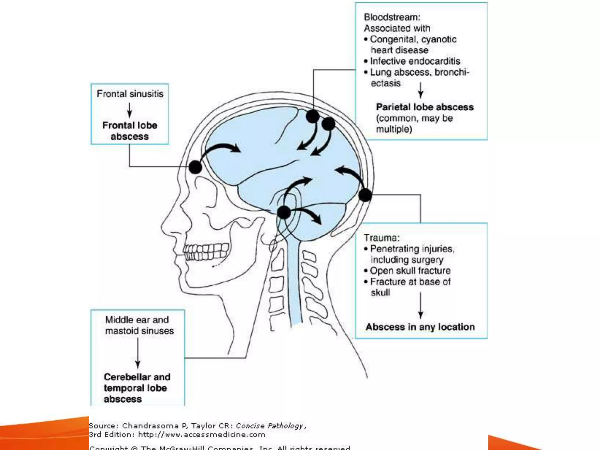
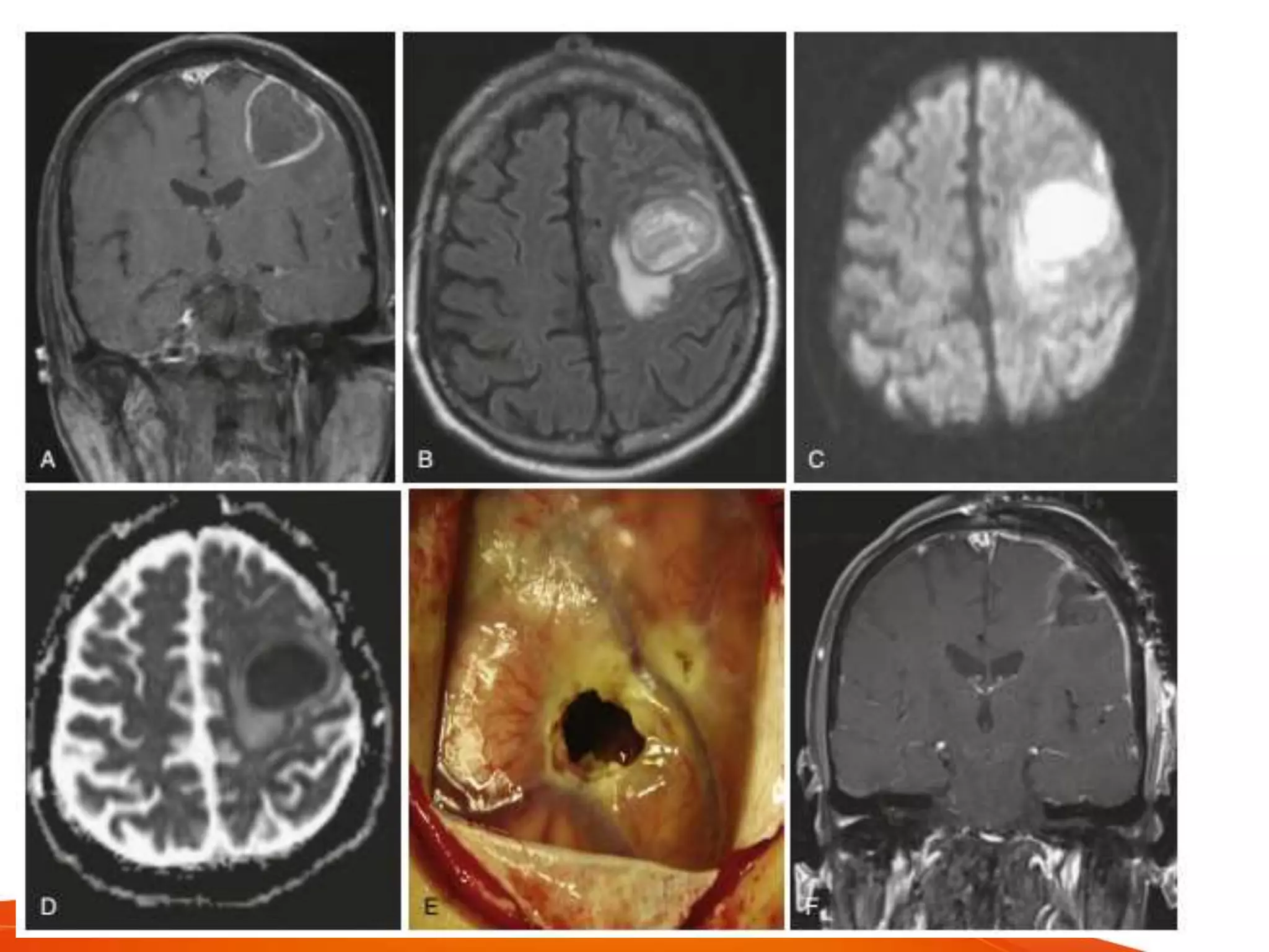
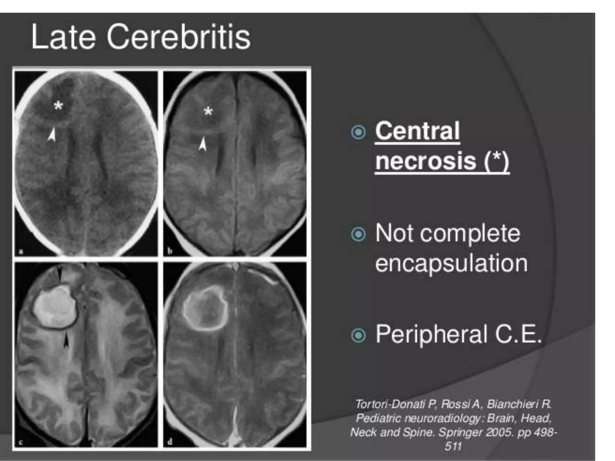
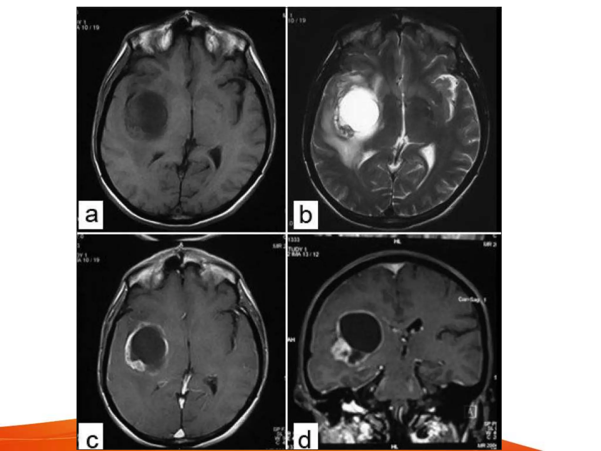
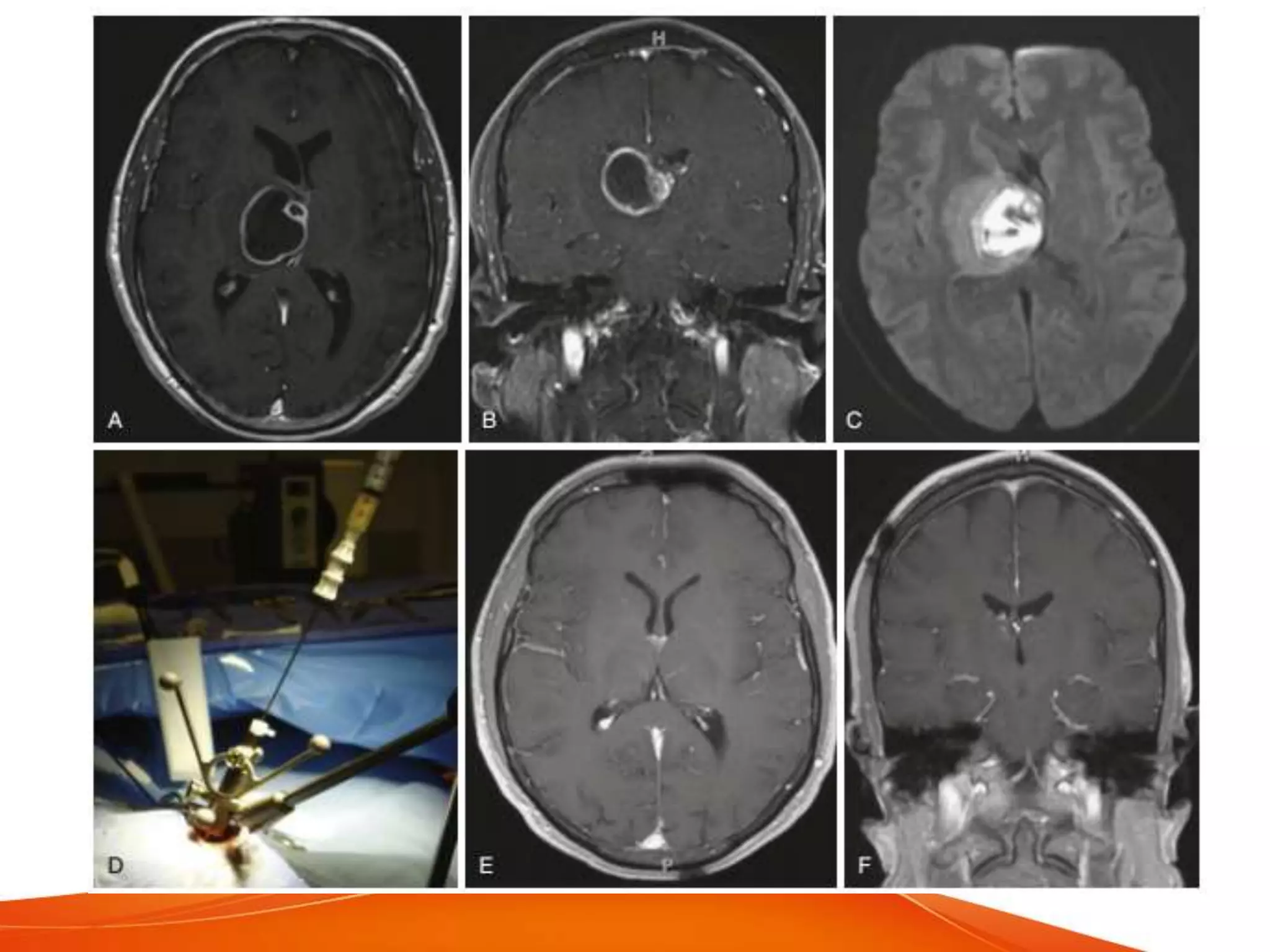
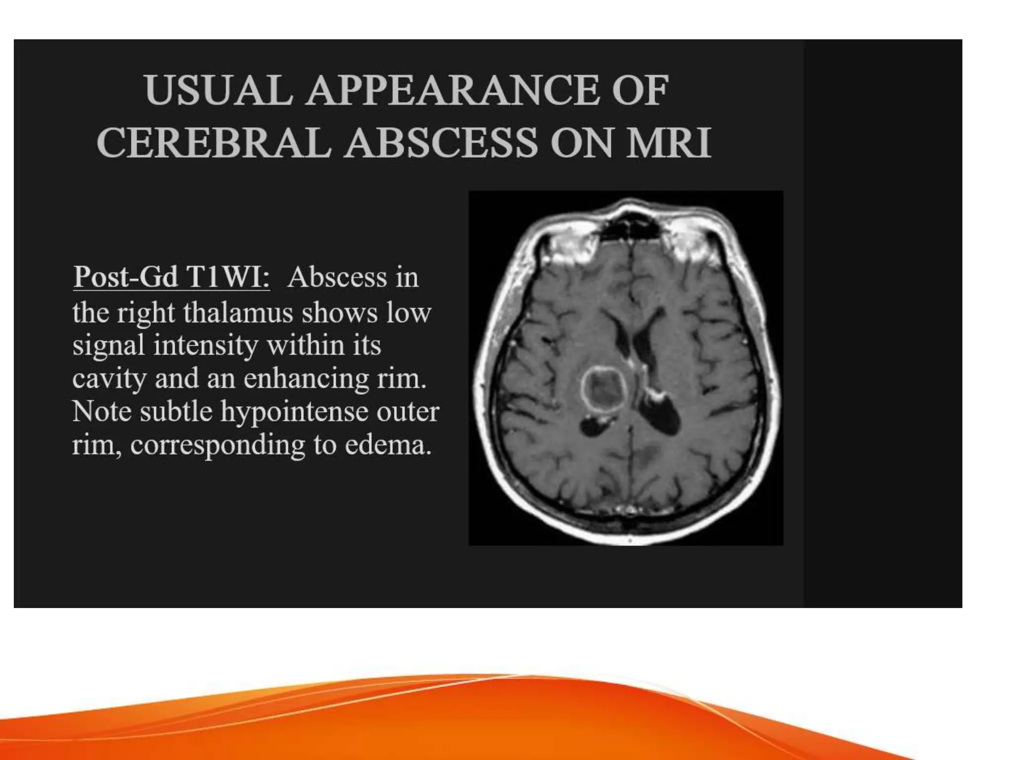
Pyogenic Brain Abscess
Dr Himanshu Soni
pathogenesis
• pyogenic brain abscess is a focal
collection of pus withing thebrain.
• the incidence is 8% of intracranial
masses.
• largely because of the brain natural
resistance to infection, mediated by
– abundant blood supply
– relative impermeability of blood brain barrier
– improvement in the treatment of ear, sinus
and orofacial infections
• peak ages vary
• pediatric peak - 4 to 7 years, found in
congenital heart disease
• causes :
– trauma
– contiguous spread from a suppurative focus
– hematogenous dissemination from a distant
infection
• 40-60% are result of contiguous infection
from middle ear, oropharynx and sinuses
• these are usually solitary lesions
• seeding via te valveless emissary veins
draining the contiguous areas, allowing the
entry into venous sinus system.
• Temporal lobe / Cerebellar abscesses
– chronic otitis media, mastoiditis
• frontal or temporal lobe
– paranasal sinusitis
• penetrating cranial trauma with dural
tear account for 10% of the abscesses
post operative brain abscess
• infrequent, approx 0.1% after clean
procedure
• at the time of surgery, infect the wound /
bone flap
metastatic brain abscess
• seeding from a distant site - 25% of brain
abscesses
• locations
– in the distribution of MCA
– parietal-occipital junction
– corticomedullary junction where the capillary flow is
slowest
• sources
– pulmonary lesions - AV fistulas occuring in hereditary
telangiectasis
– infective endocarditis
– deep seated infections such as osteomyelitis,
pulmonary empyema, pelvic infections &
intraabdominal infections
other causes
• iv drug users
• cyanotic heart diseases - in 2 to 6% cases
• Tetralogy of Fallot and transposition of
great vessels most causes
• intrasellar abscesses
– pituitary / sellar adenomas,
craniopharyngiomas, Rathke's cysts or a
complication of TS surgery
– sphenoid sinusitis
• determined by the site of infection
• Streptococci ( milleri and viridans )
– most common cause, nearly 2/3rd cases,
mostly extending from naso/oropharynx,
viridans in endocarditis
• Staphylococcus aureus - 10 to 21%
– trauma postoperative, endocarditis
– MRSA in hospitalized patients
• Actinomyeces
• pulmonary and odontogenic infections
• GNB like proteus, klebsiella,
pseudomonas, E. coli, serratia
– genitourinary or intraabdominal infections
• pseudomonas
– otitis media / externa
• clostridium
– underlying malignancy or hemolytic-uremic
syndrome
• propionibacterium
– post neurosurgical patient
• bacillus like salmonella
– intracerebral hematoma
• Nocardia (asteroides and farcinica) - 1 to
2%, carry 31% mortality rate
– immunicompromised patients
– isolated or disseminated pathology
• fungal (candida, cryptococcus, dimorphic
fungus or molds)
– immunicompromised states
• protozoa / helminths
– cat exposure or endemic areas
• Neonatal meningitis caused by Proteus
and Citrobacter, result in abscess in 40-
75% cases
PATHOLOGY
• evolution studied using a-
streptococci in canine model
• early cerebritis - day 1 to 3
– poorly circumscribed lesion
– acute inflammation and cerebral
edema associated with bacterial
invasion
• Expansion - day 4 to 9
– zone of cerebritis expands
– necrosis develops with pus
formation
– CT scan reveals some ring
enhancement with diffusion of
contrast into the necrotic center
• early capsule state - days 10 to 13
– establishment and maturation of well formed
collagenous capsule
– reduction in degree of ceribritis and edema
• Late capsule stage - day 14 and beyond
– continued maturation of thick capsule with
extracapsular gliosis
– dense ring enhancement on CT contrast
CAPSULE CHARACTERISTICS
• capsule formation and ring enhancement -
thinner and less complete on the
ventricular side
• related to poor vascularity of the deep
white matter and reduced migration of
fibroblasts
• tinner area predisposis to ventricular
rupture
• nature of the organism influences
encapsulation
• bacteroides - delayed capsule formation
with multiple daughter abscesses
• S. aureus - larger, delayed healing with
markec extracapsular abnormalities
• contiguous spread - better encapsulation
• hematogenous spread - less extensive
• immunisuppresion - incomplete
encapsulation
CLINICAL PRESENTATION
• symptoms < 2 weeks
• depend on
– size & location
– virulence of the agent
– immunologic status of the host
– cerebral edema
• triad - fever, headache & focal neurological
deficit - present in < 50%
• headache
– dull and poorly localised
– 50-70% cases and is nonspecific
– sudden worsening of a prexisting headache in
a ptient with abscess, with meningeal signs,
suggest herniation or intraventricular rupture
• fever
– 25-50% cases, more common in children
• deficits
– altered sensorium often present
– frontal and parietal abscesses
• hemiparesis, aphasia
– temporal lobe
• aphasia, visual field defects
– intrasellar
• mimic pituitary tumors
– cerebellar
– ataxia and nystagmus
LABORATORY FINDINGS
• moderate leucocytosis
• blood cultures are only 10% positive
• ESR and CRP are elevated, help in
monitoring response
• LP is strongly contraindicated
• tissue and pus samples - best during
surgery
• bacterial r-DNA PCR - crucial, more
sensitive, esp in patient's already on
antibiotics
CT SCAN
• hypodense lesion, surrounded by ring
enhancement with variable zone of edema
• sensitivity is 95-99%
• specificity is compromised
• cant differentiate with metastatic tumor or
some vascular lesions
• Indium - 111 labelled leucocyte scanning
helpful, radioleucocytes accumulate in the
area of active inflammation
• delayed contrast - value in cerebritis
MRI
• more sensitive in
– early cerebritis
– extent of central liquefaction necrosis
– early satellite lesions
– extraparenchymal extension (subdural
empyema)
• insentive to differentiate
– cystic or necrotic high grade glioma
– metastasis
• T1 - pus is hypointense
• T2 - pus is hyperintense
• FLAIR - nullifies free water signal, caused
by increased protein content
– reported in brain tumors, abscesses and
vascular insults
• Diffusion weighted imaging (DWI) and
apparent diffusion coefficient (ADC) -
based on movements of water molecules
– to measure degree of water movements, ADC
maps are useful
– abscess contains bacteria, proteins and pus.
Hence shows restricted water motion
– DWI shows incresed signal with low ADC
values
– most necrotic tumors have serous fluid and
fewer inflammatory cells. Hence shows low-
intermediate intensity on DWI and high ADC
values due to serous fluid
MR SPECTROSCOPY
• helps differentiate cystic tumors and
abscesses
• detects the metabolic profile of teh brain
• cytosolic amino acids (leucine, isoleucine
and valine) are usually detected in
cerebral abscesses and absent in tumors
• however their abscence does not rule out
pyogenic abscess.
• acetate with/out succinate supports an
anaerobic abscess
• in fungal abscess, amino acids are low
• response to treatment is followed by serial
changes in metabolite patterns
• lactate and amino acids - present
regardless of teh treatment
• acetate and pyruvate - disappeared after 1
week of therapy
PET
• the mechanism of FDG uptake is related
to the degree of inflammatory cells in the
abscess
• increased uptake corresponded to the
enhanced area
• after treatment the area showed
decreased uptake
MEDICAL MANAGEMENT
• empiric antibiotic - as early as possible
• cover gram positive, gram negative &
anerobes.
• 3rd or 4th Gen cephalosporin,
metronidazole and vancomycin - choice
• culture and modify accordingly
• MRSA - vancomycin and clindamycin
ANTIBIOTIC CONSIDERATIONS
• anti-infective agents should be
– acting against probable pathogens
– able to penetrate into the abscess
– achieve high levels in abscess pus
• Penicilin G at high doses, metronidazole,
cotrimoxazole, chloramphenicol - achieve
therapeutic concentrations
• metronidazole - high levels in abscess -
important component of most regimens
• use in combination with an agent active
against streptococci (eg penicillin)
• clindamycin, aminoglycosides and 1stG
cephalosporins - poor penetration
• potentially effective
– ceftriaxone, ceftazidime or 3rd G cephalosp
– quinolones
– monobactams
– carbapenems
• duration depends on
– causative pathogen
– adequacy of drainage
• 6-8 week is recommended
• complete resolution may take 3-4 months
radiologically
• residual contrast enhancement may take
more than 6 months
STEROIDS
• controvertial
• suggested role
– diminish microbe entry into the CNS
– reduce the elimination of viable organisms from the
abscess cavity
– inhibit effective, ring enhancing, host inflammatory
response
– delay in encapsulation
• preponderance of evidence weighs against the
routine use of steroids as an adjunct except
when signs of raised ICP are marked
SEIZURE CONTROL
• incidence of seizures - 13 to 25 %
• recommended perioperative use of AED
and continue after surgery
• long term use depends on neurologic
evaluation after the abscess has resolved
MEDICAL MANAGEMENT
• determinants of medical management are
– neurologically stable with cerebritis or small
lesions (less than 1.5cm)
– severe concomitant medical conditions
– severe bleeding diathesis
– multiple abscesses
– surgically inaccesible, dominant or disparate
location
• limitations
– lack of diagnostic specimen
– empirical long term antimicrobial therapy
– potential for drug adv effects
– risk of ventricular rupture
– frequent imaging until radiographic resolution
SURGICAL MANAGEMENT
• diagnostic and therapeutic
• larger than 2.5cm - surgery must
• provides
– pathologic diagnosis
– bacteriological profile
– reduction in mass effect
– improves milieu for antimicrobial therapy
– removes toxic necrotic material
• options
– aspiration of the abscess
– excision of brain abscess
ASPIRATION
• provides specimens
• low surgery related morbidity and mortality
• post aspiration recurrence - upto 32%
• CT guided aspiration accurate within a few
mm with yield of 95%
• highly effective in definitive drainage of
abscess
• preferred treatment for deep seated
lesions or eloquent areas
• suitable for
– brain stem
– thalamus
– basal ganglia
• stereotactic drainage - can drain multiple
abscesses, with prolonged medical
therapy is effective
• frameless neuronavigation - best
technique to localise
• less chance of seizures and other
sequelae
• Ultrasound guidance
– real time
– reliable
– fewer risks
– minimally invasive and accurate
• other methods
– endoscoic stereotactic aspiration and
irrigation
• precise localisation, minimal craniotomy, multiple
lesions addressed
SURGICAL EXCISION
• useful in
– large (>2.5cm) abscess
– superficial abscess
– refractory aspirations
– posterior fossa lesions
– fungal abscesses
– post traumatic abscess with retained bones or
foreign bodies
– gas containing abscesses
• not choice in
– cerebritis stage
– deep seated abscess
– eloquent areas
• emergency surgery in
– obtunded patient
– severe neurologic deficit
– encapsulated lesion
– aim to decompress and diagnose
• choice procedure
– image guided keyhole approach
– small incision
– limited craniotomy
– limited brain retraction
– minimal intraop trauma
– better cosmesis
• large craniotomy and decompression
– failed conservative management
– dangerios location like posterior fossa
– extensive edema, mass effect
– impending or actual hydrocephalus
intraventricular rupture
• as pus increases - abscess expands - may
rupture into the ventricle
• sudden, catastrophic deterioration of the
patient
• diagnosis
– hydrocephalus
– enhancement of teh ventricular walls
• management
– immidiate ventricular drainage
– intraventricular instillation of antibiotics
– evacuation of teh remaining abscess
– systemic antibiotic therapy
• mortality greater than 80%
• if multiple lesions
– those > 2.5cm should be aspirated
– the largest or most accessible should be
aspirated for culture
OSTEOMYELITIS OF THE SKULL
• results from
– paranasal sinusitis
– otogen infection
– odontogenic infection
– penetrating truama or craniotomy
Pyogenic brain abscess

Pyogenic brain abscess

  • 1.
  • 2.
    pathogenesis • pyogenic brainabscess is a focal collection of pus withing thebrain. • the incidence is 8% of intracranial masses. • largely because of the brain natural resistance to infection, mediated by – abundant blood supply – relative impermeability of blood brain barrier – improvement in the treatment of ear, sinus and orofacial infections • peak ages vary
  • 3.
    • pediatric peak- 4 to 7 years, found in congenital heart disease • causes : – trauma – contiguous spread from a suppurative focus – hematogenous dissemination from a distant infection • 40-60% are result of contiguous infection from middle ear, oropharynx and sinuses
  • 4.
    • these areusually solitary lesions • seeding via te valveless emissary veins draining the contiguous areas, allowing the entry into venous sinus system. • Temporal lobe / Cerebellar abscesses – chronic otitis media, mastoiditis • frontal or temporal lobe – paranasal sinusitis • penetrating cranial trauma with dural tear account for 10% of the abscesses
  • 6.
    post operative brainabscess • infrequent, approx 0.1% after clean procedure • at the time of surgery, infect the wound / bone flap
  • 7.
    metastatic brain abscess •seeding from a distant site - 25% of brain abscesses • locations – in the distribution of MCA – parietal-occipital junction – corticomedullary junction where the capillary flow is slowest • sources – pulmonary lesions - AV fistulas occuring in hereditary telangiectasis – infective endocarditis – deep seated infections such as osteomyelitis, pulmonary empyema, pelvic infections & intraabdominal infections
  • 8.
    other causes • ivdrug users • cyanotic heart diseases - in 2 to 6% cases • Tetralogy of Fallot and transposition of great vessels most causes • intrasellar abscesses – pituitary / sellar adenomas, craniopharyngiomas, Rathke's cysts or a complication of TS surgery – sphenoid sinusitis
  • 11.
    • determined bythe site of infection • Streptococci ( milleri and viridans ) – most common cause, nearly 2/3rd cases, mostly extending from naso/oropharynx, viridans in endocarditis • Staphylococcus aureus - 10 to 21% – trauma postoperative, endocarditis – MRSA in hospitalized patients • Actinomyeces • pulmonary and odontogenic infections
  • 12.
    • GNB likeproteus, klebsiella, pseudomonas, E. coli, serratia – genitourinary or intraabdominal infections • pseudomonas – otitis media / externa • clostridium – underlying malignancy or hemolytic-uremic syndrome • propionibacterium – post neurosurgical patient
  • 13.
    • bacillus likesalmonella – intracerebral hematoma • Nocardia (asteroides and farcinica) - 1 to 2%, carry 31% mortality rate – immunicompromised patients – isolated or disseminated pathology • fungal (candida, cryptococcus, dimorphic fungus or molds) – immunicompromised states • protozoa / helminths – cat exposure or endemic areas
  • 14.
    • Neonatal meningitiscaused by Proteus and Citrobacter, result in abscess in 40- 75% cases
  • 15.
    PATHOLOGY • evolution studiedusing a- streptococci in canine model • early cerebritis - day 1 to 3 – poorly circumscribed lesion – acute inflammation and cerebral edema associated with bacterial invasion • Expansion - day 4 to 9 – zone of cerebritis expands – necrosis develops with pus formation – CT scan reveals some ring enhancement with diffusion of contrast into the necrotic center
  • 16.
    • early capsulestate - days 10 to 13 – establishment and maturation of well formed collagenous capsule – reduction in degree of ceribritis and edema • Late capsule stage - day 14 and beyond – continued maturation of thick capsule with extracapsular gliosis – dense ring enhancement on CT contrast
  • 20.
    CAPSULE CHARACTERISTICS • capsuleformation and ring enhancement - thinner and less complete on the ventricular side • related to poor vascularity of the deep white matter and reduced migration of fibroblasts • tinner area predisposis to ventricular rupture • nature of the organism influences encapsulation
  • 22.
    • bacteroides -delayed capsule formation with multiple daughter abscesses • S. aureus - larger, delayed healing with markec extracapsular abnormalities • contiguous spread - better encapsulation • hematogenous spread - less extensive • immunisuppresion - incomplete encapsulation
  • 23.
    CLINICAL PRESENTATION • symptoms< 2 weeks • depend on – size & location – virulence of the agent – immunologic status of the host – cerebral edema
  • 25.
    • triad -fever, headache & focal neurological deficit - present in < 50% • headache – dull and poorly localised – 50-70% cases and is nonspecific – sudden worsening of a prexisting headache in a ptient with abscess, with meningeal signs, suggest herniation or intraventricular rupture
  • 26.
    • fever – 25-50%cases, more common in children • deficits – altered sensorium often present – frontal and parietal abscesses • hemiparesis, aphasia – temporal lobe • aphasia, visual field defects – intrasellar • mimic pituitary tumors – cerebellar – ataxia and nystagmus
  • 27.
    LABORATORY FINDINGS • moderateleucocytosis • blood cultures are only 10% positive • ESR and CRP are elevated, help in monitoring response • LP is strongly contraindicated • tissue and pus samples - best during surgery • bacterial r-DNA PCR - crucial, more sensitive, esp in patient's already on antibiotics
  • 28.
    CT SCAN • hypodenselesion, surrounded by ring enhancement with variable zone of edema • sensitivity is 95-99% • specificity is compromised • cant differentiate with metastatic tumor or some vascular lesions • Indium - 111 labelled leucocyte scanning helpful, radioleucocytes accumulate in the area of active inflammation • delayed contrast - value in cerebritis
  • 30.
    MRI • more sensitivein – early cerebritis – extent of central liquefaction necrosis – early satellite lesions – extraparenchymal extension (subdural empyema) • insentive to differentiate – cystic or necrotic high grade glioma – metastasis
  • 31.
    • T1 -pus is hypointense • T2 - pus is hyperintense • FLAIR - nullifies free water signal, caused by increased protein content – reported in brain tumors, abscesses and vascular insults • Diffusion weighted imaging (DWI) and apparent diffusion coefficient (ADC) - based on movements of water molecules – to measure degree of water movements, ADC maps are useful
  • 32.
    – abscess containsbacteria, proteins and pus. Hence shows restricted water motion – DWI shows incresed signal with low ADC values – most necrotic tumors have serous fluid and fewer inflammatory cells. Hence shows low- intermediate intensity on DWI and high ADC values due to serous fluid
  • 36.
    MR SPECTROSCOPY • helpsdifferentiate cystic tumors and abscesses • detects the metabolic profile of teh brain • cytosolic amino acids (leucine, isoleucine and valine) are usually detected in cerebral abscesses and absent in tumors • however their abscence does not rule out pyogenic abscess. • acetate with/out succinate supports an anaerobic abscess
  • 37.
    • in fungalabscess, amino acids are low • response to treatment is followed by serial changes in metabolite patterns • lactate and amino acids - present regardless of teh treatment • acetate and pyruvate - disappeared after 1 week of therapy
  • 38.
    PET • the mechanismof FDG uptake is related to the degree of inflammatory cells in the abscess • increased uptake corresponded to the enhanced area • after treatment the area showed decreased uptake
  • 39.
    MEDICAL MANAGEMENT • empiricantibiotic - as early as possible • cover gram positive, gram negative & anerobes. • 3rd or 4th Gen cephalosporin, metronidazole and vancomycin - choice • culture and modify accordingly • MRSA - vancomycin and clindamycin
  • 40.
    ANTIBIOTIC CONSIDERATIONS • anti-infectiveagents should be – acting against probable pathogens – able to penetrate into the abscess – achieve high levels in abscess pus • Penicilin G at high doses, metronidazole, cotrimoxazole, chloramphenicol - achieve therapeutic concentrations • metronidazole - high levels in abscess - important component of most regimens
  • 41.
    • use incombination with an agent active against streptococci (eg penicillin) • clindamycin, aminoglycosides and 1stG cephalosporins - poor penetration • potentially effective – ceftriaxone, ceftazidime or 3rd G cephalosp – quinolones – monobactams – carbapenems
  • 42.
    • duration dependson – causative pathogen – adequacy of drainage • 6-8 week is recommended • complete resolution may take 3-4 months radiologically • residual contrast enhancement may take more than 6 months
  • 44.
    STEROIDS • controvertial • suggestedrole – diminish microbe entry into the CNS – reduce the elimination of viable organisms from the abscess cavity – inhibit effective, ring enhancing, host inflammatory response – delay in encapsulation • preponderance of evidence weighs against the routine use of steroids as an adjunct except when signs of raised ICP are marked
  • 45.
    SEIZURE CONTROL • incidenceof seizures - 13 to 25 % • recommended perioperative use of AED and continue after surgery • long term use depends on neurologic evaluation after the abscess has resolved
  • 46.
    MEDICAL MANAGEMENT • determinantsof medical management are – neurologically stable with cerebritis or small lesions (less than 1.5cm) – severe concomitant medical conditions – severe bleeding diathesis – multiple abscesses – surgically inaccesible, dominant or disparate location
  • 47.
    • limitations – lackof diagnostic specimen – empirical long term antimicrobial therapy – potential for drug adv effects – risk of ventricular rupture – frequent imaging until radiographic resolution
  • 48.
    SURGICAL MANAGEMENT • diagnosticand therapeutic • larger than 2.5cm - surgery must • provides – pathologic diagnosis – bacteriological profile – reduction in mass effect – improves milieu for antimicrobial therapy – removes toxic necrotic material • options – aspiration of the abscess – excision of brain abscess
  • 49.
    ASPIRATION • provides specimens •low surgery related morbidity and mortality • post aspiration recurrence - upto 32% • CT guided aspiration accurate within a few mm with yield of 95% • highly effective in definitive drainage of abscess • preferred treatment for deep seated lesions or eloquent areas
  • 50.
    • suitable for –brain stem – thalamus – basal ganglia • stereotactic drainage - can drain multiple abscesses, with prolonged medical therapy is effective • frameless neuronavigation - best technique to localise • less chance of seizures and other sequelae
  • 51.
    • Ultrasound guidance –real time – reliable – fewer risks – minimally invasive and accurate • other methods – endoscoic stereotactic aspiration and irrigation • precise localisation, minimal craniotomy, multiple lesions addressed
  • 52.
    SURGICAL EXCISION • usefulin – large (>2.5cm) abscess – superficial abscess – refractory aspirations – posterior fossa lesions – fungal abscesses – post traumatic abscess with retained bones or foreign bodies – gas containing abscesses
  • 53.
    • not choicein – cerebritis stage – deep seated abscess – eloquent areas • emergency surgery in – obtunded patient – severe neurologic deficit – encapsulated lesion – aim to decompress and diagnose
  • 54.
    • choice procedure –image guided keyhole approach – small incision – limited craniotomy – limited brain retraction – minimal intraop trauma – better cosmesis • large craniotomy and decompression – failed conservative management – dangerios location like posterior fossa – extensive edema, mass effect – impending or actual hydrocephalus
  • 55.
    intraventricular rupture • aspus increases - abscess expands - may rupture into the ventricle • sudden, catastrophic deterioration of the patient • diagnosis – hydrocephalus – enhancement of teh ventricular walls • management – immidiate ventricular drainage – intraventricular instillation of antibiotics
  • 56.
    – evacuation ofteh remaining abscess – systemic antibiotic therapy • mortality greater than 80% • if multiple lesions – those > 2.5cm should be aspirated – the largest or most accessible should be aspirated for culture
  • 57.
    OSTEOMYELITIS OF THESKULL • results from – paranasal sinusitis – otogen infection – odontogenic infection – penetrating truama or craniotomy