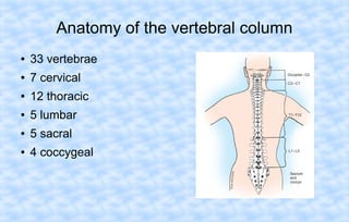
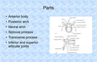
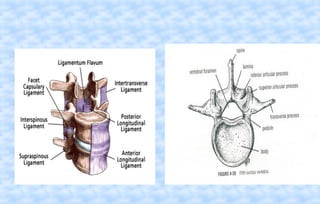
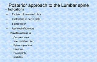
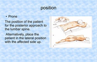
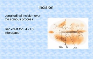
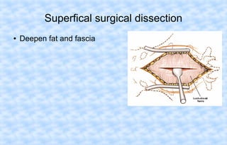
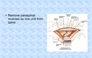
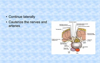
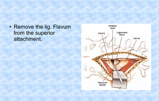
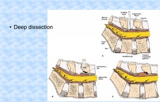
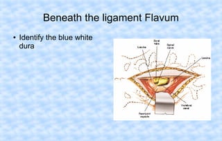
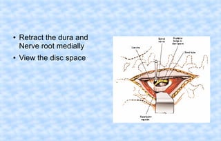
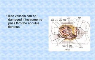
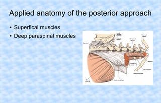
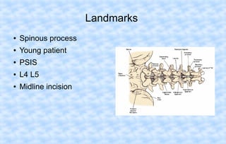
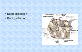
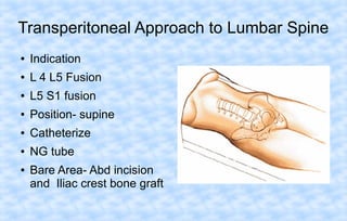
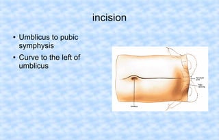
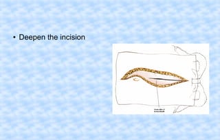
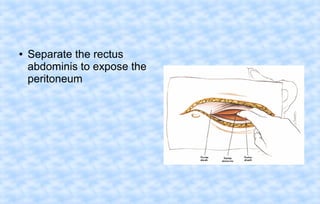
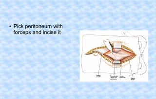
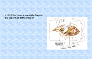
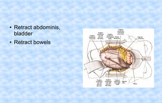
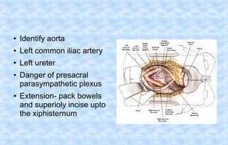
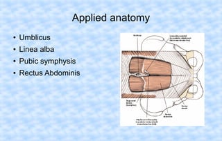
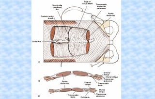
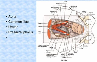
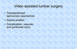
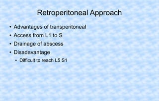
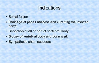
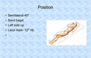
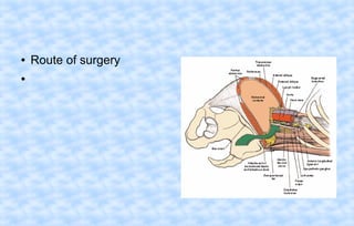
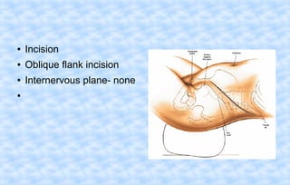
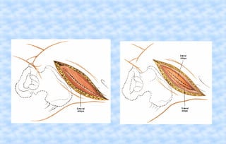
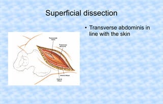
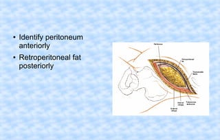
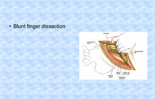
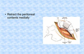
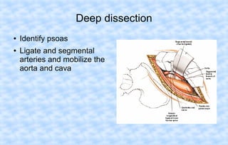
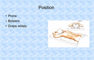
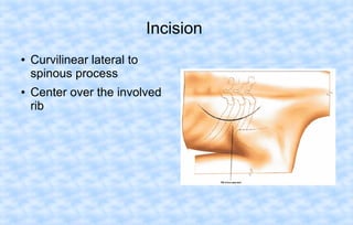
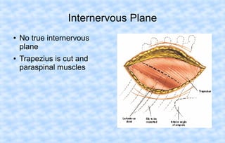
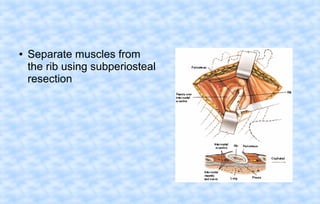
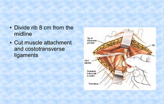
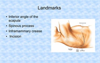
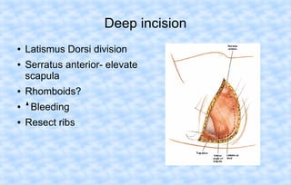
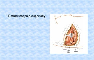
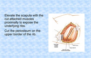
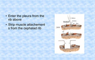
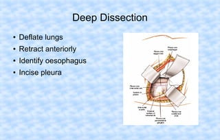
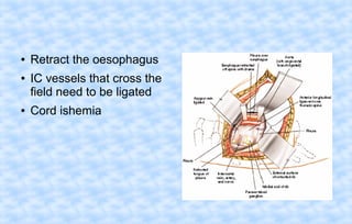
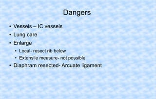
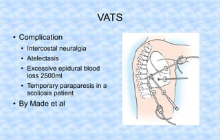
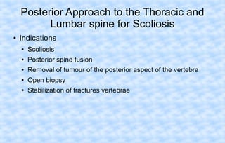
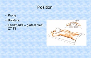
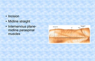
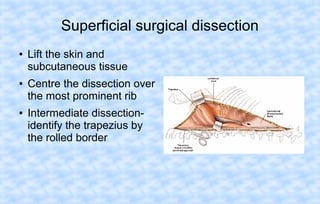
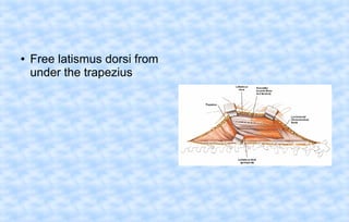
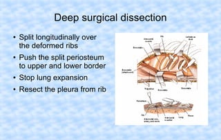
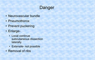
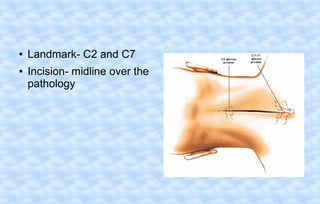
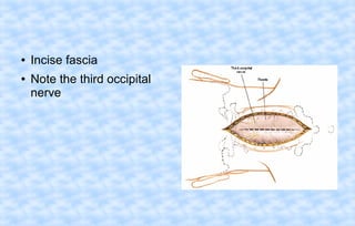
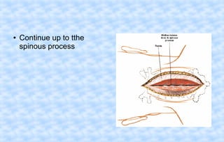
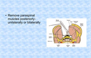
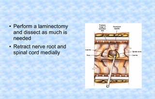
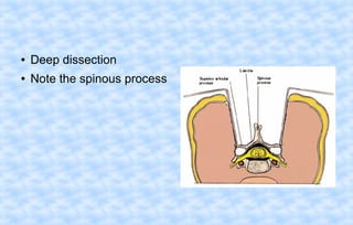
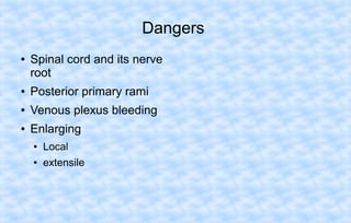
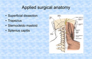
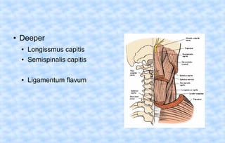
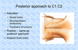
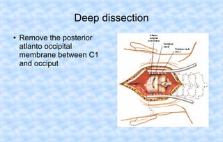
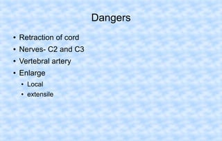
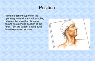
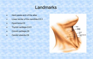
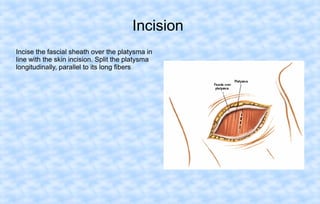
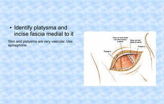
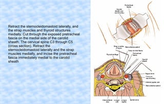
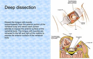
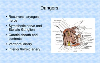
Seminar
on
“Approaches to Spine”
01 – 12 - 2010

Moderator:

Presenter:

Dr. Muralidhar N

Dr. Somnath Machani

Professor and HOD
VIMS & RC.

Post Graduate
VIMS & RC
.
Anatomy of the vertebral column
●

33 vertebrae

●

7 cervical

●

12 thoracic

●

5 lumbar

●

5 sacral

●

4 coccygeal
Parts
●

Anterior body

●

Posterior arch

●

Neural arch

●

Spinous process

●

Transverse process

●

Inferior and superior
articular joints
Cross section of the Spinal Cord
Pedicle anatomy
Pedicle screw insertion
Posterior approach to the Lumbar spine

Indications

●

•

Excision of herniated discs

•

Exploration of nerve roots

•

Spinal fusion

•

Removal of tumours

•

Provides access to
•

Cauda equina

•

Intervertebral disc

•

Spinous process

•

Laminae

•

Facet joints

•

pedicles
position
●

Prone

The position of the patient
for the posterior approach to
the lumbar spine.
Alternatively, place the
patient in the lateral position
with the affected side up.
Incision
Longitudinal incision over
the spinous process
Iliac crest for L4 - L5
interspace
Superfical surgical dissection
●

Deepen fat and fascia
●

Remove paraspinal
muscles as one unit from
spine
●
●

Continue laterally
Cauterize the nerves and
arteries
●

Remove the lig. Flavum
from the superior
attachment.
●

Deep dissection
Beneath the ligament Flavum
●

Identify the blue white
dura
●

●

Retract the dura and
Nerve root medially
View the disc space
●

Iliac vessels can be
damaged if instruments
pass thro the annulus
fibrosus
Enlarging measures
●

For Dura and Nerve root

●

For posterior spine

●

skin
Applied anatomy of the posterior approach
●

Superfical muscles

●

Deep paraspinal muscles
Landmarks
●

Spinous process

●

Young patient

●

PSIS

●

L4 L5

●

Midline incision
●

Deep dissection

●

Dura protection
Transperitoneal Approach to Lumbar Spine
●

Indication

●

L 4 L5 Fusion

●

L5 S1 fusion

●

Position- supine

●

Catheterize

●

NG tube

●

Bare Area- Abd incision
and Iliac crest bone graft
incision
●

●

Umblicus to pubic
symphysis
Curve to the left of
umblicus
●

Deepen the incision
●

Separate the rectus
abdominis to expose the
peritoneum
●

Pick peritoneum with
forceps and incise it
protect the viscera, carefully deepen
the upper half of the incision
●

●

Retract abdominis,
bladder
Retract bowels
●

Identify aorta

●

Left common iliac artery

●

Left ureter

●

●

Danger of presacral
parasympathetic plexus
Extension- pack bowels
and superioly incise upto
the xiphisternum
Applied anatomy
●

Umblicus

●

Linea alba

●

Pubic symphysis

●

Rectus Abdominis
●

Aorta

●

Common iliac

●

Ureter

●

Presacral plexus

●
Video assisted lumbar surgery
●

●
●

Transperitoneal
laproscopic approaches
Supine position
Complication- vascular
and peritoneal injury
Retroperitoneal Approach
●

Advantages of transperitoneal

●

Access from L1 to S

●

Drainage of abscess

●

Disadavantage
●

Difficult to reach L5 S1
Indications
●
●

Spinal fusion
Drainage of psoas abscess and curetting the infected
body

●

Resection of all or part of vertebral body

●

Biopsy of vertebral body and bone graft

●

Sympathetic chain exposure
Position
●

Semilateral 45º

●

Sand bagsl

●

Left side up

●

Land mark- 12th rib

●
●
●

Route of surgery
●

Incision

●

Oblique flank incision

●

Internervous plane- none

●
Superficial dissection
●

Transverse abdominis in
line with the skin
●

●

Identify peritoneum
anteriorly
Retroperitoneal fat
posteriorly
●

Blunt finger dissection
●

Retract the peritoneal
contents medially
Deep dissection
●
●

Identify psoas
Ligate and segmental
arteries and mobilize the
aorta and cava
Dangers
●

Sympathetic chain

●

Genitofemoral nerve

●

IVC

●

Ureters

●

Extension●

Posterioly

●

Upper lumbar vertebrae rib may need to be excised
Costotransversectomy approch to the
Thoracic Spine
●

Indication
●
●

Vertebral body biopsy

●

Partial verterbral body resection

●

Limited anterior spinal fusion

●

●

Abscess drainage

Ant. Lateral decompression of the spinal cord

Advantage
●

Need not enter the thoracic cavity
originally used to draining tubercular abscess
Position
●

Prone

●

Bolsters

●

Drape widely
Incision
●

●

Curvilinear lateral to
spinous process
Center over the involved
rib
Internervous Plane
●

●

No true internervous
plane
Trapezius is cut and
paraspinal muscles
●

●

Cut onto the posterior
aspect of the rib to be
resected
Incise the periosteum over
the rib
●

Separate muscles from
the rib using subperiosteal
resection
●

●

Divide rib 8 cm from the
midline
Cut muscle attachment
and costotransverse
ligaments
●

●

Enter the retropleural
space by blunt dissection
and digital palpation
Safe in disease only
Dangers
●

Nerves- Dura

●

IC vessels

●

Lungs- pneumo thorax

●

Extension- only resect the
adjacent ribs
Transthoracic Approach to the Thoracic
Spine
●

Indication

•

Treatment of infections, such as tuberculosis of the thoracic vertebral bodies20

•

Fusion of the vertebral bodies

•

Resection of the vertebral bodies for tumor and reconstruction with bone grafting

•

Correction of scoliosis (Dwyer instrumentation technique and rods)

•

Correction of kyphosis

•

Osteotomy of the spine

•

Anterior spinal cord decompression

•

Biopsy
Position
●

On the side

●

Move arm above his head

●

Approach from right side
Landmarks
●

Inferior angle of the
scapula

●

Spinous process

●

Inframammary crease

●

Incision
Deep incision
●
●

●

Latismus Dorsi division
Serratus anterior- elevate
scapula
Rhomboids?

●

Bleeding

●

Resect ribs
●
●

Retract scapula superiorly
Elevate the scapula with the
cut attached muscles
proximally to expose the
underlying ribs.
Cut the periosteum on the
upper border of the rib.
●

●

Enter the pleura from the
rib above
Strip muscle attachement
s from the cephalad rib
Deep Dissection
●

Deflate lungs

●

Retract anteriorly

●

Identify oesophagus

●

Incise pleura
●
●

●

Retract the oesophagus
IC vessels that cross the
field need to be ligated
Cord ishemia
Dangers
●

Vessels – IC vessels

●

Lung care

●

Enlarge
●
●

●

Local- resect rib below
Extensile measure- not possible

Diaphram resected- Arcuate ligament
VATS
●

Complication
●

Intercostal neuralgia

●

Atelectasis

●

●

●

Excessive epidural blood
loss 2500ml
Temporary paraparesis in a
scoliosis patient

By Made et al
Posterior Approach to the Thoracic and
Lumbar spine for Scoliosis
●

Indications
●

Scoliosis

●

Posterior spine fusion

●

Removal of tumour of the posterior aspect of the vertebra

●

Open biopsy

●

Stabilization of fractures vertebrae
Position
●

Prone

●

Bolsters

●

Landmarks – gluteal cleft,
C7 T1
●

Incision

●

Midline straight

●

Internervous planemidline paraspinal
muscles
Superficial Dissection
●

Rotation in scoliosis

●

Midline incision only
Deep dissection

●

Paraspinal muscles from spinous process

●

Keep dissection open
Dangers
●

Post primary rami

●

Segmental Vessels

●

Enlarge●

●

Local- widen exposure
using self retaining
retractor
Extensile- from cervical
spine to coccyx
Applied anatomy
●

●

Superficial mooring
muscle
Intermediate- accessory
muscles of respiration

●

Deep- paraspinal muscle

●

Landmarks
●

Superficial dissection dangers
●

Thoracic spine- more bleeding

●

Vertebral body rotation – convex side of curve

●

Intermediate surgical dissection

●

Deep portion●

lumbar facet joints are larger

●

Traumatic arthritis
Approach to the Posterio- lateral thorax for
excision of Ribs
●

After scoliosis surgery- removal of parts of ribs

●

Position- prone with bolsters

●

Land mark- prominent ribs

●

Incision- same like scoliosis surgery

●

Internervous plane- between Trapezius and Latismus
dorsi
Superficial surgical dissection
●

●

●

Lift the skin and
subcutaneous tissue
Centre the dissection over
the most prominent rib
Intermediate dissectionidentify the trapezius by
the rolled border
●

Free latismus dorsi from
under the trapezius
Deep surgical dissection
●

●

Split longitudinally over
the deformed ribs
Push the split periosteum
to upper and lower border

●

Stop lung expansion

●

Resect the pleura from rib
Danger
●

Neurovascular bundle

●

Pneumothorox

●

Prevent puckering

●

Enlarge●

●

●

Local continue
subcutaneous dissection
laterally
Extensile- not possible

Removal of ribs
Posterior Approach to cervical spine
•

Posterior cervical spine fusion

•

Excision of herniated discs

•

Treatment of tumors

•

Treatment of facet joint dislocations

•

Nerve root exploration
Position
●

Prone

●

Few degrees of flexion

●

Upright- less venous
bleeding but air emboli
●
●

Landmark- C2 and C7
Incision- midline over the
pathology
●
●

Incise fascia
Note the third occipital
nerve
●

Continue up to tthe
spinous process
●

Remove paraspinal
muscles posteriorlyunilaterally or bilaterally
●

●

Perform a laminectomy
and dissect as much is
needed
Retract nerve root and
spinal cord medially
●

Deep dissection

●

Note the spinous process
Dangers
●

Spinal cord and its nerve
root

●

Posterior primary rami

●

Venous plexus bleeding

●

Enlarging
●

Local

●

extensile
Applied surgical anatomy
●

Superficial dissection

●

Trapezius

●

Sternocleido mastoid

●

Splenius capitis
●

Deeper
●

Longissmus capitis

●

Semispinalis capitis

●

Ligamentum flavum
Posterior approach to C1 C2
●

Indication
●
●

●

●

●

Spinal fusion
Decompression
lamiectomy
Treatment of tumours

Position – same as
posterior approach
Incision from inion
●

Deepen in the midline
Superficial dissection
Incise the nuchal ligament
down onto the large spinous
processes of C2. Lateral
view (inset). Note that the
ring of C1 is further anterior
than the spinous process of
C2.
Remove the paracervical muscles
from the posterior elements of C1 and
C2. Carry the dissection up to the
base of the occiput
Deep dissection
●

Remove the posterior
atlanto occipital
membrane between C1
and occiput
Dangers
●

Retraction of cord

●

Nerves- C2 and C3

●

Vertebral artery

●

Enlarge
●

Local

●

extensile
Anterior approach to the Cervical Spine
●

Indication

•

Excision of herniated discs

•

Interbody fusion

•

Removal of osteophytes from the uncinate processes and from either the anterior or
the posterior lip of the vertebral bodies

•

Excision of tumors and associated bone grafting

•

Treatment of osteomyelitis

•

Biopsy of vertebral bodies and disc spaces

•

Drainage of abscesses
Position
Place the patient supine on the
operating table with a small sandbag
between the shoulder blades to
ensure an extended position of the
neck. Turn the patient's head away
from the planned incision
Landmarks
•

Hard palate-arch of the atlas

•

Lower border of the mandible-C2-3

•

Hyoid bone-C3

•

Thyroid cartilage-C4-5

•

Cricoid cartilage-C6

•

Carotid tubercle-C6
Incision
Incise the fascial sheath over the platysma in
line with the skin incision. Split the platysma
longitudinally, parallel to its long fibers
●

Identify platysma and
incise fascia medial to it

Skin and platysma are very vascular. Use
epinephrine
Retract the sternocleidomastoid laterally, and
the strap muscles and thyroid structures
medially. Cut through the exposed pretracheal
fascia on the medial side of the carotid
sheath. The cervical spine C3 through C5
(cross section). Retract the
sternocleidomastoid laterally and the strap
muscles medially, and incise the pretracheal
fascia immediately medial to the carotid
sheath
Deep dissection

Dissect the longus colli muscle
subperiosteally from the anterior portion of the
vertebral body and retract each portion
laterally to expose the anterior surface of the
vertebral body. The longus colli muscles are
retracted to the left and right of the midline to
expose the anterior surface of the vertebral
body
Dangers
●

●

●

Recurrent laryngeal
nerve
Symathetic nerve and
Stellate Ganglion
Carotid sheath and
contents

●

Vertebral artery

●

Inferior thyroid artery
Anterior Approaches
●

Necessity
●
●

●

Anterior spinal for cord decompression
Failed laminectomy

Relative indications
●

Traumatic

●

Infection

●

Degenerative

●

Neoplastic

●

deformity
Anterior Transoral Approach
●

Indications
●
●

Odointecomy

●

●

TB abscess
Skull base surgery

Complication
●

infection
Anterior Retropharyngeal Approach
●

Upper cerivcal spine and graft

●

Extramucosal- less chance of infection

●

Extended sub total maxillectomy
●

Alternate to transoral

●

For exposure and removal of tumour from the base of skull
Low Anterior cervical approach
●

Same as Anterior cervical approach

●

From left side 1 finger breath above the clavicle

●

Extending across the midline

●
High transthoracic Approach
●
●

●

Uses C6 to T4
Kyphosis forces the
cervical spine in to the
chest
Incision- Periscapular
Modified Anterior Thoracic Approach
●

Supine

●

Stable neck

●

●

Ligate and divide thyroid
vessels
Dont damage RLN or
Superior laryngeal nerve
Thankyou

Approaches to spine

Editor's Notes

  • #2 In 1989, Siliski, Mahring, and Hofer evaluated 52 intercondylar femoral fractures (AO type C) treated predominantly with blade plates. Three quarters of the fractures were caused by high-energy mechanisms, and 39% were open fractures. Overall, good or excellent results were obtained in 81% of fractures, and range of motion averaged 107 degrees. Results were better in type C1 fractures (92% good or excellent results) than in type C2 and type C3 fractures (77% good or excellent results). Only three (5.8%) fractures had malalignment in the sagittal plane; however, shortening of 1 to 3 cm occurred in 15 patients. Shortening was intentional to improve stability in 11 older patients (average age 60 years), but it was unintentional in four younger patients (average age 30 years). Infection occurred in four patients (7.7%) and accounted for three of the four poor results. Two fractures complicated by infection required amputation, and one required arthrodesis to treat the infection. Perioperative antibiotics were not used in closed and type I open fractures.