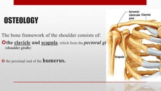
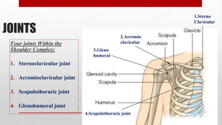
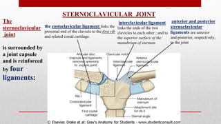
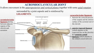
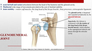
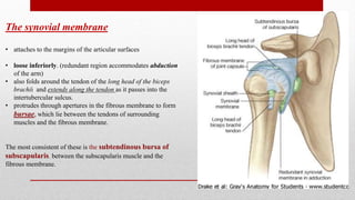
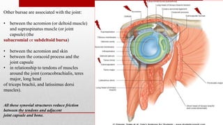
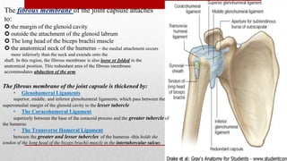
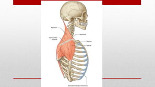
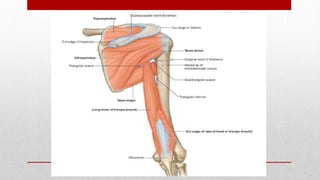
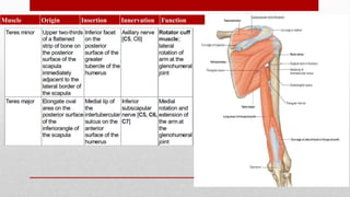
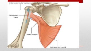
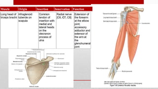
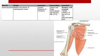
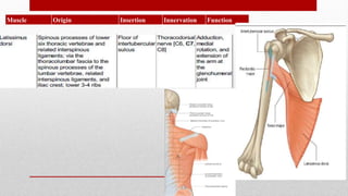
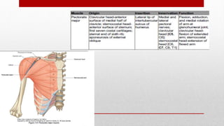
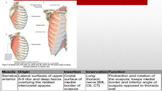
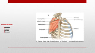
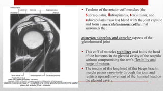
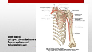
ANATOMY
& APPLIED ASPECTS OF SHOULDER JOINT
PMR PG Teaching- August 2016
Dr. Jimy Jose
Resident, Deptt. Of PMR
SMS Medical College, Jaipur
OSTEOLOGY
The bone framework of the shoulder consists of:
the clavicle and scapula, which form the pectoral girdle
(shoulder girdle)
 the proximal end of the humerus.
Clavicle
• The clavicle is the only bony attachment between the trunk and the upper limb.
• It is palpable along its entire length and has a gentle S-shaped contour
• forward-facing convex part medial and
• forward-facing concave part lateral.
The acromial (lateral) end of the clavicle is flat
The sternal (medial) end is more robust and somewhat quadrangular in shape.
the surfaces and margins of the clavicle are roughened by the attachment of muscles that
connect the clavicle to the thorax, neck, and upper limb.
 The superior surface is smoother than the inferior surface.
Scapula
The scapula is a large, flat triangular bone with:
• three angles (lateral, superior, and inferior);
• three borders (superior, lateral, and medial);
• two surfaces (costal and posterior); and
• three processes (acromion, spine, and coracoid process)
Posterior view of right
scapula.
Anterior view
of scapula
• A large triangular-shaped roughening (the infraglenoid tubercle) inferior to
the glenoid cavity is the site of attachment for the long head of the triceps
brachii muscle.
• The lateral angle of the scapula is marked by a shallow, somewhat comma-
shaped glenoid cavity,
• which articulates with the head of the humerus to form the glenohumeral joint
Lateral view
of scapula
• supraglenoid tubercle is located superior to the glenoid cavity and is the site
of attachment for the long head of the biceps brachii muscle.
• A prominent spine subdivides the posterior surface of the scapula into a
small, superior supraspinous fossa and a much larger, inferior infraspinous
fossa.
• The acromion, which is an anterolateral projection of the spine, arches over
the glenohumeral joint and articulates, via a small oval facet on its distal end,
with the clavicle.
• Unlike the posterior surface, the costal surface of the scapula is unremarkable,
being characterized by a shallow concave subscapular fossa over much of its
extent .
• The costal surface and margins provide for muscle attachment, and the costal
surface, together with its related muscle (subscapularis), moves freely over the
underlying thoracic wall.
• The lateral border of the scapula is strong and thick for muscle attachment,
• medial border and much of the superior border is thin and sharp.
 The superior border is marked on its lateral end by:
• the coracoid process, a hook-like structure that projects anterolaterally and is
positioned directly inferior to the lateral part of the clavicle
• the small but distinct suprascapular notch, which lies immediately medial to
the root of the coracoid process.
JOINTS
Four joints Within the
Shoulder Complex:
1. Sternoclavicular joint
2. Acromioclavicular joint
3. Scapulothoracic joint
4. Glenohumeral joint
1.Sterno
Clavicular
2.Acromio
clavicular
3.Gleno
humeral
4.Scapulothoracic joint
The
sternoclavicular
joint
is surrounded by
a joint capsule
and is reinforced
by four
ligaments:
anterior and posterior
sternoclavicular
ligaments are anterior
and posterior, respectively,
to the joint
the costoclavicular ligament links the
proximal end of the clavicle to the first rib
and related costal cartilage.
interclavicular ligament
links the ends of the two
clavicles to each other ; and to
the superior surface of the
manubrium of sternum
STERNOCLAVICULAR JOINT
ACROMIOCLAVICULAR JOINT
It allows movement in the anteroposterior and vertical planes together with some axial rotation.
surrounded by a joint capsule and is reinforced by
LIGAMENTS:
acromioclavicular
ligament passing
between adjacent
regions of the
clavicle and
acromion
coracoclavicular ligament-
• between the coracoid process
of the scapula and the inferior
surface of the acromial end of
the clavicle
• anterior trapezoid ligament
(which attaches to the
trapezoid line on the clavicle)
& a posterior conoid
ligament (which attaches to
the related conoid tubercle)
GLENOHUMERAL
JOINT
 synovial ball and socket articulation between the head of the humerus and the glenoid cavity.
 Multiaxial wide range of movements provided at the cost of skeletal stability
 Joint stability - rotator cuff muscles, the long head of the biceps brachii , bony processes, extracapsular ligaments
The glenoid cavity is deepened
and expanded peripherally by the
glenoid labrum
Superiorly, this labrum is
continuous with the tendon of
the long head of the biceps
brachii muscle, which attaches
to the supraglenoid tubercle and
passes through the articular
cavity
The synovial membrane
• attaches to the margins of the articular surfaces
• loose inferiorly. (redundant region accommodates abduction
of the arm)
• also folds around the tendon of the long head of the biceps
brachii and extends along the tendon as it passes into the
intertubercular sulcus.
• protrudes through apertures in the fibrous membrane to form
bursae, which lie between the tendons of surrounding
muscles and the fibrous membrane.
The most consistent of these is the subtendinous bursa of
subscapularis, between the subscapularis muscle and the
fibrous membrane.
Other bursae are associated with the joint:
• between the acromion (or deltoid muscle)
and supraspinatus muscle (or joint
capsule) (the
subacromial or subdeltoid bursa)
• between the acromion and skin
• between the coracoid process and the
joint capsule
• in relationship to tendons of muscles
around the joint (coracobrachialis, teres
major, long head
of triceps brachii, and latissimus dorsi
muscles).
All these synovial structures reduce friction
between the tendons and adjacent
joint capsule and bone.
The fibrous membrane of the joint capsule attaches
to:
 the margin of the glenoid cavity
 outside the attachment of the glenoid labrum
 The long head of the biceps brachii muscle
 the anatomical neck of the humerus – the medial attachment occurs
more inferiorly than the neck and extends onto the
shaft. In this region, the fibrous membrane is also loose or folded in the
anatomical position. This redundant area of the fibrous membrane
accommodates abduction of the arm.
The fibrous membrane of the joint capsule is thickened by:
• Glenohumeral Ligaments
superior, middle, and inferior glenohumeral ligaments, which pass between the
superomedial margin of the glenoid cavity to the lesser tubercle
• The Coracohumeral Ligament
superiorly between the base of the coracoid process and the greater tubercle of
the humerus
• The Transverse Humeral Ligament
between the greater and lesser tubercles of the humerus -this holds the
tendon of the long head of the biceps brachii muscle in the intertubercular sulcus
Scapulothoracic Joint
 not a true joint - rather a point of contact
between the anterior surface of the scapula and
the posterior-lateral wall of the thorax
the scapula is typically positioned between the
second and the seventh rib, with the medial border
located about six (6) cm lateral to the spine
SURFACE ANATOMY
SURFACE ANATOMY
SURFACE ANATOMY
SURFACE ANATOMY
 Some muscles of the
shoulder connect the
scapula and clavicle to the
trunk.
• The trapezius
• Levator scapulae
• Rhomboids
 muscles connect the
clavicle, scapula, and body
wall to the proximal end of
the humerus.
• pectoralis major
• pectoralis minor
• latissimus dorsi
• teres major
• deltoid
TRAPEZIUS AND DELTOID
• The two most superficial muscles of the shoulder
• Together, they provide the characteristic contour of the shoulder
• the trapezius attaches the scapula and clavicle to the trunk.
• the deltoid attaches the scapula and clavicle to the humerus.
• Both the trapezius and deltoid are attached to opposing surfaces and margins of the
spine of the scapula, acromion, and clavicle.
• The scapula, acromion, and clavicle can be palpated between the attachments of
trapezius and deltoid.
Muscle Origin Insertion Innervation Function
• Together, the left and right trapezius muscles form a diamond or trapezoid shape, from
which the name is derived.
• The accessory nerve can be evaluated by testing the function of the trapezius muscle. This
is done by asking patients to shrug their shoulders against resistance.
Muscle Origin Insertion Innervation Function
four rotator cuff muscles-
supraspinatus
Infraspinatus
Teres minor
Subscapularis
• Tendons of the rotator cuff muscles (the
supraspinatus, infraspinatus, teres minor, and
subscapularis muscles) blend with the joint capsule
and form a musculotendinous collar that
surrounds the :
posterior, superior, and anterior aspects of the
glenohumeral joint
• This cuff of muscles stabilizes and holds the head
of the humerus in the glenoid cavity of the scapula
without compromising the arm's flexibility and
range of motion.
• The tendon of the long head of the biceps brachii
muscle passes superiorly through the joint and
restricts upward movement of the humeral head on
the glenoid cavity.
Blood supply:
ant & post circumflex humeral vessel.
Suprascapular vessel.
Subscapular vessel
Vascular supply of Glenohumeral joint
branches of the anterior and posterior circumflex humeral and suprascapular
arteries.
innervated by branches from the posterior cord of the brachial plexus, and
from the suprascapular, axillary nerves.
Nerve supply:
axillary nerve
musculo cutaneous nerve
suprascapular nerve
Internal rotation
Subscapularis
latissimus dorsi
anterior fiber of the deltoid
pectoralis major
teres major.
External rotators -
Infraspinatus
teres minor
posterior fibers of the deltoid.
Abductors
Deltoid
Supraspinatus
trapezius,
serratus anterior.
Adduction
Subscapularis
Infraspinatus
teres minor
Pectoralis
Latissimus dorsi
teres major
Flexion of the arm
Pectoralis major
biceps brachii
and anterior deltoid
Extension
posterior deltoid
teres major
Latissimus dorsi
The muscles noted previously can be divided into functional groups
References
Clinical Anatomy Snell’s
Gray’s Anatomy
Thank you

applied aspects of shoulder joint

  • 1.
    ANATOMY & APPLIED ASPECTSOF SHOULDER JOINT PMR PG Teaching- August 2016 Dr. Jimy Jose Resident, Deptt. Of PMR SMS Medical College, Jaipur
  • 2.
    OSTEOLOGY The bone frameworkof the shoulder consists of: the clavicle and scapula, which form the pectoral girdle (shoulder girdle)  the proximal end of the humerus.
  • 3.
    Clavicle • The clavicleis the only bony attachment between the trunk and the upper limb. • It is palpable along its entire length and has a gentle S-shaped contour • forward-facing convex part medial and • forward-facing concave part lateral. The acromial (lateral) end of the clavicle is flat The sternal (medial) end is more robust and somewhat quadrangular in shape. the surfaces and margins of the clavicle are roughened by the attachment of muscles that connect the clavicle to the thorax, neck, and upper limb.  The superior surface is smoother than the inferior surface.
  • 5.
    Scapula The scapula isa large, flat triangular bone with: • three angles (lateral, superior, and inferior); • three borders (superior, lateral, and medial); • two surfaces (costal and posterior); and • three processes (acromion, spine, and coracoid process)
  • 6.
    Posterior view ofright scapula.
  • 7.
  • 8.
    • A largetriangular-shaped roughening (the infraglenoid tubercle) inferior to the glenoid cavity is the site of attachment for the long head of the triceps brachii muscle. • The lateral angle of the scapula is marked by a shallow, somewhat comma- shaped glenoid cavity, • which articulates with the head of the humerus to form the glenohumeral joint
  • 9.
  • 10.
    • supraglenoid tubercleis located superior to the glenoid cavity and is the site of attachment for the long head of the biceps brachii muscle. • A prominent spine subdivides the posterior surface of the scapula into a small, superior supraspinous fossa and a much larger, inferior infraspinous fossa. • The acromion, which is an anterolateral projection of the spine, arches over the glenohumeral joint and articulates, via a small oval facet on its distal end, with the clavicle.
  • 11.
    • Unlike theposterior surface, the costal surface of the scapula is unremarkable, being characterized by a shallow concave subscapular fossa over much of its extent . • The costal surface and margins provide for muscle attachment, and the costal surface, together with its related muscle (subscapularis), moves freely over the underlying thoracic wall. • The lateral border of the scapula is strong and thick for muscle attachment, • medial border and much of the superior border is thin and sharp.
  • 12.
     The superiorborder is marked on its lateral end by: • the coracoid process, a hook-like structure that projects anterolaterally and is positioned directly inferior to the lateral part of the clavicle • the small but distinct suprascapular notch, which lies immediately medial to the root of the coracoid process.
  • 13.
    JOINTS Four joints Withinthe Shoulder Complex: 1. Sternoclavicular joint 2. Acromioclavicular joint 3. Scapulothoracic joint 4. Glenohumeral joint 1.Sterno Clavicular 2.Acromio clavicular 3.Gleno humeral 4.Scapulothoracic joint
  • 14.
    The sternoclavicular joint is surrounded by ajoint capsule and is reinforced by four ligaments: anterior and posterior sternoclavicular ligaments are anterior and posterior, respectively, to the joint the costoclavicular ligament links the proximal end of the clavicle to the first rib and related costal cartilage. interclavicular ligament links the ends of the two clavicles to each other ; and to the superior surface of the manubrium of sternum STERNOCLAVICULAR JOINT
  • 15.
    ACROMIOCLAVICULAR JOINT It allowsmovement in the anteroposterior and vertical planes together with some axial rotation. surrounded by a joint capsule and is reinforced by LIGAMENTS: acromioclavicular ligament passing between adjacent regions of the clavicle and acromion coracoclavicular ligament- • between the coracoid process of the scapula and the inferior surface of the acromial end of the clavicle • anterior trapezoid ligament (which attaches to the trapezoid line on the clavicle) & a posterior conoid ligament (which attaches to the related conoid tubercle)
  • 16.
    GLENOHUMERAL JOINT  synovial balland socket articulation between the head of the humerus and the glenoid cavity.  Multiaxial wide range of movements provided at the cost of skeletal stability  Joint stability - rotator cuff muscles, the long head of the biceps brachii , bony processes, extracapsular ligaments The glenoid cavity is deepened and expanded peripherally by the glenoid labrum Superiorly, this labrum is continuous with the tendon of the long head of the biceps brachii muscle, which attaches to the supraglenoid tubercle and passes through the articular cavity
  • 17.
    The synovial membrane •attaches to the margins of the articular surfaces • loose inferiorly. (redundant region accommodates abduction of the arm) • also folds around the tendon of the long head of the biceps brachii and extends along the tendon as it passes into the intertubercular sulcus. • protrudes through apertures in the fibrous membrane to form bursae, which lie between the tendons of surrounding muscles and the fibrous membrane. The most consistent of these is the subtendinous bursa of subscapularis, between the subscapularis muscle and the fibrous membrane.
  • 18.
    Other bursae areassociated with the joint: • between the acromion (or deltoid muscle) and supraspinatus muscle (or joint capsule) (the subacromial or subdeltoid bursa) • between the acromion and skin • between the coracoid process and the joint capsule • in relationship to tendons of muscles around the joint (coracobrachialis, teres major, long head of triceps brachii, and latissimus dorsi muscles). All these synovial structures reduce friction between the tendons and adjacent joint capsule and bone.
  • 19.
    The fibrous membraneof the joint capsule attaches to:  the margin of the glenoid cavity  outside the attachment of the glenoid labrum  The long head of the biceps brachii muscle  the anatomical neck of the humerus – the medial attachment occurs more inferiorly than the neck and extends onto the shaft. In this region, the fibrous membrane is also loose or folded in the anatomical position. This redundant area of the fibrous membrane accommodates abduction of the arm. The fibrous membrane of the joint capsule is thickened by: • Glenohumeral Ligaments superior, middle, and inferior glenohumeral ligaments, which pass between the superomedial margin of the glenoid cavity to the lesser tubercle • The Coracohumeral Ligament superiorly between the base of the coracoid process and the greater tubercle of the humerus • The Transverse Humeral Ligament between the greater and lesser tubercles of the humerus -this holds the tendon of the long head of the biceps brachii muscle in the intertubercular sulcus
  • 20.
    Scapulothoracic Joint  nota true joint - rather a point of contact between the anterior surface of the scapula and the posterior-lateral wall of the thorax the scapula is typically positioned between the second and the seventh rib, with the medial border located about six (6) cm lateral to the spine
  • 21.
  • 22.
  • 23.
  • 24.
  • 25.
     Some musclesof the shoulder connect the scapula and clavicle to the trunk. • The trapezius • Levator scapulae • Rhomboids  muscles connect the clavicle, scapula, and body wall to the proximal end of the humerus. • pectoralis major • pectoralis minor • latissimus dorsi • teres major • deltoid
  • 26.
    TRAPEZIUS AND DELTOID •The two most superficial muscles of the shoulder • Together, they provide the characteristic contour of the shoulder • the trapezius attaches the scapula and clavicle to the trunk. • the deltoid attaches the scapula and clavicle to the humerus. • Both the trapezius and deltoid are attached to opposing surfaces and margins of the spine of the scapula, acromion, and clavicle. • The scapula, acromion, and clavicle can be palpated between the attachments of trapezius and deltoid.
  • 27.
    Muscle Origin InsertionInnervation Function
  • 29.
    • Together, theleft and right trapezius muscles form a diamond or trapezoid shape, from which the name is derived. • The accessory nerve can be evaluated by testing the function of the trapezius muscle. This is done by asking patients to shrug their shoulders against resistance.
  • 31.
    Muscle Origin InsertionInnervation Function
  • 32.
    Muscle Origin InsertionInnervation Function
  • 35.
    Muscle Origin InsertionInnervation Function
  • 37.
    Muscle Origin InsertionInnervation Function
  • 39.
    Muscle Origin InsertionInnervation Function
  • 43.
    four rotator cuffmuscles- supraspinatus Infraspinatus Teres minor Subscapularis
  • 44.
    • Tendons ofthe rotator cuff muscles (the supraspinatus, infraspinatus, teres minor, and subscapularis muscles) blend with the joint capsule and form a musculotendinous collar that surrounds the : posterior, superior, and anterior aspects of the glenohumeral joint • This cuff of muscles stabilizes and holds the head of the humerus in the glenoid cavity of the scapula without compromising the arm's flexibility and range of motion. • The tendon of the long head of the biceps brachii muscle passes superiorly through the joint and restricts upward movement of the humeral head on the glenoid cavity.
  • 46.
    Blood supply: ant &post circumflex humeral vessel. Suprascapular vessel. Subscapular vessel
  • 47.
    Vascular supply ofGlenohumeral joint branches of the anterior and posterior circumflex humeral and suprascapular arteries. innervated by branches from the posterior cord of the brachial plexus, and from the suprascapular, axillary nerves.
  • 48.
    Nerve supply: axillary nerve musculocutaneous nerve suprascapular nerve
  • 49.
    Internal rotation Subscapularis latissimus dorsi anteriorfiber of the deltoid pectoralis major teres major. External rotators - Infraspinatus teres minor posterior fibers of the deltoid. Abductors Deltoid Supraspinatus trapezius, serratus anterior. Adduction Subscapularis Infraspinatus teres minor Pectoralis Latissimus dorsi teres major Flexion of the arm Pectoralis major biceps brachii and anterior deltoid Extension posterior deltoid teres major Latissimus dorsi The muscles noted previously can be divided into functional groups
  • 50.
  • 51.

Editor's Notes

  • #21 Movement of the Scapulothoracic Joint is A Composite of the Sternoclavicular and Acromioclavicular Joint Movements
  • #48 Vascular supply of Glenohumeral joint branches of the anterior and posterior circumflex humeral and suprascapular arteries. innervated by branches from the posterior cord of the brachial plexus, and from the suprascapular, axillary nerves.