Biomechanics of elbow joint
•Download as PPTX, PDF•
15 likes•692 views

Dr. Amrit parihar IKDRC ITS college of physiotherapy, Ahmedabad amritparihar94@yahoo.com 8233341883

Recommended
More Related Content
What's hot
What's hot (20)
Similar to Biomechanics of elbow joint
Similar to Biomechanics of elbow joint (20)
More from Dr Amrit Parihar
Recently uploaded
Recently uploaded (20)
Biomechanics of elbow joint
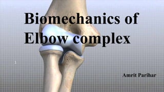
- 1. Biomechanics of Elbow complex 1 Amrit Parihar
- 2. The elbow complex includes the elbow joint (humeroulnar and humeroradial joints) and the proximal and distal radioulnar joints. The elbow joint is considered to be a compound joint that functions as a modified or loose hinge joint. One degree of freedom is possible at the elbow, permitting the motions of flexion and extension, which occur in the sagittal plane around a coronal axis. A slight bit of axial rotation and side-to-side motion of the ulna occurs during flexion and extension. (that is why considered to be a modified or loose hinge joint rather than a pure hinge joint) 2
- 3. Two major ligaments and five muscles are directly associated with the elbow joint. Three muscles are flexors and two muscles are extensors. The proximal and distal radioulnar joints are linked and function as one joint. The two joints acting together produce rotation of the forearm and have 1 degree of freedom of motion. The radioulnar joints are diarthrodial uniaxial joints of the pivot type and permit rotation which occurs in the transverse plane around a longitudinal axis. Six ligaments and four muscles are associated with these joints. Two muscles are for supination, and two are for pronation. 3
- 4. Articulating Surfaces on the Humerus: The articulating surfaces on the anterior aspect of the distal humerus are the hourglass-shaped trochlea and the spherical capitulum The trochlea, which forms part of the humeroulnar articulation A groove called the trochlear groove spirals obliquely around the trochlea and divides it into medial and lateral portions. The medial portion of the trochlea projects distally more than the lateral portion and results in a valgus angulation of the forearm. 4
- 5. The indentation in the humerus located just above the trochlea is called the coronoid fossa and is designed to receive the coronoid process of the ulna at the end of elbow flexion range of motion. The indentation located on the humerus just above the capitulum is called the radial fossa and is designed to receive the head of the radius in elbow flexion. Posteriorly, the distal humerus is indented by a deep fossa called the olecranon fossa, which is designed to receive the olecranon process of the ulna at the end of elbow extension ROM. 5
- 6. 6
- 7. Articulating Surfaces on the Radius and Ulna : The ulnar articulating surface of the humeroulnar joint is a deep semicircular concave surface called the trochlear notch. The proximal portion of the notch is divided into two unequal parts by the trochlear ridge, which corresponds to the trochlear groove on the humerus. 7
- 8. The radial articulating surface of the humeroradial joint is composed of the proximal end of the radius, known as the head of the radius The radial head has a slightly cup-shaped concave surface called the fovea that is surrounded by a rim. The radial head’s convex rim fits into the capitulotrochlear groove. 8
- 9. Articulation : Articulation between the ulna and humerus at the humeroulnar joint occurs primarily as a sliding motion of the ulnar trochlear ridge on the humeral trochlear groove. Articulation between the radial head and the capitulum at the humeroradial joint involves sliding of the shallow concave radial head over the convex surface of the capitulum. In full extension, no contact occurs between the humeroradial joint. In flexion, the rim of the radial head slides in the capitulotrochlear groove and enters the radial fossa as the end of the flexion range is reached. 9
- 10. 10
- 11. Joint Capsule : The humeroulnar and humeroradial joints and the superior radioulnar joint are enclosed in a single joint capsule. Anteriorly, the proximal attachment of the capsule is just above the coronoid and radial fossae, and distally it is inserted into the ulna on the margin of the coronoid process. The capsule blends with the proximal border of the annular ligament except posteriorly, where the capsule passes deep below the annular ligament to attach to the posterior and inferior margins of the neck of the radius. Laterally, the capsule’s attachment to the radius blends with the fibers of the lateral collateral ligament (LCL). 11
- 12. 12
- 13. Medially, the capsule blends with fibers of the medial collateral ligament (MCL). Posteriorly, the capsule is attached to the humerus along the upper edge of the olecranon fossa. The capsule is fairly large, loose, and weak anteriorly and posteriorly, and it contains folds that are able to unfold to allow for a full range of elbow motion. Laterally and medially, the capsule is reinforced by the collateral ligaments. 13
- 14. The capsule’s synovial membrane lines the coronoid, radial, and olecranon fossae. It also lines the flat medial trochlear surface and the lower part of the annular ligament. A triangular synovial fold inserted between the proximal radius and ulna partly divides the elbow joint into two joints. 14
- 15. Ligaments : 15
- 16. Medial (Ulnar) Collateral Ligament : The MCL is described as consisting of three parts (anterior, transverse, and posterior) The anterior part of the MCL extends from the anterior aspect, tip, and medial edge of the medial epicondyle of the humerus to attach on the ulnar coronoid process. The anterior portion of the MCL is considered to be the primary restraint of valgus stress from 20 to 120 of elbow flexion. The posterior part of the MCL is not as distinct as the anterior part, and sometimes its fibers blend with the fibers from the medial portion of the joint capsule. 16
- 17. The posterior portion of the MCL extends from the posterior aspect of the medial epicondyle of the humerus to attach to the ulnar coronoid and olecranon processes. The posterior MCL limits elbow extension but plays a less significant role than the anterior MCL in providing valgus stability for the elbow. The oblique (transverse) fibers of the MCL extend between the olecranon and ulnar coronoid processes. This portion of the ligament assists in providing valgus stability and helps to keep the joint surfaces in approximation. 17
- 18. Lateral (Radial) Collateral Ligamentous Complex : The lateral collateral ligamentous complex includes the LCL, the lateral ulnar collateral ligament (LUCL) and the annular ligament. The LCL is a fan-shaped structure that extends from the inferior aspect of the lateral epicondyle of the humerus to attach to the annular ligament and to the olecranon process. Ligamentous tissue extending from the lateral epicondyle to the lateral aspect of the ulnar and the annular ligament is referred to as the LUCL 18
- 19. The LUCL adheres closely to the supinator, extensor, and anconeus muscles and lies just posterior to the LCL. The LCL provides reinforcement for the humeroradial articulation, offers some protection against varus stress in some positions of the elbow, and assists in providing resistance to longitudinal distraction of the joint surfaces. Some fibers of the LCL remain taut throughout the flexion ROM when either a varus or valgus moment is applied. 19
- 20. Muscles : Nine muscles cross the anterior aspect of the elbow joint, but only three of these muscles (the brachialis, biceps brachii, and brachioradialis) have primary functions at the elbow joint. The supinator teres and pronator teres have major functions at the radiolunar joints. The remaining four muscles (flexor carpi radialis, flexor carpi ulnaris, flexor digitorum superficialis, and palmaris longus), which arise by a common tendon from the medial epicondyle of the humerus, have primary functions at wrist, hand, and fingers, but are considered to be weak flexors of the elbow. 20
- 21. The major flexors of the elbow are the brachialis, the biceps brachii, and the brachioradialis. The brachialis muscle arises from the anterior surface of the lower portion of the humeral shaft and attaches by a thick, broad tendon to the ulnar tuberosity and coronoid process. The biceps brachii arises from two heads, one short and the other long. The short head arises as a thick, flat tendon from the coracoid process of the scapula, and the long head arises as a long, narrow tendon from the scapula’s supraglenoid tubercle. 21
- 22. The muscle fibers arising from the two tendons unite in the middle of the upper arm to form the prominent muscle bulk of the upper arm. Muscle fibers from both heads insert by way of the strong flattened tendon on the rough posterior area of the tuberosity of the radius. Other fibers of the biceps brachii insert into the bicipital aponeurosis that extends medially to blend with the fascia that lies over the forearm flexors. The brachioradialis muscle arises from the lateral supracondylar ridge of the humerus and inserts into the distal end of the radius just proximal to the radial styloid process. 22
- 23. 23
- 24. The two extensors of the elbow are the triceps and the anconeus. The triceps has three heads, (long, medial, and lateral). The long head crosses both the glenohumeral joint at the shoulder as well as the elbow joint. The long head arises from the infraglenoid tubercle of the scapula by a flattened tendon that blends with the glenohumeral joint capsule. The medial and lateral heads cross only the elbow joint. 24
- 25. The medial head covers an extensive area as it arises from the entire posterior surface of the humerus. In contrast, the lateral head arises from only a narrow ridge on the posterior humeral surface. The three heads insert via a common tendon into the olecranon process. The anconeus is a small triangular muscle that arises from the posterior surface of the lateral epicondyle of the humerus and extends medially to attach to the lateral aspect of the olecranon process and the adjacent proximal quarter of the posterior surface of the ulna. 25
- 26. 26
- 27. In addition to the anconeus muscle, a number of muscles with primary actions at the wrist and fingers insert into the lateral humeral epicondyle by way of common extensor tendon. These muscles include : Extensor carpi radialis longus Extensor carpi radialis brevis Extensor digitorum communis Extensor carpi ulnaris Extensor digiti minimi 27
- 28. 28
- 29. Thank you 29