1. Animal reproduction can occur through asexual or sexual reproduction. Asexual reproduction produces offspring without fusion of gametes but maintains parental traits, while sexual reproduction allows for genetic variability through gamete fusion.
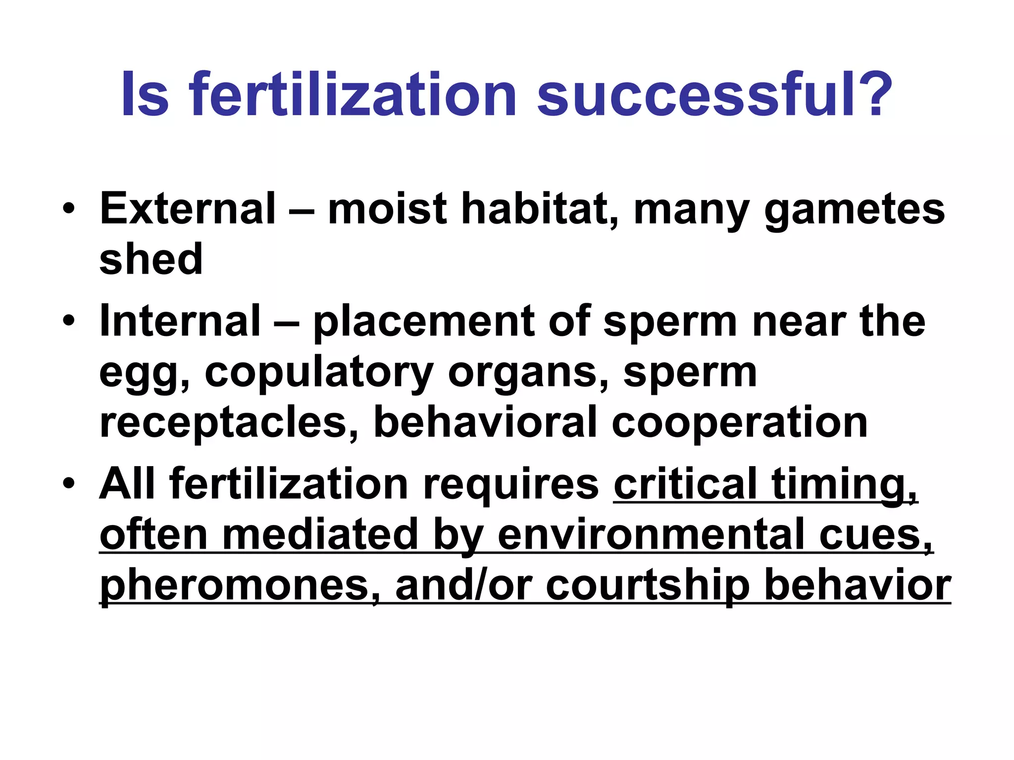
2. Sexual reproduction in animals involves complex reproductive systems with gonads that produce gametes and accessory structures for gamete transport and nourishment. Fertilization can be internal or external.
3. After fertilization, the embryo undergoes cleavage and then gastrulation to form the germ layers and begin organ development. Hormones coordinate the reproductive cycles.