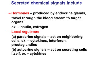
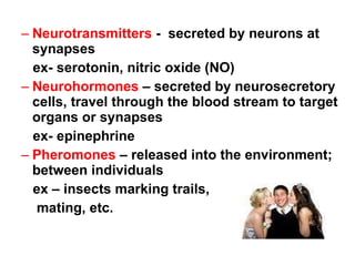
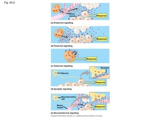
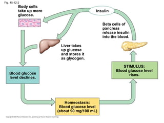
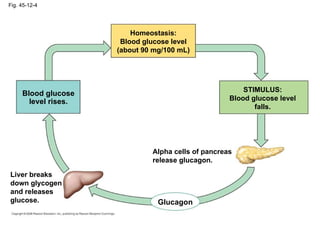
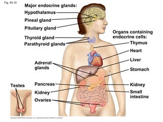
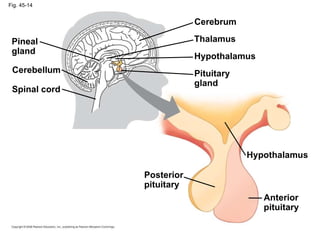
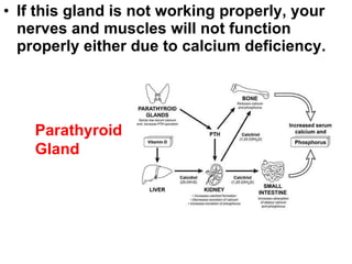
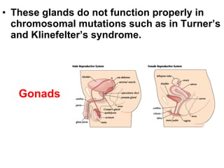
The Endocrine System AP Chapter 45
The endocrine system, along with the nervous system, is responsible for  coordinating our responses .  The endocrine system is a slower system and the nervous system is a faster response.
Chemical signals Chemicals found in both systems and also as part of other signaling mechanisms bind to specific receptor proteins on or in target cells.
Secreted chemical signals include   Hormones  – produced by endocrine glands, travel through the blood stream to target organs ex – insulin, estrogen Local regulators (a) paracrine signals – act on neighboring cells, ex. – cytokines, interferon, prostaglandins (b) autocrine signals – act on secreting cells itself, ex – cytokines
Neurotransmitters  -  secreted by neurons at synapses ex- serotonin, nitric oxide (NO) Neurohormones  – secreted by neurosecretory cells, travel through the blood stream to target organs or synapses ex- epinephrine Pheromones  – released into the environment; between individuals ex – insects marking trails,  mating, etc.
 
Fig. 45-2 Blood vessel Response Response Response Response (a) Endocrine signaling (b) Paracrine signaling (c) Autocrine signaling (d) Synaptic signaling Neuron Neurosecretory cell (e) Neuroendocrine signaling Blood vessel Synapse Response
Chemical Classes of Hormones Three major classes of molecules function as hormones in vertebrates: Polypeptides (proteins and peptides) Amines derived from amino acids Steroid hormones
Fig. 45-3 Water-soluble Lipid-soluble Steroid: Cortisol Polypeptide: Insulin Amine: Epinephrine Amine: Thyroxine 0.8 nm
Lipid-soluble hormones (steroid hormones)  pass easily  through cell membranes, while water-soluble hormones (polypeptides and amines) do not The solubility of a hormone correlates with the location of receptors inside or on the surface of target cells
Water-soluble hormones are secreted by exocytosis, travel freely in the bloodstream, and bind to cell-surface receptors Lipid-soluble hormones diffuse across cell membranes, travel in the bloodstream bound to transport proteins, and diffuse through the membrane of target cells
Fig. 45-5-1 NUCLEUS Signal receptor (a) (b) TARGET CELL Signal receptor Transport protein Water- soluble hormone Fat-soluble hormone
Fig. 45-5-2 Signal receptor TARGET CELL Signal receptor Transport protein Water- soluble hormone Fat-soluble hormone Gene regulation Cytoplasmic response Gene regulation Cytoplasmic response OR (a) NUCLEUS (b)
Multiple Effects of Hormones The same hormone may have different effects on target cells that have Different receptors for the hormone Different signal transduction pathways Different proteins for carrying out the response due to different transcription factors they activate A hormone can also have different effects in different species
Fig. 45-8-1 Glycogen deposits    receptor Vessel dilates. Epinephrine (a) Liver cell Epinephrine    receptor Glycogen breaks down and glucose is released. (b) Skeletal muscle blood vessel Same receptors but different intracellular proteins (not shown)
Fig. 45-8-2 Glycogen deposits    receptor Vessel dilates. Epinephrine (a) Liver cell Epinephrine    receptor Glycogen breaks down and glucose is released. (b) Skeletal muscle blood vessel Same receptors but different intracellular proteins (not shown) Epinephrine    receptor Different receptors Epinephrine    receptor Vessel constricts. (c) Intestinal blood vessel
Negative feedback and antagonistic hormone pairs are common features of the endocrine system Hormones are assembled into regulatory pathways A  negative feedback  loop inhibits a response by reducing the initial stimulus Negative feedback regulates many hormonal pathways involved in homeostasis
Fig. 45-11 Pathway Example Stimulus Low pH in duodenum S cells of duodenum secrete secretin (  ) Endocrine cell Blood vessel Pancreas Target cells Response Bicarbonate release Negative feedback –
Insulin and Glucagon: Control of Blood Glucose – an example of antagonistic hormone pairs The  pancreas   has clusters of endocrine cells called  islets of Langerhans   with alpha cells that produce glucagon and beta cells that produce insulin Insulin  reduces  blood glucose levels by Promoting the cellular uptake of glucose Slowing glycogen breakdown in the liver Promoting fat storage
Glucagon  increases  blood glucose levels by Stimulating conversion of glycogen to glucose in the liver Stimulating breakdown of fat and protein into glucose Remember:  Glucagon – “Glucose ON!”
Fig. 45-12-2 Homeostasis: Blood glucose level (about 90 mg/100 mL) Insulin Beta cells of pancreas release insulin into the blood. STIMULUS: Blood glucose level rises. Liver takes up glucose and stores it as glycogen. Blood glucose level declines. Body cells take up more glucose.
Fig. 45-12-4 Homeostasis: Blood glucose level (about 90 mg/100 mL) Glucagon STIMULUS: Blood glucose level falls. Alpha cells of pancreas release glucagon. Liver breaks down glycogen and releases glucose. Blood glucose level rises.
Diabetes Mellitus Diabetes mellitus  is perhaps the best-known endocrine disorder It is caused by a deficiency of insulin or a decreased response to insulin in target tissues It is marked by elevated blood glucose levels
Type I diabetes mellitus  (insulin-dependent) is an autoimmune disorder in which the immune system destroys pancreatic beta cells  Type II diabetes mellitus  (non-insulin-dependent) involves insulin deficiency or reduced response of target cells due to change in insulin receptors
Fig. 45-10 Major endocrine glands: Adrenal glands Hypothalamus Pineal gland Pituitary gland Thyroid gland Parathyroid glands Pancreas Kidney Ovaries Testes Organs containing endocrine cells: Thymus Heart Liver Stomach Kidney Small intestine
Coordination of Endocrine and Nervous Systems in Vertebrates The  hypothalamus  receives information from the nervous system and initiates responses through the endocrine system Attached to the hypothalamus is the  pituitary gland  composed of the posterior pituitary and anterior pituitary
The  posterior pituitary  stores and secretes hormones that are made in the hypothalamus The  anterior pituitary  makes and releases hormones under regulation of the hypothalamus
Fig. 45-14 Spinal cord Posterior pituitary Cerebellum Pineal gland Anterior pituitary Hypothalamus Pituitary gland Hypothalamus Thalamus Cerebrum
Hypothalamus The  hypothalamus  secretes two hormones which are stored in the posterior pituitary. 1)  oxytocin  – induces uterine contractions during birth and milk production 2)  ADH  – which decreases urine volume
Fig. 45-15 Posterior pituitary Anterior pituitary Neurosecretory cells of the hypothalamus Hypothalamus Axon HORMONE Oxytocin ADH Kidney tubules TARGET Mammary glands, uterine muscles
The anterior pituitary gland secretes releasing hormones and inhibiting hormones. TSH – thyroid stimulating FSH and LH – stimulates gonads ACTH - stimulates adrenal cortex Prolactin – milk production MSH – stimulates production of melanocytes (skin pigments) GH – growth hormone
Fig. 45-17 Hypothalamic releasing and inhibiting hormones Neurosecretory cells of the hypothalamus HORMONE TARGET Posterior pituitary Portal vessels Endocrine cells of the anterior pituitary Pituitary hormones Tropic effects only: FSH LH TSH ACTH Nontropic effects only: Prolactin MSH Nontropic and tropic effects: GH Testes or ovaries Thyroid FSH and LH TSH Adrenal cortex Mammary glands ACTH Prolactin MSH GH Melanocytes Liver, bones, other tissues
Tropic Hormones A  tropic hormone  regulates the function of endocrine cells or glands The four strictly tropic hormones are Thyroid-stimulating hormone (TSH)  Follicle-stimulating hormone (FSH) Luteinizing hormone (LH) Adrenocorticotropic hormone (ACTH)
Thyroid Gland T 3  and T 4 , regulates metabolism (needs dietary iodine to function properly – goiter) Calcitonin  – lowers calcium in blood – deposition in bones and secretion into kidney filtrate
Parathyroid Gland PTH parathormone  – raises calcium levels in blood – from bones and reuptake in kidneys
Fig. 45-20-2 PTH Parathyroid gland (behind thyroid) STIMULUS: Falling blood Ca 2+  level Homeostasis: Blood Ca 2+  level (about 10 mg/100 mL) Blood Ca 2+   level rises. Stimulates Ca 2+ uptake in kidneys Stimulates  Ca 2+  release  from bones Increases  Ca 2+  uptake  in intestines Active vitamin D
Adrenal medulla Epinephrine (adrenaline ) – raises metabolic rate, “fight or flight” Norepinephrine (noradrenaline ) controls blood pressure
Adrenal cortex Glucocorticoids  – glucose from noncarb sources, such as muscles Mineralocorticoids (aldosterone ) – induces kidneys to reabsorb water and salts Both of these deal with  long-term stress
Fig. 45-21c (b) Long-term stress response Effects of mineralocorticoids: Effects of glucocorticoids: 1. Retention of sodium   ions and water by   kidneys 2. Increased blood   volume and blood   pressure 2. Possible suppression of   immune system 1. Proteins and fats broken down   and converted to glucose, leading   to increased blood glucose Adrenal gland Kidney Adrenal cortex
Testes Androgens (testosterone) – gender, male secondary sex characteristics
Ovaries Estrogen –  maintenance of female reproductive system and development of secondary female characteristics Progesterone  – prepares uterus for child
Pineal Gland Melatonin – biological clock
Hormonal pathways work with the hypothalamus and anterior pituitary to coordinate responses Ex – in the gonads GnRH (hypothalamus) affects FSH and LH (anterior pituitary) which affects estrogens and androgens (ovaries/testes)
 
Which endocrine gland? Too little of my hormone and you will feel tired and sluggish and probably gain weight. THYROID
A malfunction in this gland can result in a giant. Anterior Pituitary
This gland prepares me for an emergency situation by increasing my heartrate. Adrenal Glands
This gland is also used in the digestive system.  It also comes into play when I eat lots of M and M’s! PANCREAS
This gland is called the “master gland” because it secretes nine hormones many of which control other endocrine glands by feedback control. Pituitary Gland
If this gland is not working properly, diabetes can result. Pancreas
If this gland is not working properly, your nerves and muscles will not function properly either due to calcium deficiency. Parathyroid Gland
These glands do not function properly in chromosomal mutations such as in Turner’s and Klinefelter’s syndrome. Gonads
This gland makes me wake up in the morning and ready to go! Pineal Gland

Ap chap 45 hormones and

  • 1.
    The Endocrine SystemAP Chapter 45
  • 2.
    The endocrine system,along with the nervous system, is responsible for coordinating our responses . The endocrine system is a slower system and the nervous system is a faster response.
  • 3.
    Chemical signals Chemicalsfound in both systems and also as part of other signaling mechanisms bind to specific receptor proteins on or in target cells.
  • 4.
    Secreted chemical signalsinclude Hormones – produced by endocrine glands, travel through the blood stream to target organs ex – insulin, estrogen Local regulators (a) paracrine signals – act on neighboring cells, ex. – cytokines, interferon, prostaglandins (b) autocrine signals – act on secreting cells itself, ex – cytokines
  • 5.
    Neurotransmitters - secreted by neurons at synapses ex- serotonin, nitric oxide (NO) Neurohormones – secreted by neurosecretory cells, travel through the blood stream to target organs or synapses ex- epinephrine Pheromones – released into the environment; between individuals ex – insects marking trails, mating, etc.
  • 6.
  • 7.
    Fig. 45-2 Bloodvessel Response Response Response Response (a) Endocrine signaling (b) Paracrine signaling (c) Autocrine signaling (d) Synaptic signaling Neuron Neurosecretory cell (e) Neuroendocrine signaling Blood vessel Synapse Response
  • 8.
    Chemical Classes ofHormones Three major classes of molecules function as hormones in vertebrates: Polypeptides (proteins and peptides) Amines derived from amino acids Steroid hormones
  • 9.
    Fig. 45-3 Water-solubleLipid-soluble Steroid: Cortisol Polypeptide: Insulin Amine: Epinephrine Amine: Thyroxine 0.8 nm
  • 10.
    Lipid-soluble hormones (steroidhormones) pass easily through cell membranes, while water-soluble hormones (polypeptides and amines) do not The solubility of a hormone correlates with the location of receptors inside or on the surface of target cells
  • 11.
    Water-soluble hormones aresecreted by exocytosis, travel freely in the bloodstream, and bind to cell-surface receptors Lipid-soluble hormones diffuse across cell membranes, travel in the bloodstream bound to transport proteins, and diffuse through the membrane of target cells
  • 12.
    Fig. 45-5-1 NUCLEUSSignal receptor (a) (b) TARGET CELL Signal receptor Transport protein Water- soluble hormone Fat-soluble hormone
  • 13.
    Fig. 45-5-2 Signalreceptor TARGET CELL Signal receptor Transport protein Water- soluble hormone Fat-soluble hormone Gene regulation Cytoplasmic response Gene regulation Cytoplasmic response OR (a) NUCLEUS (b)
  • 14.
    Multiple Effects ofHormones The same hormone may have different effects on target cells that have Different receptors for the hormone Different signal transduction pathways Different proteins for carrying out the response due to different transcription factors they activate A hormone can also have different effects in different species
  • 15.
    Fig. 45-8-1 Glycogendeposits  receptor Vessel dilates. Epinephrine (a) Liver cell Epinephrine  receptor Glycogen breaks down and glucose is released. (b) Skeletal muscle blood vessel Same receptors but different intracellular proteins (not shown)
  • 16.
    Fig. 45-8-2 Glycogendeposits  receptor Vessel dilates. Epinephrine (a) Liver cell Epinephrine  receptor Glycogen breaks down and glucose is released. (b) Skeletal muscle blood vessel Same receptors but different intracellular proteins (not shown) Epinephrine  receptor Different receptors Epinephrine  receptor Vessel constricts. (c) Intestinal blood vessel
  • 17.
    Negative feedback andantagonistic hormone pairs are common features of the endocrine system Hormones are assembled into regulatory pathways A negative feedback loop inhibits a response by reducing the initial stimulus Negative feedback regulates many hormonal pathways involved in homeostasis
  • 18.
    Fig. 45-11 PathwayExample Stimulus Low pH in duodenum S cells of duodenum secrete secretin ( ) Endocrine cell Blood vessel Pancreas Target cells Response Bicarbonate release Negative feedback –
  • 19.
    Insulin and Glucagon:Control of Blood Glucose – an example of antagonistic hormone pairs The pancreas has clusters of endocrine cells called islets of Langerhans with alpha cells that produce glucagon and beta cells that produce insulin Insulin reduces blood glucose levels by Promoting the cellular uptake of glucose Slowing glycogen breakdown in the liver Promoting fat storage
  • 20.
    Glucagon increases blood glucose levels by Stimulating conversion of glycogen to glucose in the liver Stimulating breakdown of fat and protein into glucose Remember: Glucagon – “Glucose ON!”
  • 21.
    Fig. 45-12-2 Homeostasis:Blood glucose level (about 90 mg/100 mL) Insulin Beta cells of pancreas release insulin into the blood. STIMULUS: Blood glucose level rises. Liver takes up glucose and stores it as glycogen. Blood glucose level declines. Body cells take up more glucose.
  • 22.
    Fig. 45-12-4 Homeostasis:Blood glucose level (about 90 mg/100 mL) Glucagon STIMULUS: Blood glucose level falls. Alpha cells of pancreas release glucagon. Liver breaks down glycogen and releases glucose. Blood glucose level rises.
  • 23.
    Diabetes Mellitus Diabetesmellitus is perhaps the best-known endocrine disorder It is caused by a deficiency of insulin or a decreased response to insulin in target tissues It is marked by elevated blood glucose levels
  • 24.
    Type I diabetesmellitus (insulin-dependent) is an autoimmune disorder in which the immune system destroys pancreatic beta cells Type II diabetes mellitus (non-insulin-dependent) involves insulin deficiency or reduced response of target cells due to change in insulin receptors
  • 25.
    Fig. 45-10 Majorendocrine glands: Adrenal glands Hypothalamus Pineal gland Pituitary gland Thyroid gland Parathyroid glands Pancreas Kidney Ovaries Testes Organs containing endocrine cells: Thymus Heart Liver Stomach Kidney Small intestine
  • 26.
    Coordination of Endocrineand Nervous Systems in Vertebrates The hypothalamus receives information from the nervous system and initiates responses through the endocrine system Attached to the hypothalamus is the pituitary gland composed of the posterior pituitary and anterior pituitary
  • 27.
    The posteriorpituitary stores and secretes hormones that are made in the hypothalamus The anterior pituitary makes and releases hormones under regulation of the hypothalamus
  • 28.
    Fig. 45-14 Spinalcord Posterior pituitary Cerebellum Pineal gland Anterior pituitary Hypothalamus Pituitary gland Hypothalamus Thalamus Cerebrum
  • 29.
    Hypothalamus The hypothalamus secretes two hormones which are stored in the posterior pituitary. 1) oxytocin – induces uterine contractions during birth and milk production 2) ADH – which decreases urine volume
  • 30.
    Fig. 45-15 Posteriorpituitary Anterior pituitary Neurosecretory cells of the hypothalamus Hypothalamus Axon HORMONE Oxytocin ADH Kidney tubules TARGET Mammary glands, uterine muscles
  • 31.
    The anterior pituitarygland secretes releasing hormones and inhibiting hormones. TSH – thyroid stimulating FSH and LH – stimulates gonads ACTH - stimulates adrenal cortex Prolactin – milk production MSH – stimulates production of melanocytes (skin pigments) GH – growth hormone
  • 32.
    Fig. 45-17 Hypothalamicreleasing and inhibiting hormones Neurosecretory cells of the hypothalamus HORMONE TARGET Posterior pituitary Portal vessels Endocrine cells of the anterior pituitary Pituitary hormones Tropic effects only: FSH LH TSH ACTH Nontropic effects only: Prolactin MSH Nontropic and tropic effects: GH Testes or ovaries Thyroid FSH and LH TSH Adrenal cortex Mammary glands ACTH Prolactin MSH GH Melanocytes Liver, bones, other tissues
  • 33.
    Tropic Hormones A tropic hormone regulates the function of endocrine cells or glands The four strictly tropic hormones are Thyroid-stimulating hormone (TSH) Follicle-stimulating hormone (FSH) Luteinizing hormone (LH) Adrenocorticotropic hormone (ACTH)
  • 34.
    Thyroid Gland T3 and T 4 , regulates metabolism (needs dietary iodine to function properly – goiter) Calcitonin – lowers calcium in blood – deposition in bones and secretion into kidney filtrate
  • 35.
    Parathyroid Gland PTHparathormone – raises calcium levels in blood – from bones and reuptake in kidneys
  • 36.
    Fig. 45-20-2 PTHParathyroid gland (behind thyroid) STIMULUS: Falling blood Ca 2+ level Homeostasis: Blood Ca 2+ level (about 10 mg/100 mL) Blood Ca 2+ level rises. Stimulates Ca 2+ uptake in kidneys Stimulates Ca 2+ release from bones Increases Ca 2+ uptake in intestines Active vitamin D
  • 37.
    Adrenal medulla Epinephrine(adrenaline ) – raises metabolic rate, “fight or flight” Norepinephrine (noradrenaline ) controls blood pressure
  • 38.
    Adrenal cortex Glucocorticoids – glucose from noncarb sources, such as muscles Mineralocorticoids (aldosterone ) – induces kidneys to reabsorb water and salts Both of these deal with long-term stress
  • 39.
    Fig. 45-21c (b)Long-term stress response Effects of mineralocorticoids: Effects of glucocorticoids: 1. Retention of sodium ions and water by kidneys 2. Increased blood volume and blood pressure 2. Possible suppression of immune system 1. Proteins and fats broken down and converted to glucose, leading to increased blood glucose Adrenal gland Kidney Adrenal cortex
  • 40.
    Testes Androgens (testosterone)– gender, male secondary sex characteristics
  • 41.
    Ovaries Estrogen – maintenance of female reproductive system and development of secondary female characteristics Progesterone – prepares uterus for child
  • 42.
    Pineal Gland Melatonin– biological clock
  • 43.
    Hormonal pathways workwith the hypothalamus and anterior pituitary to coordinate responses Ex – in the gonads GnRH (hypothalamus) affects FSH and LH (anterior pituitary) which affects estrogens and androgens (ovaries/testes)
  • 44.
  • 45.
    Which endocrine gland?Too little of my hormone and you will feel tired and sluggish and probably gain weight. THYROID
  • 46.
    A malfunction inthis gland can result in a giant. Anterior Pituitary
  • 47.
    This gland preparesme for an emergency situation by increasing my heartrate. Adrenal Glands
  • 48.
    This gland isalso used in the digestive system. It also comes into play when I eat lots of M and M’s! PANCREAS
  • 49.
    This gland iscalled the “master gland” because it secretes nine hormones many of which control other endocrine glands by feedback control. Pituitary Gland
  • 50.
    If this glandis not working properly, diabetes can result. Pancreas
  • 51.
    If this glandis not working properly, your nerves and muscles will not function properly either due to calcium deficiency. Parathyroid Gland
  • 52.
    These glands donot function properly in chromosomal mutations such as in Turner’s and Klinefelter’s syndrome. Gonads
  • 53.
    This gland makesme wake up in the morning and ready to go! Pineal Gland

Editor's Notes

  • #8 Figure 45.2 Intercellular communication by secreted molecules
  • #10 Figure 45.3 Hormones differ in form and solubility
  • #13 Figure 45.5 Receptor location varies with hormone type
  • #14 Figure 45.5 Receptor location varies with hormone type
  • #16 Figure 45.8 One hormone, different effects
  • #17 Figure 45.8 One hormone, different effects
  • #19 Figure 45.11 A simple endocrine pathway
  • #22 Figure 45.12 Maintenance of glucose homeostasis by insulin and glucagon
  • #23 Figure 45.12 Maintenance of glucose homeostasis by insulin and glucagon
  • #26 Figure 45.10 Major human endocrine glands
  • #29 Figure 45.14 Endocrine glands in the human brain
  • #31 Figure 45.15 Production and release of posterior pituitary hormones
  • #33 Figure 45.17 Production and release of anterior pituitary hormones
  • #37 Figure 45.20 The roles of parathyroid hormone (PTH) in regulating blood calcium levels in mammals
  • #40 Figure 45.21b Stress and the adrenal gland