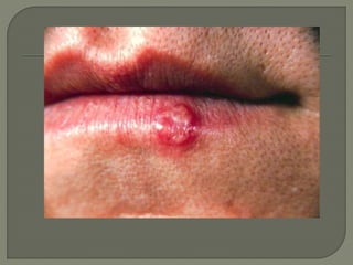
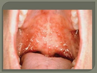
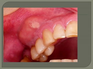
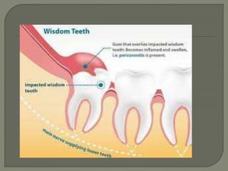
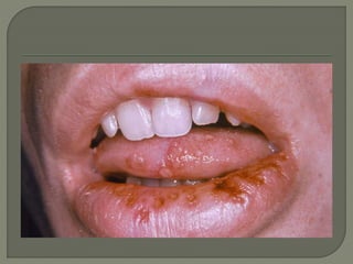
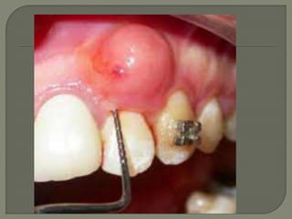
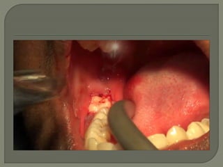
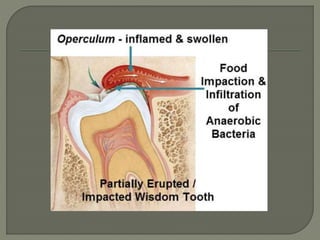
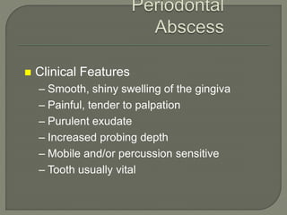
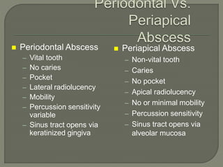
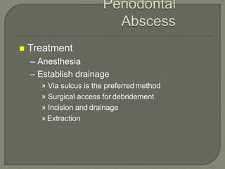
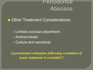
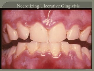
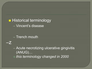
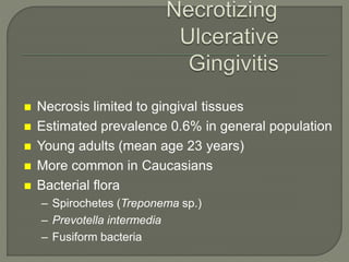
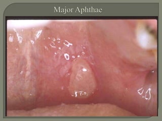
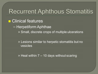
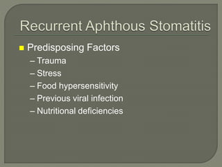
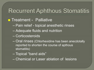
ACUTE PERIODONTAL
CONDITIONS
 Abscesses of the Periodontium
 Necrotizing Periodontal Diseases
 Gingival Diseases of Viral Origin-
Herpesvirus
 Recurrent Aphthous Stomatitis
 Allergic Reactions
Abscesses of the
Periodontium
 Gingival Abscess
 Periodontal Abscess
 Pericoronal Abscess
Gingival Abscess
A localized purulent infection that
involves the marginal gingiva or
interdental papilla
Gingivall
 Etiology
– Acute inflammatory response to foreign
substances forced into the gingiva
 Clinical Features
– Localized swelling of marginal gingiva or papilla
– A red, smooth, shiny surface
– May be painful and appear pointed
– Purulent exudate may be present
– No previous periodontal disease
 Treatment
– Elimination of foreign object
– Drainage through sulcus with probe or light
scaling
– Follow-up after 24-48 hours
 A localized purulent infection within the
tissues adjacent to the periodontal
pocket that may lead to the destruction
of periodontal ligament and alveolar
bone
 Usually pre-existing chronic periodontitis present!!!
 Factors associated with abscess development
– Occlusion of pocket orifice (by healing of marginal gingiva
following supragingival scaling)
– Furcation involvement
– Systemic antibiotic therapy (allowing overgrowth of resistant
bacteria)
– Diabetes Mellitus
 Clinical Features
– Smooth, shiny swelling of the gingiva
– Painful, tender to palpation
– Purulent exudate
– Increased probing depth
– Mobile and/or percussion sensitive
– Tooth usually vital
 Periodontal Abscess
– Vital tooth
– No caries
– Pocket
– Lateral radiolucency
– Mobility
– Percussion sensitivity
variable
– Sinus tract opens via
keratinized gingiva
 Periapical Abscess
– Non-vital tooth
– Caries
– No pocket
– Apical radiolucency
– No or minimal mobility
– Percussion sensitivity
– Sinus tract opens via
alveolar mucosa
 Treatment
– Anesthesia
– Establish drainage
» Via sulcus is the preferred method
» Surgical access for debridement
» Incision and drainage
» Extraction
 Other Treatment Considerations:
– Limited occlusal adjustment
– Antimicrobials
– Culture and sensitivity
A periodontal evaluation following resolution of
acute symptoms is essential!!!
 Antibiotics (if indicated due to fever, malaise,
lymphadenopathy, or inability to obtain drainage)
– Without penicillin allergy
» Penicillin
– With penicillin allergy
» Azithromycin
» Clindamycin
– Alter therapy if indicated by
culture/sensitivity
 A localized purulent infection within the
tissue surrounding the crown of a
partially erupted tooth.
Most common adjacent to mandibular
third molars in young adults; usually
caused by impaction of debris under the
soft tissue flap
 Clinical Features
– Operculum (soft tissue flap)
– Localized red, swollen tissue
– Area painful to touch
– Tissue trauma from opposing tooth common
– Purulent exudate, trismus,
lymphadenopathy, fever, and malaise may
be present
 Treatment Options
– Debride/irrigate under pericoronal flap
– Tissue recontouring (removing tissue flap)
– Extraction of involved and/or opposing
tooth
– Antimicrobials (local and/or systemic as
needed)
– Culture and sensitivity
– Follow-up
 Necrotizing Ulcerative Gingivitis (NUG)
 Necrotizing Ulcerative Periodontitis
(NUP)
 An infection characterized by gingival
necrosis presenting as “punched-out”
papillae, with gingival bleeding and pain
 Historical terminology
– Vincent’s disease
– Trench mouth
–z
– Acute necrotizing ulcerative gingivitis
(ANUG)…
 this terminology changed in 2000
 Necrosis limited to gingival tissues
 Estimated prevalence 0.6% in general population
 Young adults (mean age 23 years)
 More common in Caucasians
 Bacterial flora
– Spirochetes (Treponema sp.)
– Prevotella intermedia
– Fusiform bacteria
 Clinical Features
– Gingival necrosis, especially tips of
papillae
– Gingival bleeding
– Pain
– Fetid breath
– Pseudomembrane formation
 Predisposing Factors
– Emotional stress
– Poor oral hygiene
– Cigarette smoking
– Poor nutrition
– Immunosuppression
***Necrotizing Periodontal diseases are common in
immunocompromised patients, especially those who
are HIV (+) or have AIDS
 An infection characterized by necrosis
of gingival tissues, periodontal ligament,
and alveolar bone
 Clinical Features
– Clinical appearance of NUG
– Severe deep aching pain
– Very rapid rate of bone destruction
– Deep pocket formation not evident
 Treatment
– Local debridement
– Oral hygiene instructions
– Oral rinses
– Pain control
– Antibiotics
– Modify predisposing factors
– Proper follow-up
 Treatment
– Local debridement
» Most cases adequately treated by debridement
and sc/rp
» Anesthetics as needed
» Consider avoiding ultrasonic instrumentation due
to risk of HIV transmission
– Oral hygiene instructions
 Treatment
– Oral rinses – (frequent, at least until pain subsides
allowing effective OH)
» Chlorhexidine gluconate 0.12%; 1/2 oz 2 x daily
» Hydrogen peroxide/water
» Povidone iodine
– Pain control
 Treatment
– Antibiotics (systemic or severe involvement)
» Metronidazole
» Avoid broad spectrum antibiotics in AIDS patients
– Modify predisposing factors
– Follow-up
» Frequent until resolution of symptoms
» Comprehensive periodontal evaluation
following acute phase!!!!
 Acute manifestations of viral infections
of the oral mucosa, characterized by
redness and multiple vesicles that
easily rupture to form painful ulcers
affecting the gingiva.
 Classic initial infection of herpes simplex
type 1
 Mainly in young children
 90% of primary oral infections are
asymptomatic
 Clinical Features
– Painful severe gingivitis with ulcerations,
edema, and stomatitis
– Vesicles rupture, coalesce and form ulcers
– Fever and lymphadenopathy are classic
features
– Lesions usually resolve in 7-14 days
 Treatment
– Bed rest
– Fluids – forced
– Nutrition
– Antipyretics
» Acetaminophen, not ASA due to risk of Reye’s
Syndrome
 Treatment
– Pain relief
» Viscous lidocaine
» Benadryl elixir
» 50% Benadryl elixir/50% Maalox
– Antiviral medications
» Immunocompromised patients
 “Fever blisters” or “cold sores”
 Oral lesions usually herpes simplex virus
type 1
 Recurrent infections in 20-40% of those
with primary infection
 Herpes labialis common
 Recurrent infections less severe than
primary
 Clinical Features
– Prodromal syndrome
– Lesions start as vesicles, rupture and
leave ulcers
– A cluster of small painful ulcers on
attached gingiva or lip is characteristic
– Can cause post-operative pain following
dental treatment
 Virus reactivation
– Fever
– Systemic infection
– Ultraviolet radiation
– Stress
– Immune system changes
– Trauma
– Unidentified causes
 Treatment
– Palliative
– Antiviral medications
» Consider for treatment of immunocompromised
patients, but not for periodic recurrence in
healthy patients
 “Canker sores”
 Etiology unknown
 Prevalence 10 to 20% of general
population
 Usually begins in childhood
 Outbreaks sporadic, decreasing with
age
 Clinical features
– Affects mobile mucosa
– Most common oral ulcerative condition
– Three forms
» Minor
» Major
» Herpetiform
 Clinical features
– Minor Aphthae
» Mostcommon
» Small, shallow ulcerations with slightly raised
erythematous borders
» Central area covered by yellow-white
pseudomembrane
» Heals without scarring in 10 –14 days
 Clinical features
– Major Aphthae
» Usually larger than 0.5cm in diameter
» May persist for months
» Frequently heal with scarring
 Clinical features
– Herpetiform Aphthae
» Small, discrete crops of multiple ulcerations
» Lesions similar to herpetic stomatitis but no
vesicles
» Heal within 7 – 10 days without scaring
 Predisposing Factors
– Trauma
– Stress
– Food hypersensitivity
– Previous viral infection
– Nutritional deficiencies
 Treatment - Palliative
– Pain relief - topical anesthetic rinses
– Adequate fluids and nutrition
– Corticosteroids
– Oral rinses (Chlorhexidine has been anecdotally
reported to shorten the course of apthous
stomatitis)
– Topical “band aids”
– Chemical or Laser ablation of lesions
 Intraoral occurrence uncommon
– Higher concentrations of allergen required for
allergic reaction to occur in the oral mucosa
than in skin and other surfaces
 Examples
– Dental restorative materials
» Mercury, nickel, gold, zinc, chromium, and
acrylics
– Toothpastes and mouthwashes
» Flavor additives (cinnamon) or preservatives
– Foods
» Peanuts, red peppers, etc.
 Clinical Features – Variable
– Resemble oral lichen planus or leukoplakia
– Ulcerated lesions
– Fiery red edematous gingivitis
 Treatment
– Comprehensive history and interview
– Lesions resolve after elimination of offending agent
 Abscesses of the Periodontium
 Necrotizing Periodontal Diseases
 Gingival Diseases of Viral Origin
 Recurrent Aphthous Stomatitis

Acute periodontal coditions / Dr.Afaq

  • 1.
  • 11.
     Abscesses ofthe Periodontium  Necrotizing Periodontal Diseases  Gingival Diseases of Viral Origin- Herpesvirus  Recurrent Aphthous Stomatitis  Allergic Reactions
  • 12.
  • 13.
     Gingival Abscess Periodontal Abscess  Pericoronal Abscess
  • 14.
  • 15.
    A localized purulentinfection that involves the marginal gingiva or interdental papilla
  • 17.
  • 18.
     Etiology – Acuteinflammatory response to foreign substances forced into the gingiva  Clinical Features – Localized swelling of marginal gingiva or papilla – A red, smooth, shiny surface – May be painful and appear pointed – Purulent exudate may be present – No previous periodontal disease
  • 19.
     Treatment – Eliminationof foreign object – Drainage through sulcus with probe or light scaling – Follow-up after 24-48 hours
  • 20.
     A localizedpurulent infection within the tissues adjacent to the periodontal pocket that may lead to the destruction of periodontal ligament and alveolar bone
  • 22.
     Usually pre-existingchronic periodontitis present!!!  Factors associated with abscess development – Occlusion of pocket orifice (by healing of marginal gingiva following supragingival scaling) – Furcation involvement – Systemic antibiotic therapy (allowing overgrowth of resistant bacteria) – Diabetes Mellitus
  • 23.
     Clinical Features –Smooth, shiny swelling of the gingiva – Painful, tender to palpation – Purulent exudate – Increased probing depth – Mobile and/or percussion sensitive – Tooth usually vital
  • 24.
     Periodontal Abscess –Vital tooth – No caries – Pocket – Lateral radiolucency – Mobility – Percussion sensitivity variable – Sinus tract opens via keratinized gingiva  Periapical Abscess – Non-vital tooth – Caries – No pocket – Apical radiolucency – No or minimal mobility – Percussion sensitivity – Sinus tract opens via alveolar mucosa
  • 25.
     Treatment – Anesthesia –Establish drainage » Via sulcus is the preferred method » Surgical access for debridement » Incision and drainage » Extraction
  • 26.
     Other TreatmentConsiderations: – Limited occlusal adjustment – Antimicrobials – Culture and sensitivity A periodontal evaluation following resolution of acute symptoms is essential!!!
  • 27.
     Antibiotics (ifindicated due to fever, malaise, lymphadenopathy, or inability to obtain drainage) – Without penicillin allergy » Penicillin – With penicillin allergy » Azithromycin » Clindamycin – Alter therapy if indicated by culture/sensitivity
  • 28.
     A localizedpurulent infection within the tissue surrounding the crown of a partially erupted tooth.
  • 29.
    Most common adjacentto mandibular third molars in young adults; usually caused by impaction of debris under the soft tissue flap
  • 31.
     Clinical Features –Operculum (soft tissue flap) – Localized red, swollen tissue – Area painful to touch – Tissue trauma from opposing tooth common – Purulent exudate, trismus, lymphadenopathy, fever, and malaise may be present
  • 32.
     Treatment Options –Debride/irrigate under pericoronal flap – Tissue recontouring (removing tissue flap) – Extraction of involved and/or opposing tooth – Antimicrobials (local and/or systemic as needed) – Culture and sensitivity – Follow-up
  • 33.
     Necrotizing UlcerativeGingivitis (NUG)  Necrotizing Ulcerative Periodontitis (NUP)
  • 34.
     An infectioncharacterized by gingival necrosis presenting as “punched-out” papillae, with gingival bleeding and pain
  • 37.
     Historical terminology –Vincent’s disease – Trench mouth –z – Acute necrotizing ulcerative gingivitis (ANUG)…  this terminology changed in 2000
  • 38.
     Necrosis limitedto gingival tissues  Estimated prevalence 0.6% in general population  Young adults (mean age 23 years)  More common in Caucasians  Bacterial flora – Spirochetes (Treponema sp.) – Prevotella intermedia – Fusiform bacteria
  • 39.
     Clinical Features –Gingival necrosis, especially tips of papillae – Gingival bleeding – Pain – Fetid breath – Pseudomembrane formation
  • 40.
     Predisposing Factors –Emotional stress – Poor oral hygiene – Cigarette smoking – Poor nutrition – Immunosuppression ***Necrotizing Periodontal diseases are common in immunocompromised patients, especially those who are HIV (+) or have AIDS
  • 41.
     An infectioncharacterized by necrosis of gingival tissues, periodontal ligament, and alveolar bone
  • 43.
     Clinical Features –Clinical appearance of NUG – Severe deep aching pain – Very rapid rate of bone destruction – Deep pocket formation not evident
  • 44.
     Treatment – Localdebridement – Oral hygiene instructions – Oral rinses – Pain control – Antibiotics – Modify predisposing factors – Proper follow-up
  • 45.
     Treatment – Localdebridement » Most cases adequately treated by debridement and sc/rp » Anesthetics as needed » Consider avoiding ultrasonic instrumentation due to risk of HIV transmission – Oral hygiene instructions
  • 46.
     Treatment – Oralrinses – (frequent, at least until pain subsides allowing effective OH) » Chlorhexidine gluconate 0.12%; 1/2 oz 2 x daily » Hydrogen peroxide/water » Povidone iodine – Pain control
  • 47.
     Treatment – Antibiotics(systemic or severe involvement) » Metronidazole » Avoid broad spectrum antibiotics in AIDS patients – Modify predisposing factors – Follow-up » Frequent until resolution of symptoms » Comprehensive periodontal evaluation following acute phase!!!!
  • 48.
     Acute manifestationsof viral infections of the oral mucosa, characterized by redness and multiple vesicles that easily rupture to form painful ulcers affecting the gingiva.
  • 49.
     Classic initialinfection of herpes simplex type 1  Mainly in young children  90% of primary oral infections are asymptomatic
  • 51.
     Clinical Features –Painful severe gingivitis with ulcerations, edema, and stomatitis – Vesicles rupture, coalesce and form ulcers – Fever and lymphadenopathy are classic features – Lesions usually resolve in 7-14 days
  • 52.
     Treatment – Bedrest – Fluids – forced – Nutrition – Antipyretics » Acetaminophen, not ASA due to risk of Reye’s Syndrome
  • 53.
     Treatment – Painrelief » Viscous lidocaine » Benadryl elixir » 50% Benadryl elixir/50% Maalox – Antiviral medications » Immunocompromised patients
  • 54.
     “Fever blisters”or “cold sores”  Oral lesions usually herpes simplex virus type 1  Recurrent infections in 20-40% of those with primary infection  Herpes labialis common  Recurrent infections less severe than primary
  • 56.
     Clinical Features –Prodromal syndrome – Lesions start as vesicles, rupture and leave ulcers – A cluster of small painful ulcers on attached gingiva or lip is characteristic – Can cause post-operative pain following dental treatment
  • 57.
     Virus reactivation –Fever – Systemic infection – Ultraviolet radiation – Stress – Immune system changes – Trauma – Unidentified causes
  • 58.
     Treatment – Palliative –Antiviral medications » Consider for treatment of immunocompromised patients, but not for periodic recurrence in healthy patients
  • 59.
     “Canker sores” Etiology unknown  Prevalence 10 to 20% of general population  Usually begins in childhood  Outbreaks sporadic, decreasing with age
  • 60.
     Clinical features –Affects mobile mucosa – Most common oral ulcerative condition – Three forms » Minor » Major » Herpetiform
  • 61.
     Clinical features –Minor Aphthae » Mostcommon » Small, shallow ulcerations with slightly raised erythematous borders » Central area covered by yellow-white pseudomembrane » Heals without scarring in 10 –14 days
  • 63.
     Clinical features –Major Aphthae » Usually larger than 0.5cm in diameter » May persist for months » Frequently heal with scarring
  • 65.
     Clinical features –Herpetiform Aphthae » Small, discrete crops of multiple ulcerations » Lesions similar to herpetic stomatitis but no vesicles » Heal within 7 – 10 days without scaring
  • 66.
     Predisposing Factors –Trauma – Stress – Food hypersensitivity – Previous viral infection – Nutritional deficiencies
  • 67.
     Treatment -Palliative – Pain relief - topical anesthetic rinses – Adequate fluids and nutrition – Corticosteroids – Oral rinses (Chlorhexidine has been anecdotally reported to shorten the course of apthous stomatitis) – Topical “band aids” – Chemical or Laser ablation of lesions
  • 68.
     Intraoral occurrenceuncommon – Higher concentrations of allergen required for allergic reaction to occur in the oral mucosa than in skin and other surfaces
  • 69.
     Examples – Dentalrestorative materials » Mercury, nickel, gold, zinc, chromium, and acrylics – Toothpastes and mouthwashes » Flavor additives (cinnamon) or preservatives – Foods » Peanuts, red peppers, etc.
  • 70.
     Clinical Features– Variable – Resemble oral lichen planus or leukoplakia – Ulcerated lesions – Fiery red edematous gingivitis  Treatment – Comprehensive history and interview – Lesions resolve after elimination of offending agent
  • 72.
     Abscesses ofthe Periodontium  Necrotizing Periodontal Diseases  Gingival Diseases of Viral Origin  Recurrent Aphthous Stomatitis