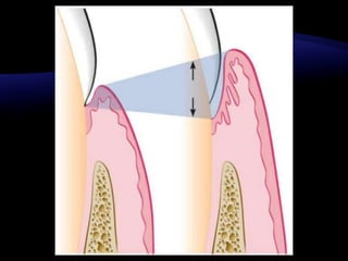
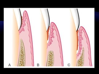
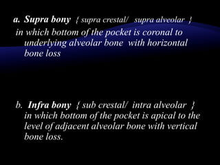
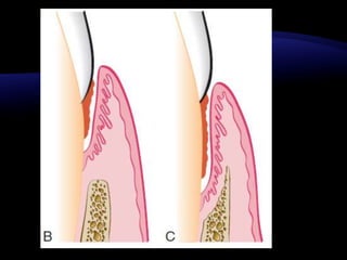
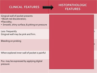
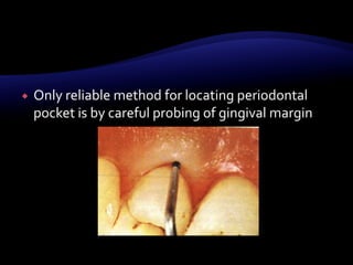
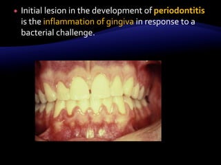
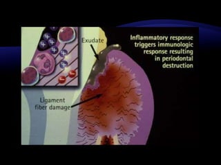
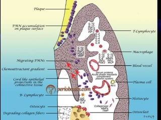
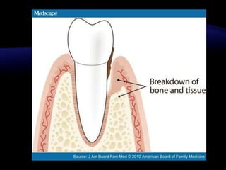
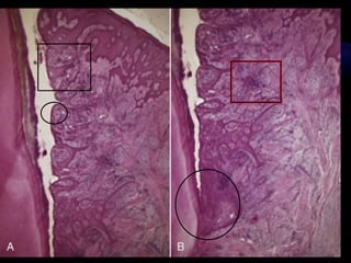
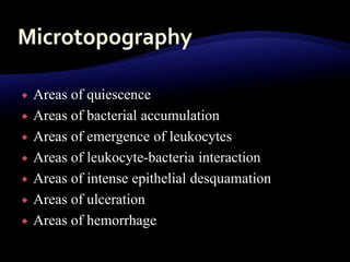
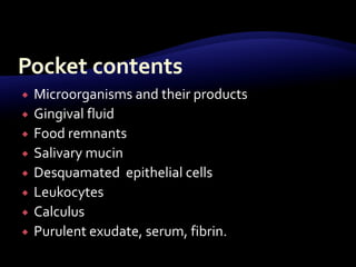
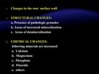
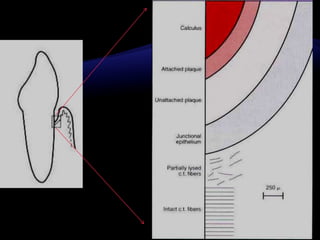
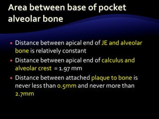
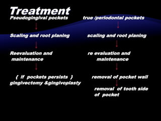
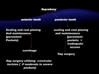
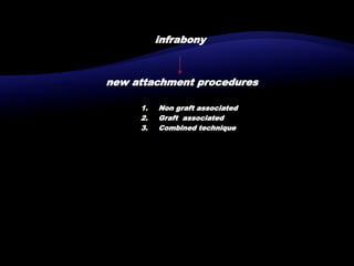
 Pathologically deepened gingival
sulcus.
CLASSIFICATION
Pocket
Periodontal
True pocket
Suprobony
Supra crestal
supraalveolar
Intrabony
Infrabony
Subcrestal
Intraalveolar
Gingival
pseudopocket
1. Gingival pockets { pseudo pockets/ relative
pockets }:
Is formed by gingival enlargement without
destruction of underlying periodontal tissue
2. Periodontal pocket { true pocket / absolute
pocket }:
is formed by destruction of underlying
periodontal tissues leading to loosening and
exfoliation of teeth
a. Supra bony { supra crestal/ supra alveolar }
in which bottom of the pocket is coronal to
underlying alveolar bone with horizontal
bone loss
b. Infra bony { sub crestal/ intra alveolar }
in which bottom of the pocket is apical to the
level of adjacent alveolar bone with vertical
bone loss.
• It can also be classified as
a. simple pocket:
involving one tooth surface.
b. Compound pocket:
involving 2 or more tooth surfaces
c. Complex / spiral pocket:
originating on one tooth surface and
twisting around the tooth to involve one or
more additional surfaces
 Signs:
a. thickened / ‘rolled edge’ marginal gingiva
b. Bluish red vertical zone from the marginal
gingiva to the alveolar mucosa.
c. Gingival bleeding ,suppuration or both
d. diastema formation
 Localized gnawing pain (dull and continuous)
 Radiating pain deep in the jaws.
 A sensation of pressure after eating gradually diminishes.
 foul taste in localized areas.
 tendency to suck material from the interproximal spaces.
 feeling of itchiness in the gums.
 urge to dig a pointed instrumentinto the gums
with relief obtained form the resultant
bleeding.
 Sensitivity to heat and cold, toothache in the
absence of caries.
CLINICAL FEATURES
HISTOPATHOLOGIC
FEATURES
Gingival wall of pocket presents
• Bluish red discoloration;
•Flaccidity;
• Smooth, shiny surface; & pitting on pressure
Discoloration  circulatory stagnation
flaccidity destruction of gingival fibers
Smooth shiny surface, pitting on pressure 
Edema atrophy of epithelium degeneration
Less frequently
Gingival wall may be pink and firm.
Fibrotic changes predominate over exudation
and degeneration (external wall)
Bleeding on probing Vascularity thinning , degeneration of
epithelium , proximity of engorged vessels to
inner surface.
When explored inner wall of pocket is painful Pain on tactile stimulation is caused by
ulceration of inner aspect of pocket wall
Pus may be expressed by applying digital
pressure
Suppurative inflammation of inner wall
 Only reliable method for locating periodontal
pocket is by careful probing of gingival margin
 On the basis of depth only it is difficult to
differentiate between deep normal sulcus and
shallow periodontal pocket.
 In such borderline case pathological changes in
gingiva distinguish the two conditions
 Initial lesion in the development of periodontitis
is the inflammation of gingiva in response to a
bacterial challenge.
 First event
inflammation { due to lying down of Gram
positive bacteria on supra gingival tooth surface }
changes in junctional epithelium
proliferates along root surface
coronal portion apical portion of junctional
detatches from the epithelium migrates
root
Due to bacterial enzymes replaced by pocket
And physical forces epithelium
Exerted by them.
 Second event:
aggressive growth and action of Gram negative
bacteria.
Emigration of neutrophils in large no.
Disruption of epithelial barrier causing open
communication.
loss of chemotactic gradient
Tissue destruction due to products released by
neutrophills as well as bacteria
resorption of alveolar bone
periodontal pocket is established
1. Changes in the soft tissue wall
a. Blood vessels are increased in number , dilated and enlarged
b. Connective tissue is edematous and densely infiltrated with
plasma cells (80%) , lymphocytes , scattering of PMN’s .
c. JE becomes shorter
d. Degeneration and necrosis of the epithelium leading to
ulceration of the epithelium and exposure of the underlying
connective tissue.
 Proliferation of endothelial cells capillaries
fibroblast collagen fibers
 Bacterial invasion along the lateral and apical
areas of the pocket.
 Areas of quiescence
 Areas of bacterial accumulation
 Areas of emergence of leukocytes
 Areas of leukocyte-bacteria interaction
 Areas of intense epithelial desquamation
 Areas of ulceration
 Areas of hemorrhage
 Balance between Constructive and destructive
changes
 Edematous pocket wall
 Fibrotic pocket wall
 Microorganisms and their products
 Gingival fluid
 Food remnants
 Salivary mucin
 Desquamated epithelial cells
 Leukocytes
 Calculus
 Purulent exudate, serum, fibrin.
2. Changes in the root surface wall
A. STRUCTURAL CHANGES:
a. Presence of pathologic granules
b. Areas of increased mineralization
c. Areas of demineralization
B. CHEMICAL CHANGES:
following minerals are increased
a. Calcium
b. Magnesium
c. Phosphate
d. Fluoride
e. others
C. Cytotoxic changes:
bacterial penetration into the cementum can be found as deep as
cemento-dentinal junction in addition bacterial products such as
endotoxins have also been detected
D. Zone seen:
Changes in the root surface wall
A.STRUCTURAL
CHANGES:
B.a. Pathologic
C. granules
b. Areas of
increased
mineralization
c. Areas of
demineralization
B.CHEMICAL
CHANGES:
C.following minerals
are increased
D.a. Calcium
b. Magnesium
c. Phosphate
d. Fluoride
e. others
C.CYTOTOXIC
CHANGES:
D.Bacterial
penetration as deep
as cemento-dentinal
junction
E.Endotoxins have
also been detected
 Slow but continuous
 Periods of exacerbation and quiescence
 Distance between apical end of JE and alveolar
bone is relatively constant
 Distance between apical end of calculus and
alveolar crest = 1.97 mm
 Distance between attached plaque to bone is
never less than 0.5mm and never more than
2.7mm
 Lateral periodontal cyst
• Detection of pocket : clinical examination and probing
are more efficient.
a. Radiographic method: pockets are not detected by
radiographic method alone
guttapercha points or calibrated silver points can be
used along with radiograph
Pseudogingival pockets true /periodontal pockets
Scaling and root planing scaling and root planing
Reevaluation and re evaluation and
maintenance maintenance
{ if pockets persists } removal of pocket wall
gingivectomy &gingivoplasty
removal of tooth side
of pocket
Suprabony
anterior teeth posterior teeth
Scaling and root planing scaling and root planing
And maintenance and maintenance
{persistent {persistent
Pockets} pockets +
inadequate
access
curettage
flap surgery
flap surgery utilizing crevicular
incision { if moderate to severe
pockets}
infrabony
new attachment procedures
1. Non graft associated
2. Graft associated
3. Combined technique

Periodontal pockets.pptx

  • 2.
  • 4.
  • 5.
  • 6.
    1. Gingival pockets{ pseudo pockets/ relative pockets }: Is formed by gingival enlargement without destruction of underlying periodontal tissue 2. Periodontal pocket { true pocket / absolute pocket }: is formed by destruction of underlying periodontal tissues leading to loosening and exfoliation of teeth
  • 9.
    a. Supra bony{ supra crestal/ supra alveolar } in which bottom of the pocket is coronal to underlying alveolar bone with horizontal bone loss b. Infra bony { sub crestal/ intra alveolar } in which bottom of the pocket is apical to the level of adjacent alveolar bone with vertical bone loss.
  • 12.
    • It canalso be classified as a. simple pocket: involving one tooth surface. b. Compound pocket: involving 2 or more tooth surfaces c. Complex / spiral pocket: originating on one tooth surface and twisting around the tooth to involve one or more additional surfaces
  • 14.
     Signs: a. thickened/ ‘rolled edge’ marginal gingiva b. Bluish red vertical zone from the marginal gingiva to the alveolar mucosa. c. Gingival bleeding ,suppuration or both d. diastema formation
  • 15.
     Localized gnawingpain (dull and continuous)  Radiating pain deep in the jaws.  A sensation of pressure after eating gradually diminishes.  foul taste in localized areas.  tendency to suck material from the interproximal spaces.  feeling of itchiness in the gums.
  • 16.
     urge todig a pointed instrumentinto the gums with relief obtained form the resultant bleeding.  Sensitivity to heat and cold, toothache in the absence of caries.
  • 18.
    CLINICAL FEATURES HISTOPATHOLOGIC FEATURES Gingival wallof pocket presents • Bluish red discoloration; •Flaccidity; • Smooth, shiny surface; & pitting on pressure Discoloration  circulatory stagnation flaccidity destruction of gingival fibers Smooth shiny surface, pitting on pressure  Edema atrophy of epithelium degeneration Less frequently Gingival wall may be pink and firm. Fibrotic changes predominate over exudation and degeneration (external wall) Bleeding on probing Vascularity thinning , degeneration of epithelium , proximity of engorged vessels to inner surface. When explored inner wall of pocket is painful Pain on tactile stimulation is caused by ulceration of inner aspect of pocket wall Pus may be expressed by applying digital pressure Suppurative inflammation of inner wall
  • 19.
     Only reliablemethod for locating periodontal pocket is by careful probing of gingival margin
  • 20.
     On thebasis of depth only it is difficult to differentiate between deep normal sulcus and shallow periodontal pocket.  In such borderline case pathological changes in gingiva distinguish the two conditions
  • 22.
     Initial lesionin the development of periodontitis is the inflammation of gingiva in response to a bacterial challenge.
  • 33.
     First event inflammation{ due to lying down of Gram positive bacteria on supra gingival tooth surface } changes in junctional epithelium proliferates along root surface coronal portion apical portion of junctional detatches from the epithelium migrates root
  • 34.
    Due to bacterialenzymes replaced by pocket And physical forces epithelium Exerted by them.  Second event: aggressive growth and action of Gram negative bacteria. Emigration of neutrophils in large no.
  • 35.
    Disruption of epithelialbarrier causing open communication. loss of chemotactic gradient Tissue destruction due to products released by neutrophills as well as bacteria resorption of alveolar bone periodontal pocket is established
  • 36.
    1. Changes inthe soft tissue wall a. Blood vessels are increased in number , dilated and enlarged b. Connective tissue is edematous and densely infiltrated with plasma cells (80%) , lymphocytes , scattering of PMN’s . c. JE becomes shorter d. Degeneration and necrosis of the epithelium leading to ulceration of the epithelium and exposure of the underlying connective tissue.
  • 38.
     Proliferation ofendothelial cells capillaries fibroblast collagen fibers  Bacterial invasion along the lateral and apical areas of the pocket.
  • 39.
     Areas ofquiescence  Areas of bacterial accumulation  Areas of emergence of leukocytes  Areas of leukocyte-bacteria interaction  Areas of intense epithelial desquamation  Areas of ulceration  Areas of hemorrhage
  • 40.
     Balance betweenConstructive and destructive changes  Edematous pocket wall  Fibrotic pocket wall
  • 41.
     Microorganisms andtheir products  Gingival fluid  Food remnants  Salivary mucin  Desquamated epithelial cells  Leukocytes  Calculus  Purulent exudate, serum, fibrin.
  • 43.
    2. Changes inthe root surface wall A. STRUCTURAL CHANGES: a. Presence of pathologic granules b. Areas of increased mineralization c. Areas of demineralization B. CHEMICAL CHANGES: following minerals are increased a. Calcium b. Magnesium c. Phosphate d. Fluoride e. others
  • 44.
    C. Cytotoxic changes: bacterialpenetration into the cementum can be found as deep as cemento-dentinal junction in addition bacterial products such as endotoxins have also been detected D. Zone seen:
  • 45.
    Changes in theroot surface wall A.STRUCTURAL CHANGES: B.a. Pathologic C. granules b. Areas of increased mineralization c. Areas of demineralization B.CHEMICAL CHANGES: C.following minerals are increased D.a. Calcium b. Magnesium c. Phosphate d. Fluoride e. others C.CYTOTOXIC CHANGES: D.Bacterial penetration as deep as cemento-dentinal junction E.Endotoxins have also been detected
  • 47.
     Slow butcontinuous  Periods of exacerbation and quiescence
  • 50.
     Distance betweenapical end of JE and alveolar bone is relatively constant  Distance between apical end of calculus and alveolar crest = 1.97 mm  Distance between attached plaque to bone is never less than 0.5mm and never more than 2.7mm
  • 54.
  • 57.
    • Detection ofpocket : clinical examination and probing are more efficient. a. Radiographic method: pockets are not detected by radiographic method alone guttapercha points or calibrated silver points can be used along with radiograph
  • 59.
    Pseudogingival pockets true/periodontal pockets Scaling and root planing scaling and root planing Reevaluation and re evaluation and maintenance maintenance { if pockets persists } removal of pocket wall gingivectomy &gingivoplasty removal of tooth side of pocket
  • 60.
    Suprabony anterior teeth posteriorteeth Scaling and root planing scaling and root planing And maintenance and maintenance {persistent {persistent Pockets} pockets + inadequate access curettage flap surgery flap surgery utilizing crevicular incision { if moderate to severe pockets}
  • 61.
    infrabony new attachment procedures 1.Non graft associated 2. Graft associated 3. Combined technique