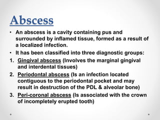
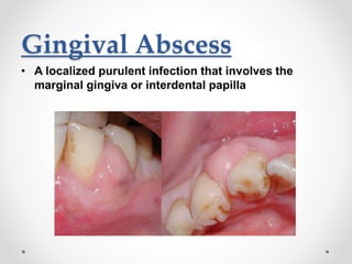
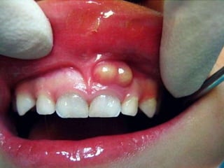
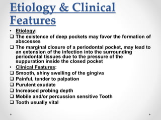
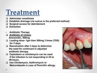
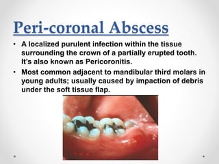
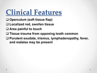
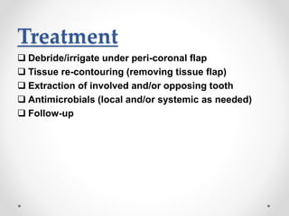
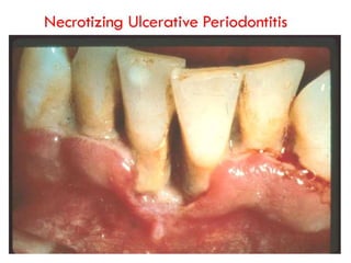
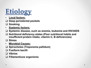
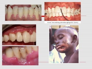
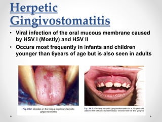
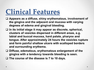
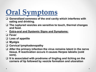
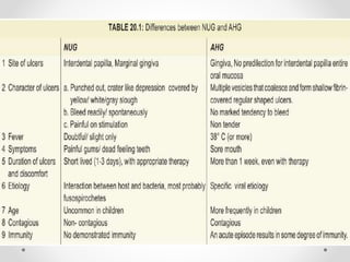
Acute Gingival
Infections
By: Dr. Muhammad Ali Arshad
Department: Periodontology
Date: 10-03-2020
Introduction
• Acute lesions have a rapid onset & is accompanied
by pain.
• Acute lesions are by definition, of sudden onset,
limited duration & with well defined features in
contrast with chronic lesions. Some of the common
acute gingival infections are as follows:
1. Abscess
2. NUG (Necrotizing Ulcerative Gingivitis)
3. Herpetic Gingivostomatitis
Abscess
• An abscess is a cavity containing pus and
surrounded by inflamed tissue, formed as a result of
a localized infection.
• It has been classified into three diagnostic groups:
1. Gingival abscess (Involves the marginal gingival
and interdental tissues)
2. Periodontal abscess (Is an infection located
contiguous to the periodontal pocket and may
result in destruction of the PDL & alveolar bone)
3. Peri-coronal abscess (Is associated with the crown
of incompletely erupted tooth)
Gingival Abscess
• A localized purulent infection that involves the
marginal gingiva or interdental papilla
Etiology & Clinical
Features
• Etiology:
 Acute inflammatory response to foreign substances
forced into the gingiva
• Clinical Features:
 Localized swelling of marginal gingiva or papilla
 A red, smooth, shiny surface
 May be painful and appear pointed
 Purulent exudate may be present
 No previous periodontal disease
Treatment
 Topical or local anesthesia by infiltration is administered
to establish drainage and remove microbial deposits.
 In acute cases, the fluctuant area is incised with a #15
scalpel blade and exudate may be expressed by gentle
digital pressure.
 Any foreign material is removed.
 The area is irrigated with warm saline water and covered
with moist gauze under light pressure.
 Once bleeding has stopped, patient is dismissed with
instructions to rinse with warm saline water every 2 hrs.
 After 24 hrs, the area is reassessed, and if resolution is
sufficient, scaling not previously completed is
undertaken.
 If the lesion is large or poorly accessible, surgical access
may be required.
Periodontal Abscess
• A periodontal abscess is defined as a localized
purulent infection affecting the tissues surrounding
a periodontal pocket that can lead to the destruction
of supporting structures
• It is also known as a lateral abscess or parietal
abscess.
• It is the third most frequent dental emergency,
representing 7–14% of all dental emergencies, and
affecting 6–7% of all patients seen in a dental clinic.
• It is typically found in patients with untreated
periodontitis and in association with moderate to
deep periodontal pockets.
Etiology & Clinical
Features
• Etiology:
 The existence of deep pockets may favor the formation of
abscesses
 The marginal closure of a periodontal pocket, may lead to
an extension of the infection into the surrounding
periodontal tissues due to the pressure of the
suppuration inside the closed pocket
• Clinical Features:
 Smooth, shiny swelling of the gingiva
 Painful, tender to palpation
 Purulent exudate
 Increased probing depth
 Mobile and/or percussion sensitive Tooth
 Tooth usually vital
Treatment
 Administer anesthesia
 Establish drainage (via sulcus is the preferred method)
 Surgical access for debridement
 Extraction
• Antibiotic Therapy
 Antibiotic of choice:
Amoxicillin, 500mg
 Loading dose- 1gm then 500mg 3 times (TDS)
for 3 days.
 Reevaluation after 3 days to determine
the need for continued or adjusted
antibiotic therapy.
 Cephalexin or clindamycin can be used
if the infection is not responding in 24 to
48 hours.
 Use Clindamycin, Azithromycin or
Metronidazole in case of Penicillin allergy.
Peri-coronal Abscess
• A localized purulent infection within the tissue
surrounding the crown of a partially erupted tooth.
It’s also known as Pericoronitis.
• Most common adjacent to mandibular third molars in
young adults; usually caused by impaction of debris
under the soft tissue flap.
Clinical Features
 Operculum (soft tissue flap)
 Localized red, swollen tissue
 Area painful to touch
 Tissue trauma from opposing tooth common
 Purulent exudate, trismus, lymphadenopathy, fever,
and malaise may be present
Treatment
 Debride/irrigate under peri-coronal flap
 Tissue re-contouring (removing tissue flap)
 Extraction of involved and/or opposing tooth
 Antimicrobials (local and/or systemic as needed)
 Follow-up
NUG (Necrotizing
Ulcerative Gingivitis)
• Necrotizing ulcerative gingivitis (NUG) is a microbial
disease of the gingiva with impaired host response
• It manifests with the characteristic clinical signs of
necrosis and sloughing of the gingival tissues and
may be accompanied by systemic symptoms
• Also called Trench mouth, Vincent’s Stomatitis,
Vincent’s Infection, Fusospirochetal Gingivitis, Acute
Ulcerative Gingivitis, Necrotizing Gingivitis
• NUG occurs at all ages, with the highest incidence
reported between ages 20 & 30 yrs & ages 15 -20 yrs
• The disease seems to occur slightly more among
HIV infected individuals
Etiology
• Local factors:
 Deep periodontal pockets
 Smoking
• Systemic factors:
 Systemic disease, such as anemia, leukemia and HIV/AIDS
 Nutritional deficiency states (Poor nutritional habits and
insufficient protein intake, vitamin C, B deficiencies)
 Stress
• Microbial Causes:
 Spirochetes (Treponema pallidum)
 Fusiform bacilli
 Vibrios
 Fillamentiuos organisms
Clinical Features
• NG – an inflammatory destructive gingival condition, characterized by
punched out crater like depressions at the crest of the interdental papillae,
subsequently extending to the marginal gingiva and rarely to the attached
gingiva & oral mucosa.
• Spontaneous gingival hemorrhage or pronounced bleeding after the slight
stimulation are characteristic clinical signs.
• The lesion is extremely sensitive to touch, & the patient may often
complain of a constant radiating, gnawing pain that is often intensified by
eating spicy or hot foods & chewing.
• There is metallic foul taste
• In mild & moderate stages of disease
• Local lymphadenopathy & slight elevation in temperature.
• In severe cases
• High fever, increased pulse rate, leucocytois, loss of appetite & general
lassitude.
• Systemic reactions are more severe in children.
• Insomnia, constipation, gastro-intestinal disorders, headache, & mental
depression sometimes accompany the condition.
Stages of oral necrotizing
disease – By
Horning & Cohen
• Stage 1- necrosis of the top of the interdental papilla.
• Stage 2- necrosis of entire papilla
• Stage 3- necrosis extending to the gingival margin.
• Stage 4- necrosis extending to the attached gingiva.
• Stage 5– necrosis extending to labial & buccal
mucosa.
• Stage 6- necrosis exposing alveolar bone.
• Stage 7– necrosis perforating skin of cheek.
Treatment Of
Ambulatory Patients
• First visit:
 Topical anesthetic
 Gently swabbed with a cotton pellet to remove pseudo-membrane
and non-attached surface debris
 Area is cleansed with warm water
 Superficial calculus is removed with ultrasonic scalers
 Antibiotics prescription
 Sub-gingival scaling and curettage are contraindicated
• Instructions to the patient:
 Avoid smoking and alcohol.
 Rinse with 3% hydrogen peroxide and warm water for every two hours.
 Confine tooth brushing to the removal of surface debris with a bland
dentifrice, use of interdental aids and chlorhexidine mouth rinse are
recommended.
Continued…
• Second visit:
 Scalers and curettes are added to the instrumentarium.
 Shrinkage of the gingiva may expose previously covered calculus which is
gently removed.
 Same instructions are reinforced.
• Third visit:
 Scaling and root planning are repeated,
 Plaque control instructions are given.
 Hydrogen peroxide rinses are discontinued.
• Fourth visit:
 Oral hygiene instructions are reinforced
 Thorough scaling and root planning are performed.
• Fifth visit:
 Appointments are fixed for treatment of chronic gingivitis, periodontal
pockets and peri-coronal flaps, and for the elimination of all local irritants.
 Patient is placed on maintenance program.
Treatment Of Non-
ambulatory Patients
• Day 1:
 gently removing the necrotic pseudo-membrane with a pellet of
cotton saturated with hydrogen peroxide (H2O2).
 Advised bed rest and rinse the mouth every 2 hours with a
diluted 3% hydrogen peroxide (H2O2).
 Systemic antibiotics like penicillin or metronidazole can be
prescribed.
• Day 2:
 After 24 hours, a bedside visit should be made. The treatment
again includes gently swab the area with hydrogen peroxide,
instructions of the previous day are repeated.
• Day 3:
 In most cases, the condition will be improved.
 Start the treatment for ambulatory patients.
Herpetic
Gingivostomatitis
• Viral infection of the oral mucous membrane caused
by HSV I (Mostly) and HSV II
• Occurs most frequently in infants and children
younger than 6years of age but is also seen in adults
Clinical Features
 Appears as a diffuse, shiny erythematous, involvement of
the gingiva and the adjacent oral mucosa with varying
degrees of edema and gingival bleeding.
 In its initial stage it may appear as discrete, spherical,
clusters of vesicles dispersed in different areas, e.g.
labial and buccal mucosa, hard palate, pharynx and
tongue. After approximately 24 hours the vesicles rupture
and form painful shallow ulcers with scalloped borders
and surrounding erythema.
 Diffuse, edematous, erythematous enlargement of the
gingiva with a tendency towards bleeding is seen.
 The course of the disease is 7 to 10 days.
Oral Symptoms
 Generalized soreness of the oral cavity which interferes with
eating and drinking.
 The ruptured vesicles are sensitive to touch, thermal changes
and food.
• Extra-oral and Systemic Signs and Symptoms:
 Fever
 Loss of appetite
 Myalgia
 Cervical lymphadenopathy
 After the primary infection the virus remains latent in the nerve
tissue. If reactivation occurs it causes Herpes labialis (cold
sore).
 It is associated with prodrome of tingling and itching on the
corners of lip followed by vesicle formation and ulceration
Treatment
 Topical lignocaine for pain relief
 Acyclovir at 15 mg/kg five times a day for 5-7 days
 Topical antiviral medications such as 5% acyclovir
cream or 3% Penciclovir
 Cream is applied three to five times a day
Acute Gingival Infections

Acute Gingival Infections

  • 1.
    Acute Gingival Infections By: Dr.Muhammad Ali Arshad Department: Periodontology Date: 10-03-2020
  • 2.
    Introduction • Acute lesionshave a rapid onset & is accompanied by pain. • Acute lesions are by definition, of sudden onset, limited duration & with well defined features in contrast with chronic lesions. Some of the common acute gingival infections are as follows: 1. Abscess 2. NUG (Necrotizing Ulcerative Gingivitis) 3. Herpetic Gingivostomatitis
  • 3.
    Abscess • An abscessis a cavity containing pus and surrounded by inflamed tissue, formed as a result of a localized infection. • It has been classified into three diagnostic groups: 1. Gingival abscess (Involves the marginal gingival and interdental tissues) 2. Periodontal abscess (Is an infection located contiguous to the periodontal pocket and may result in destruction of the PDL & alveolar bone) 3. Peri-coronal abscess (Is associated with the crown of incompletely erupted tooth)
  • 4.
    Gingival Abscess • Alocalized purulent infection that involves the marginal gingiva or interdental papilla
  • 5.
    Etiology & Clinical Features •Etiology:  Acute inflammatory response to foreign substances forced into the gingiva • Clinical Features:  Localized swelling of marginal gingiva or papilla  A red, smooth, shiny surface  May be painful and appear pointed  Purulent exudate may be present  No previous periodontal disease
  • 6.
    Treatment  Topical orlocal anesthesia by infiltration is administered to establish drainage and remove microbial deposits.  In acute cases, the fluctuant area is incised with a #15 scalpel blade and exudate may be expressed by gentle digital pressure.  Any foreign material is removed.  The area is irrigated with warm saline water and covered with moist gauze under light pressure.  Once bleeding has stopped, patient is dismissed with instructions to rinse with warm saline water every 2 hrs.  After 24 hrs, the area is reassessed, and if resolution is sufficient, scaling not previously completed is undertaken.  If the lesion is large or poorly accessible, surgical access may be required.
  • 7.
    Periodontal Abscess • Aperiodontal abscess is defined as a localized purulent infection affecting the tissues surrounding a periodontal pocket that can lead to the destruction of supporting structures • It is also known as a lateral abscess or parietal abscess. • It is the third most frequent dental emergency, representing 7–14% of all dental emergencies, and affecting 6–7% of all patients seen in a dental clinic. • It is typically found in patients with untreated periodontitis and in association with moderate to deep periodontal pockets.
  • 9.
    Etiology & Clinical Features •Etiology:  The existence of deep pockets may favor the formation of abscesses  The marginal closure of a periodontal pocket, may lead to an extension of the infection into the surrounding periodontal tissues due to the pressure of the suppuration inside the closed pocket • Clinical Features:  Smooth, shiny swelling of the gingiva  Painful, tender to palpation  Purulent exudate  Increased probing depth  Mobile and/or percussion sensitive Tooth  Tooth usually vital
  • 10.
    Treatment  Administer anesthesia Establish drainage (via sulcus is the preferred method)  Surgical access for debridement  Extraction • Antibiotic Therapy  Antibiotic of choice: Amoxicillin, 500mg  Loading dose- 1gm then 500mg 3 times (TDS) for 3 days.  Reevaluation after 3 days to determine the need for continued or adjusted antibiotic therapy.  Cephalexin or clindamycin can be used if the infection is not responding in 24 to 48 hours.  Use Clindamycin, Azithromycin or Metronidazole in case of Penicillin allergy.
  • 11.
    Peri-coronal Abscess • Alocalized purulent infection within the tissue surrounding the crown of a partially erupted tooth. It’s also known as Pericoronitis. • Most common adjacent to mandibular third molars in young adults; usually caused by impaction of debris under the soft tissue flap.
  • 12.
    Clinical Features  Operculum(soft tissue flap)  Localized red, swollen tissue  Area painful to touch  Tissue trauma from opposing tooth common  Purulent exudate, trismus, lymphadenopathy, fever, and malaise may be present
  • 13.
    Treatment  Debride/irrigate underperi-coronal flap  Tissue re-contouring (removing tissue flap)  Extraction of involved and/or opposing tooth  Antimicrobials (local and/or systemic as needed)  Follow-up
  • 14.
    NUG (Necrotizing Ulcerative Gingivitis) •Necrotizing ulcerative gingivitis (NUG) is a microbial disease of the gingiva with impaired host response • It manifests with the characteristic clinical signs of necrosis and sloughing of the gingival tissues and may be accompanied by systemic symptoms • Also called Trench mouth, Vincent’s Stomatitis, Vincent’s Infection, Fusospirochetal Gingivitis, Acute Ulcerative Gingivitis, Necrotizing Gingivitis • NUG occurs at all ages, with the highest incidence reported between ages 20 & 30 yrs & ages 15 -20 yrs • The disease seems to occur slightly more among HIV infected individuals
  • 16.
    Etiology • Local factors: Deep periodontal pockets  Smoking • Systemic factors:  Systemic disease, such as anemia, leukemia and HIV/AIDS  Nutritional deficiency states (Poor nutritional habits and insufficient protein intake, vitamin C, B deficiencies)  Stress • Microbial Causes:  Spirochetes (Treponema pallidum)  Fusiform bacilli  Vibrios  Fillamentiuos organisms
  • 17.
    Clinical Features • NG– an inflammatory destructive gingival condition, characterized by punched out crater like depressions at the crest of the interdental papillae, subsequently extending to the marginal gingiva and rarely to the attached gingiva & oral mucosa. • Spontaneous gingival hemorrhage or pronounced bleeding after the slight stimulation are characteristic clinical signs. • The lesion is extremely sensitive to touch, & the patient may often complain of a constant radiating, gnawing pain that is often intensified by eating spicy or hot foods & chewing. • There is metallic foul taste • In mild & moderate stages of disease • Local lymphadenopathy & slight elevation in temperature. • In severe cases • High fever, increased pulse rate, leucocytois, loss of appetite & general lassitude. • Systemic reactions are more severe in children. • Insomnia, constipation, gastro-intestinal disorders, headache, & mental depression sometimes accompany the condition.
  • 18.
    Stages of oralnecrotizing disease – By Horning & Cohen • Stage 1- necrosis of the top of the interdental papilla. • Stage 2- necrosis of entire papilla • Stage 3- necrosis extending to the gingival margin. • Stage 4- necrosis extending to the attached gingiva. • Stage 5– necrosis extending to labial & buccal mucosa. • Stage 6- necrosis exposing alveolar bone. • Stage 7– necrosis perforating skin of cheek.
  • 21.
    Treatment Of Ambulatory Patients •First visit:  Topical anesthetic  Gently swabbed with a cotton pellet to remove pseudo-membrane and non-attached surface debris  Area is cleansed with warm water  Superficial calculus is removed with ultrasonic scalers  Antibiotics prescription  Sub-gingival scaling and curettage are contraindicated • Instructions to the patient:  Avoid smoking and alcohol.  Rinse with 3% hydrogen peroxide and warm water for every two hours.  Confine tooth brushing to the removal of surface debris with a bland dentifrice, use of interdental aids and chlorhexidine mouth rinse are recommended.
  • 22.
    Continued… • Second visit: Scalers and curettes are added to the instrumentarium.  Shrinkage of the gingiva may expose previously covered calculus which is gently removed.  Same instructions are reinforced. • Third visit:  Scaling and root planning are repeated,  Plaque control instructions are given.  Hydrogen peroxide rinses are discontinued. • Fourth visit:  Oral hygiene instructions are reinforced  Thorough scaling and root planning are performed. • Fifth visit:  Appointments are fixed for treatment of chronic gingivitis, periodontal pockets and peri-coronal flaps, and for the elimination of all local irritants.  Patient is placed on maintenance program.
  • 23.
    Treatment Of Non- ambulatoryPatients • Day 1:  gently removing the necrotic pseudo-membrane with a pellet of cotton saturated with hydrogen peroxide (H2O2).  Advised bed rest and rinse the mouth every 2 hours with a diluted 3% hydrogen peroxide (H2O2).  Systemic antibiotics like penicillin or metronidazole can be prescribed. • Day 2:  After 24 hours, a bedside visit should be made. The treatment again includes gently swab the area with hydrogen peroxide, instructions of the previous day are repeated. • Day 3:  In most cases, the condition will be improved.  Start the treatment for ambulatory patients.
  • 24.
    Herpetic Gingivostomatitis • Viral infectionof the oral mucous membrane caused by HSV I (Mostly) and HSV II • Occurs most frequently in infants and children younger than 6years of age but is also seen in adults
  • 25.
    Clinical Features  Appearsas a diffuse, shiny erythematous, involvement of the gingiva and the adjacent oral mucosa with varying degrees of edema and gingival bleeding.  In its initial stage it may appear as discrete, spherical, clusters of vesicles dispersed in different areas, e.g. labial and buccal mucosa, hard palate, pharynx and tongue. After approximately 24 hours the vesicles rupture and form painful shallow ulcers with scalloped borders and surrounding erythema.  Diffuse, edematous, erythematous enlargement of the gingiva with a tendency towards bleeding is seen.  The course of the disease is 7 to 10 days.
  • 26.
    Oral Symptoms  Generalizedsoreness of the oral cavity which interferes with eating and drinking.  The ruptured vesicles are sensitive to touch, thermal changes and food. • Extra-oral and Systemic Signs and Symptoms:  Fever  Loss of appetite  Myalgia  Cervical lymphadenopathy  After the primary infection the virus remains latent in the nerve tissue. If reactivation occurs it causes Herpes labialis (cold sore).  It is associated with prodrome of tingling and itching on the corners of lip followed by vesicle formation and ulceration
  • 27.
    Treatment  Topical lignocainefor pain relief  Acyclovir at 15 mg/kg five times a day for 5-7 days  Topical antiviral medications such as 5% acyclovir cream or 3% Penciclovir  Cream is applied three to five times a day