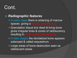
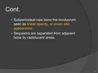
Osteomyeltis of
jaw
Instructor – Dr. Jesus George
1
Definition
 It is an inflammatory condition of bone,
that begins as an infection of medullary
spaces and harvesian systems of the
cortex & extends to involve the
periosteum of the affected area.
2
Etiology
 1-Odontogenic infections-
 Infections originating from pulpal or
periodontal tissue
 Pericoronitis
 Infected socket
 Infected cyst
 Tumor
 2.trauma-
 Compound fracture
 Surgery
3
cont.
 3-Infections of orofacial regions
 Periostitis following gingival ulceration
 Lymph nodes infected from furuncles
 Lacerations
 Peritonsillar abscess
 4-Infection from hematogenous route
 Upper respiratory tract infection
 Middle ear infection
 Systemic TB
 Furuncle of face
 Wound on the skin
4
Microbiology
 Strep.Viridans
 Peptostreptococci
 Fusobacteria
 Bacteriodes
 Klebsiella
 Pseudomonas
 Mycobacterium tuberculosis
 Actinomyces
5
Classification
 Suppurative osteomyelitis
 A/c suppurative
 C/c suppurative
 Infantile
 Nonsuppurative osteomyelitis
 C/c sclerosing
○ Focal sclerosing
○ Diffuse sclerosing
 Garre`s Sclerosing Oml
6
CONT.
 Actinomycotic Oml
 Radiation Oml
 Specific Infective Oml
○ Tb
○ Syphilis
7
A/c suppurative Oml
 Microbiology
 Staph.Aureus
 Strep.Pyogenes
 Spirochetes
 E.Coli
 Etiology
 A-Odontogenic Infections
 Periapical Pathology Secondary To Pulpal
Disease
 Periodontal Disease
8
Cont.
 Pericoronitis
 Infected Odontogenic Cyst
 Infection Of Extraction Wound Or fracture
 B-Local traumatic injuries
 injuries to gingiva
 C-Peritonsillar abscess
 D-Furuncles of skin
 E-Infected compound odontome
9
Cont.
 F-Hematogenous infection
 From minor wounds in skin
 Infection of upper respiratory tract
 Infection of middle ear
 G-Compound # of jaws
10
Cont.
 Clinical features
 Fever
 Malaise
 Nausea
 Vomiting
 Anorexia
 Deep seated boring, continuous intense pain
 Paresthesia or anesthesia of lower lip
 Facial cellulitis
11
Cont.
 Indurated swelling
 Trismus
 Involved tooth is loose & tender to
percussion
 Purulent discharge through sinus
 Fetid odour
 Regional lymphadenopathy
12
c/c suppurative oml
 Clinical features
 Pain & tenderness
 Induration of soft tissues
 Intraoral or extraoral draining fistula
 Enlargement of mandible
 Pathologic #
 Teeth in the area becomes loose & sensitive
to percussion
13
Cont.
 Radiographic features
 In early stage there is widening of marrow
spaces giving a mottled appearance
 Granulation tissue b/w dead & living bone
gives irregular lines & zones of radiolucency
resulting in moth -eaten appearance.
 In later stages the devitalized bone appears
sclerosed & called sequestrum.
 Large areas of bone destruction seen as
radiolucent areas.
14
Cont.
 Subperiosteal new bone the involucrum
seen as linear opacity, or onion skin
appearance.
 Sequestra are separated from adjacent
bone by radiolucent areas.
15
Cont.
 Management
 Conservative management
 Complete bed rest
 Supportive therapy
 Nutritional support
 Hydration
 Oral
 I/v
16
Cont.
 Blood transfusion-if RBC & Hb is low
 Analgesics
 Antimicrobial agents-
 Regimen 1-
 Aqueous penicillin 2 million units IV every 4
hrs
 Oxacillin 1gm 4th hrly
 If the patient is asymptomatic for 48 to 72hrs
penicillin v orally 500mg 4th hrly with
cloxacillin 250mg orally 4th hrly for 2-4 weeks
17
Cont.
 Regimen 2
 Oxacillin
 Dicloxacillin
 In case of allergy to penicillin
○ Clindamycin 300-600mg orally 6th hrly
○ Cefazolin 500mg 8th hrly
○ Erythromycin 2gm 6th hrly i/v then 500mg 6th
hrly orally
18
Cont.
 Specific treatment for
 Anemia
 Diabetes mellitus
 Malnutrition
 Hyperbaric o2 therapy
 It involves intermittent, inhalation of 100%
humidified o2 under pressure greater than 1
atm
 It decreases hypoxic environment
 Bactericidal action
19
Cont.
 Exotoxins of micro organisms are rendered
inert by exposure to elevated pressure of 02
 It increases vascular supply
 It aids in healing draining sinus
 Improves osteogenesis
 Surgical management
 Incision & drainage
 It relieves pressure & pain caused by pus.
20
Cont.
 Reduces absorption of toxic products &
prevents further spread of infection
 It can be done by opening the pulp chamber
 It can also be done by making a fenestration
through cortical plate over the apical area
 In an edentulous area, make an incision
over the alveolar crest
 At the angle of mandible a small incision is
made over the point of greatest tenderness.
 Extraction of offending tooth
21
Cont.
 Debridement
 Followed by incision & drainage the area is
irrigated with H2O2 & saline.
 Any foreign body ,necrotic tissue or small
sequestrum is removed
 Decortication
 A buccal flap is created by the crestal
incision
 Mucoperiosteal flap is reflected
 Tooth in the involved area is removed
22
Cont.
 Chronically infected lateral & inferior cortical
plates of bone 1 to 2 cm beyond the area of
involvement is removed
 Bone is thoroughly debrided
 Flap is closed
 Sequestrectomy
 A preoperative radiograph is taken to decide
the site of incision
 Sequestrum usually lies on the surface of
bone & can be removed easily
23
Cont.
 If sequestrum is encased by involucrum ,a
window is made to take it out
 The granulation tissue around the sequestrum
is curetted until healthy bone is exposed.
Antibiotic therapy is continued for 2weeks
 Saucerization
 It is performed when removal of sequestrum
leaves a large cavity
 The buccal cortex is reduced to the level of
unattached mucosa producing a soccer like
defect
24
Cont.
 Trephination or fenestration
 Creation of bony holes or windows in the
cortical bone adjacent to the infectious
process for decompression of medullary
compartment.
 It allows vascular communication b/w
periosteum & medullary cavity.
 Resection
 When extensive portion of bone is involved
resection is performed
25
Ont.
 Reconstruction
 Iliac crest is the graft is used
 Stabilization is achieved with titanium or
vitallium mesh.
 Complications
 Neoplastic transformation
 Discontinuity defects
 Progressive diffuse sclerosis
26
Infantile oml
 It is seen in infants commonly occuring
in maxilla
 Etiology
 Trauma caused to oral mucosa during
delivery
 Infection of maxillary sinus
 Contaminated human or artificial nipples
 Infections from nose
 Hematogenous spread
27
Cont.
 Clinical features
 Pyrexia
 Anorexia
 Dehydration
 Convulsions
 Vomiting
 Facial cellulitis centered around the orbit
 Palpebral edema
28
Cont.
 Conjunctivitis
 Proptosis
 Buccal or palatal swelling in maxillary
molar region
 Presence of fluctuation for the sweeling
 Fistulae in the alveolar mucosa
 Microbiology
 Staph.Aureus
 Streptococci
29
Cont.
 Radiographic findings
 In later stages sequestra & necrotic
tooth germs
 Complications
 Permanent Optic Damage
 Neurologic Complications
 Loss Of Tooth Buds
30
Cont.
 Treatment
 Antibiotics-penicillins,flucloxacillin or
broad spectrum antibiotics
 Incision & drainage of fluctuant areas
 Irrigation of sinus tracts
 Analgesics
 Antipyretics
 Fluids
31
Cont.
 Nuritious diet
 Sequestrectomy or removal of necrotic
tooth germs
32
Garre`s sclerosing
osteomyelitis
 First described by carl garre
 There is peripheral subperiosteal bone
deposition caused by mild irritation
Etiology
 Carious tooth
 Overlying soft tissue infection
 Clinical features
 Usually involves mandible
33
Cont.
 Disease occurs in children & young
adults
 localized hard, nontender, bony swelling
of lateral & inferior aspects of mandible
 Radiologic features
 Focal area of well calcified bone & has
an onion skin appearance.
34
Cont.
 Treatment
 Removal of infected tooth & curettage of
the socket
 Surgical recontouring
 Endodontic therapy
 Antibiotics
35
C/c sclerosing oml
 Radiographically there are 2 forms
focal & diffuse
 Focal form
 Occures before the age of 20
 It is more common in mandible
 It is associated with infected pulp of
Lower molars & premolars
36
Cont.
 It appears as a circumscribed radio-
opaque mass of sclerotic bone
associated with the tooth roots
 Treated by extraction or endodontic
therapy
 Diffuse form
 It occures both in maxilla & mandible
 Pain & suppuration may be there
37
Cont.
 Radiologically it shows dense radio-
opaque mass
 Treated by debridement,antibiotic
therapy,alveolectomy & hyperbaric o2
therapy
38
Actinomycotic oml of jaws
 Definition
 It is the c/c infection manifesting both
granulomatous & suppurative
features,usually involves soft tissues &
sometimes bone.
 Types
 Cervicofacial
 Thoracic
 Abdominal
39
Cont.
 Clinical features
 Cervicofacial type usually involves
mandible, overlying soft tissues, parotid
gland, tongue & maxillary sinus
 Appears as soft or firm tissue mass on
skin that have a purple, dark red, oily
areas with small areas of fluctuation
 Spontaneous drainage of serous fluid
containing yellowish
40
Cont.
 Granular material called sulphur
granules representing colonies of
bacteria
 Enlarged regional lymph nodes
 Trismus
 Pyrexia
 Radiologic features
 Radiolucent areas of varying sizes
41
Cont.
 Management
 Incision & drainage
 Parentral antibiotics
 Penicillin 10 to 20 million units daily for 3 to
4 months
 If allergic to penicillin tetracycline 250mg 4
times daily for 8 to 16 weeks or
erythromycin 500mg 4 times daily for 6
months
 Sequestrectomy & saucerization
42
Tuberculous osteomyelitis
of jaw bones
 It is a c/c infection caused by
mycobacterium tuberculosis
Clinical features
 The sites commonly involved are ramus
& body of mandible
 The age group is b/w 15 to 40 years
 There are 2 types of presentations
closed & open lesions
43
Cont.
 Closed lesions
 Located centrally in bone.
 It presents as swelling & no draining
sinuses.
 There is absence of oral septic focus.
 Usually ramus of mandible is involved.
 Open lesions
 There is multiple sinuses with mucopurulant
discharge.
 Oral focus may or may not be present
44
Cont.
 Diagnosis
 Aspiration & culture studies-done in
closed lesions
 Radiographs
 OPG
 PA mandible
 Lateral oblique view of mandible.
 Closed lesions are seen as small well
defined radiolucency with destruction of
buccal or medial cortical plates
 45
Cont.
 Chest radiograph
 Mantaux testing-intradermal injection of
5 tuberculous units in 0.1ml solution of
purified protien derivativeis given using
a 27 guage needle.A positive reaction is
seen after 48hrs as erythema &
induration >5 to 10 mm.
 Sputum for AFB-early morning sputum
samples are collected on 3 consecutive
days
46
Cont.
 Biopsy-
 Incisional biopsy is done for open cases.
 Aspiration is done for closed cases
 Treatment
 Antikoch`s treatment-isoniazide,
rifampicin, ethambutol,pyrizinamide for
first 4 months
 Isoniazide, rifampicin for next 4 months
47
Osteoradionecrosis of
facial bones
 Definition
 Osteradionecrosis is an exposure of
non - viable , non - healing , non -
septic lesion in the irradiated bone ,
which fails to heal without intervention .
 It is a sequelae of irradiation induced
tissue injury , in which hypocellularity ,
hypovascularity & hypoxia are the
underlying causes .
48
Cont.
 Mechanism
Therapeutic doses of irradiation

Endothelial death, thrombosis &
hyalinsation of blood vessels .

Progressive obliterative endarteteritis
hyalinisation & fibrosis &
thrombosis of vessels
 49
Cont.
Decreased microcirculation

Osteoblasts & osteocytes are destroyed
& marrow spaces in bone become filled
with fibrous tissue .

Decrease of cellularity & vascularity

Hypoxia in irradiated tissue
50
Cont.
 Clinical features .
 Severe , deep , boring pain which may
continue for weeks or months .
 Swelling of face when infection develops
 Soft tissue abscesses & persistently
draining sinuses .
 Exposed bone ; in association with
intraoral or extraoral fistulae .
 Trismus .
51
Cont.
 Foetid odour .
 Pyrexia .
 Pathological fracture
 Radiological fratures .
 Radilucent area with indefinite
nonsclerotic border
 Radioopacity usually associated with
sequestrum
52
Cont.
 Treatment
 Debridement
 Antibiotics
 Hydration of the patient
 High protien & vitamin diet
 Analgesics
 Maintenance Of Good Oral hygiene-oral
rinse
53
Cont.
 Frequent irrigation of wounds
 Loose exposed dead bone is removed
 Sequestrectomy
 Bone resection if there is persistant
infection or pathologic #
 Hyperbaric o2 therapy
54
Cont.
 Prevention
 Preirradiation dental care
 Teeth in direct beam of radiation ,
nonrestorable teeth, teeth with
periodontal disease are extracted.
 Radiation therapy is delayed for 10 to 14
days after extraction.
 Prominent interdental septa,sharp
socket margins are trimmed
55
Cont.
 Unerupted, deeply buried teeth are left
in situ
 Restorable teeth are restored
 Periodontal therapy is done
 Oral Hygiene Instructions Are Given
 Topical Flouride 0.4% Stannous Flouride
Gel, or 1% acidulated flourophosphate
gel is applied for 15 min. twice a day for
2 weeks
56
Cont.
 Dental care during radiation therapy
 Mouthwash 0.2% aqueous chlorhexidine
 Supervised cleaning of teeth
 Oral hygiene instructions with flouride
tooth paste, flouride mouthwash
 Post irradiation dental care
 Avoidance of denture for one year
 Maintenance of oral hygiene
57
Cont.
 Saliva substitutes to reduce xerostomia
induced disorders
 Restoration of teeth with post -irradiation
Pulpitis
 Extraction should be the last resort.
 Teeth should be removed atraumatically.
 Sharp bony margins should be trimmed.
 Risk of ORN is is highest in 4to 12
months.
58

17 osteomyelitis

  • 1.
  • 2.
    Definition  It isan inflammatory condition of bone, that begins as an infection of medullary spaces and harvesian systems of the cortex & extends to involve the periosteum of the affected area. 2
  • 3.
    Etiology  1-Odontogenic infections- Infections originating from pulpal or periodontal tissue  Pericoronitis  Infected socket  Infected cyst  Tumor  2.trauma-  Compound fracture  Surgery 3
  • 4.
    cont.  3-Infections oforofacial regions  Periostitis following gingival ulceration  Lymph nodes infected from furuncles  Lacerations  Peritonsillar abscess  4-Infection from hematogenous route  Upper respiratory tract infection  Middle ear infection  Systemic TB  Furuncle of face  Wound on the skin 4
  • 5.
    Microbiology  Strep.Viridans  Peptostreptococci Fusobacteria  Bacteriodes  Klebsiella  Pseudomonas  Mycobacterium tuberculosis  Actinomyces 5
  • 6.
    Classification  Suppurative osteomyelitis A/c suppurative  C/c suppurative  Infantile  Nonsuppurative osteomyelitis  C/c sclerosing ○ Focal sclerosing ○ Diffuse sclerosing  Garre`s Sclerosing Oml 6
  • 7.
    CONT.  Actinomycotic Oml Radiation Oml  Specific Infective Oml ○ Tb ○ Syphilis 7
  • 8.
    A/c suppurative Oml Microbiology  Staph.Aureus  Strep.Pyogenes  Spirochetes  E.Coli  Etiology  A-Odontogenic Infections  Periapical Pathology Secondary To Pulpal Disease  Periodontal Disease 8
  • 9.
    Cont.  Pericoronitis  InfectedOdontogenic Cyst  Infection Of Extraction Wound Or fracture  B-Local traumatic injuries  injuries to gingiva  C-Peritonsillar abscess  D-Furuncles of skin  E-Infected compound odontome 9
  • 10.
    Cont.  F-Hematogenous infection From minor wounds in skin  Infection of upper respiratory tract  Infection of middle ear  G-Compound # of jaws 10
  • 11.
    Cont.  Clinical features Fever  Malaise  Nausea  Vomiting  Anorexia  Deep seated boring, continuous intense pain  Paresthesia or anesthesia of lower lip  Facial cellulitis 11
  • 12.
    Cont.  Indurated swelling Trismus  Involved tooth is loose & tender to percussion  Purulent discharge through sinus  Fetid odour  Regional lymphadenopathy 12
  • 13.
    c/c suppurative oml Clinical features  Pain & tenderness  Induration of soft tissues  Intraoral or extraoral draining fistula  Enlargement of mandible  Pathologic #  Teeth in the area becomes loose & sensitive to percussion 13
  • 14.
    Cont.  Radiographic features In early stage there is widening of marrow spaces giving a mottled appearance  Granulation tissue b/w dead & living bone gives irregular lines & zones of radiolucency resulting in moth -eaten appearance.  In later stages the devitalized bone appears sclerosed & called sequestrum.  Large areas of bone destruction seen as radiolucent areas. 14
  • 15.
    Cont.  Subperiosteal newbone the involucrum seen as linear opacity, or onion skin appearance.  Sequestra are separated from adjacent bone by radiolucent areas. 15
  • 16.
    Cont.  Management  Conservativemanagement  Complete bed rest  Supportive therapy  Nutritional support  Hydration  Oral  I/v 16
  • 17.
    Cont.  Blood transfusion-ifRBC & Hb is low  Analgesics  Antimicrobial agents-  Regimen 1-  Aqueous penicillin 2 million units IV every 4 hrs  Oxacillin 1gm 4th hrly  If the patient is asymptomatic for 48 to 72hrs penicillin v orally 500mg 4th hrly with cloxacillin 250mg orally 4th hrly for 2-4 weeks 17
  • 18.
    Cont.  Regimen 2 Oxacillin  Dicloxacillin  In case of allergy to penicillin ○ Clindamycin 300-600mg orally 6th hrly ○ Cefazolin 500mg 8th hrly ○ Erythromycin 2gm 6th hrly i/v then 500mg 6th hrly orally 18
  • 19.
    Cont.  Specific treatmentfor  Anemia  Diabetes mellitus  Malnutrition  Hyperbaric o2 therapy  It involves intermittent, inhalation of 100% humidified o2 under pressure greater than 1 atm  It decreases hypoxic environment  Bactericidal action 19
  • 20.
    Cont.  Exotoxins ofmicro organisms are rendered inert by exposure to elevated pressure of 02  It increases vascular supply  It aids in healing draining sinus  Improves osteogenesis  Surgical management  Incision & drainage  It relieves pressure & pain caused by pus. 20
  • 21.
    Cont.  Reduces absorptionof toxic products & prevents further spread of infection  It can be done by opening the pulp chamber  It can also be done by making a fenestration through cortical plate over the apical area  In an edentulous area, make an incision over the alveolar crest  At the angle of mandible a small incision is made over the point of greatest tenderness.  Extraction of offending tooth 21
  • 22.
    Cont.  Debridement  Followedby incision & drainage the area is irrigated with H2O2 & saline.  Any foreign body ,necrotic tissue or small sequestrum is removed  Decortication  A buccal flap is created by the crestal incision  Mucoperiosteal flap is reflected  Tooth in the involved area is removed 22
  • 23.
    Cont.  Chronically infectedlateral & inferior cortical plates of bone 1 to 2 cm beyond the area of involvement is removed  Bone is thoroughly debrided  Flap is closed  Sequestrectomy  A preoperative radiograph is taken to decide the site of incision  Sequestrum usually lies on the surface of bone & can be removed easily 23
  • 24.
    Cont.  If sequestrumis encased by involucrum ,a window is made to take it out  The granulation tissue around the sequestrum is curetted until healthy bone is exposed. Antibiotic therapy is continued for 2weeks  Saucerization  It is performed when removal of sequestrum leaves a large cavity  The buccal cortex is reduced to the level of unattached mucosa producing a soccer like defect 24
  • 25.
    Cont.  Trephination orfenestration  Creation of bony holes or windows in the cortical bone adjacent to the infectious process for decompression of medullary compartment.  It allows vascular communication b/w periosteum & medullary cavity.  Resection  When extensive portion of bone is involved resection is performed 25
  • 26.
    Ont.  Reconstruction  Iliaccrest is the graft is used  Stabilization is achieved with titanium or vitallium mesh.  Complications  Neoplastic transformation  Discontinuity defects  Progressive diffuse sclerosis 26
  • 27.
    Infantile oml  Itis seen in infants commonly occuring in maxilla  Etiology  Trauma caused to oral mucosa during delivery  Infection of maxillary sinus  Contaminated human or artificial nipples  Infections from nose  Hematogenous spread 27
  • 28.
    Cont.  Clinical features Pyrexia  Anorexia  Dehydration  Convulsions  Vomiting  Facial cellulitis centered around the orbit  Palpebral edema 28
  • 29.
    Cont.  Conjunctivitis  Proptosis Buccal or palatal swelling in maxillary molar region  Presence of fluctuation for the sweeling  Fistulae in the alveolar mucosa  Microbiology  Staph.Aureus  Streptococci 29
  • 30.
    Cont.  Radiographic findings In later stages sequestra & necrotic tooth germs  Complications  Permanent Optic Damage  Neurologic Complications  Loss Of Tooth Buds 30
  • 31.
    Cont.  Treatment  Antibiotics-penicillins,flucloxacillinor broad spectrum antibiotics  Incision & drainage of fluctuant areas  Irrigation of sinus tracts  Analgesics  Antipyretics  Fluids 31
  • 32.
    Cont.  Nuritious diet Sequestrectomy or removal of necrotic tooth germs 32
  • 33.
    Garre`s sclerosing osteomyelitis  Firstdescribed by carl garre  There is peripheral subperiosteal bone deposition caused by mild irritation Etiology  Carious tooth  Overlying soft tissue infection  Clinical features  Usually involves mandible 33
  • 34.
    Cont.  Disease occursin children & young adults  localized hard, nontender, bony swelling of lateral & inferior aspects of mandible  Radiologic features  Focal area of well calcified bone & has an onion skin appearance. 34
  • 35.
    Cont.  Treatment  Removalof infected tooth & curettage of the socket  Surgical recontouring  Endodontic therapy  Antibiotics 35
  • 36.
    C/c sclerosing oml Radiographically there are 2 forms focal & diffuse  Focal form  Occures before the age of 20  It is more common in mandible  It is associated with infected pulp of Lower molars & premolars 36
  • 37.
    Cont.  It appearsas a circumscribed radio- opaque mass of sclerotic bone associated with the tooth roots  Treated by extraction or endodontic therapy  Diffuse form  It occures both in maxilla & mandible  Pain & suppuration may be there 37
  • 38.
    Cont.  Radiologically itshows dense radio- opaque mass  Treated by debridement,antibiotic therapy,alveolectomy & hyperbaric o2 therapy 38
  • 39.
    Actinomycotic oml ofjaws  Definition  It is the c/c infection manifesting both granulomatous & suppurative features,usually involves soft tissues & sometimes bone.  Types  Cervicofacial  Thoracic  Abdominal 39
  • 40.
    Cont.  Clinical features Cervicofacial type usually involves mandible, overlying soft tissues, parotid gland, tongue & maxillary sinus  Appears as soft or firm tissue mass on skin that have a purple, dark red, oily areas with small areas of fluctuation  Spontaneous drainage of serous fluid containing yellowish 40
  • 41.
    Cont.  Granular materialcalled sulphur granules representing colonies of bacteria  Enlarged regional lymph nodes  Trismus  Pyrexia  Radiologic features  Radiolucent areas of varying sizes 41
  • 42.
    Cont.  Management  Incision& drainage  Parentral antibiotics  Penicillin 10 to 20 million units daily for 3 to 4 months  If allergic to penicillin tetracycline 250mg 4 times daily for 8 to 16 weeks or erythromycin 500mg 4 times daily for 6 months  Sequestrectomy & saucerization 42
  • 43.
    Tuberculous osteomyelitis of jawbones  It is a c/c infection caused by mycobacterium tuberculosis Clinical features  The sites commonly involved are ramus & body of mandible  The age group is b/w 15 to 40 years  There are 2 types of presentations closed & open lesions 43
  • 44.
    Cont.  Closed lesions Located centrally in bone.  It presents as swelling & no draining sinuses.  There is absence of oral septic focus.  Usually ramus of mandible is involved.  Open lesions  There is multiple sinuses with mucopurulant discharge.  Oral focus may or may not be present 44
  • 45.
    Cont.  Diagnosis  Aspiration& culture studies-done in closed lesions  Radiographs  OPG  PA mandible  Lateral oblique view of mandible.  Closed lesions are seen as small well defined radiolucency with destruction of buccal or medial cortical plates  45
  • 46.
    Cont.  Chest radiograph Mantaux testing-intradermal injection of 5 tuberculous units in 0.1ml solution of purified protien derivativeis given using a 27 guage needle.A positive reaction is seen after 48hrs as erythema & induration >5 to 10 mm.  Sputum for AFB-early morning sputum samples are collected on 3 consecutive days 46
  • 47.
    Cont.  Biopsy-  Incisionalbiopsy is done for open cases.  Aspiration is done for closed cases  Treatment  Antikoch`s treatment-isoniazide, rifampicin, ethambutol,pyrizinamide for first 4 months  Isoniazide, rifampicin for next 4 months 47
  • 48.
    Osteoradionecrosis of facial bones Definition  Osteradionecrosis is an exposure of non - viable , non - healing , non - septic lesion in the irradiated bone , which fails to heal without intervention .  It is a sequelae of irradiation induced tissue injury , in which hypocellularity , hypovascularity & hypoxia are the underlying causes . 48
  • 49.
    Cont.  Mechanism Therapeutic dosesof irradiation  Endothelial death, thrombosis & hyalinsation of blood vessels .  Progressive obliterative endarteteritis hyalinisation & fibrosis & thrombosis of vessels  49
  • 50.
    Cont. Decreased microcirculation  Osteoblasts &osteocytes are destroyed & marrow spaces in bone become filled with fibrous tissue .  Decrease of cellularity & vascularity  Hypoxia in irradiated tissue 50
  • 51.
    Cont.  Clinical features.  Severe , deep , boring pain which may continue for weeks or months .  Swelling of face when infection develops  Soft tissue abscesses & persistently draining sinuses .  Exposed bone ; in association with intraoral or extraoral fistulae .  Trismus . 51
  • 52.
    Cont.  Foetid odour.  Pyrexia .  Pathological fracture  Radiological fratures .  Radilucent area with indefinite nonsclerotic border  Radioopacity usually associated with sequestrum 52
  • 53.
    Cont.  Treatment  Debridement Antibiotics  Hydration of the patient  High protien & vitamin diet  Analgesics  Maintenance Of Good Oral hygiene-oral rinse 53
  • 54.
    Cont.  Frequent irrigationof wounds  Loose exposed dead bone is removed  Sequestrectomy  Bone resection if there is persistant infection or pathologic #  Hyperbaric o2 therapy 54
  • 55.
    Cont.  Prevention  Preirradiationdental care  Teeth in direct beam of radiation , nonrestorable teeth, teeth with periodontal disease are extracted.  Radiation therapy is delayed for 10 to 14 days after extraction.  Prominent interdental septa,sharp socket margins are trimmed 55
  • 56.
    Cont.  Unerupted, deeplyburied teeth are left in situ  Restorable teeth are restored  Periodontal therapy is done  Oral Hygiene Instructions Are Given  Topical Flouride 0.4% Stannous Flouride Gel, or 1% acidulated flourophosphate gel is applied for 15 min. twice a day for 2 weeks 56
  • 57.
    Cont.  Dental careduring radiation therapy  Mouthwash 0.2% aqueous chlorhexidine  Supervised cleaning of teeth  Oral hygiene instructions with flouride tooth paste, flouride mouthwash  Post irradiation dental care  Avoidance of denture for one year  Maintenance of oral hygiene 57
  • 58.
    Cont.  Saliva substitutesto reduce xerostomia induced disorders  Restoration of teeth with post -irradiation Pulpitis  Extraction should be the last resort.  Teeth should be removed atraumatically.  Sharp bony margins should be trimmed.  Risk of ORN is is highest in 4to 12 months. 58