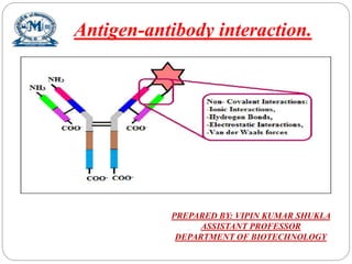
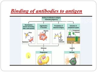
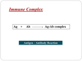
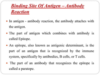
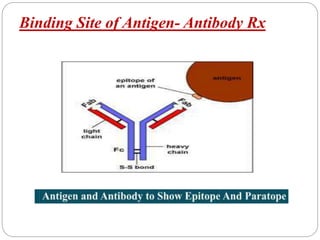
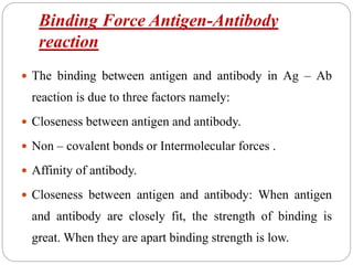
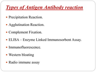
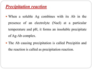
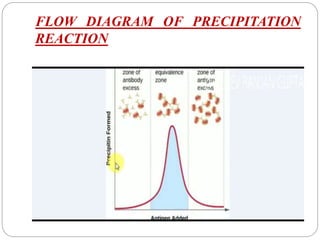
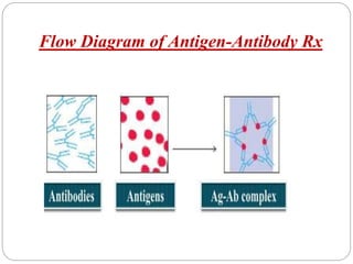
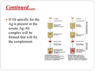
Antigen-antibody interaction.
PREPARED BY: VIPIN KUMAR SHUKLA
ASSISTANT PROFESSOR
DEPARTMENT OF BIOTECHNOLOGY
Binding of antibodies to antigen
CONTENT
 Introduction.
 Salient Features of Antigen – Antibody Reaction.
 Strength of Antigen – Antibody Reaction.
 Properties of Antigen – Antibody Reaction .
 Types of Antigen – Antibody Reaction .
 Application of Antigen – Antibody Reaction.
 Conclusion
Introduction
 The antigen and antibody combines specifically with each
other.
 This interaction between them is called Antibody- Antigen
 interaction.
 It may be abbreviated as Ag –Ab reaction.
 These form the basis for humoral immunity or antibody
mediated immunity.
 These reaction form the basis for detection of infectious disease
causing agents and also some non specific antigens like
enzymes.
 When antigen – antibody reactions occur in-vitro they are
known as Serological Test.
Salient Features of Antibody-Antigen
Reaction
 Specificity of Antigen – Antibody Reaction.
 Immune complex.
 Binding Site of Antigen – Antibody Reaction.
Specificity of antigen antibody reaction
 Specificity refers to the ability of an individual
antibody combining.
 Site to react with only one antigenic determinant or
the ability of a population of antibody molecules to
react with only one antigen.
Specificity of antigen antibody reaction
Immune complex
 An immune complex is formed from the integral
binding of an antibody to a soluble antigen.
 An immune complex is formed from the integral
binding of an antibody to a soluble antigen.
 Mechanisms of antigen-antibody interaction leading
to inflammation.
 Antigen-antibody immune complex formation results
in complement activation, Opsonization of target
cells, assembly of membrane attack complexes and
release of complement activators .
Immune Complex
Binding Site Of Antigen – Antibody
Reaction
 In antigen - antibody reaction, the antibody attaches with
the antigen.
 The part of antigen which combines with antibody is
called Epitope.
 An epitope, also known as antigenic determinant, is the
part of an antigen that is recognized by the immune
system, specifically by antibodies, B cells, or T cells.
 The part of an antibody that recognizes the epitope is
called a paratope.
Binding Site of Antigen- Antibody Rx
Binding Force Antigen-Antibody
reaction
 The binding between antigen and antibody in Ag – Ab
reaction is due to three factors namely:
 Closeness between antigen and antibody.
 Non – covalent bonds or Intermolecular forces .
 Affinity of antibody.
 Closeness between antigen and antibody: When antigen
and antibody are closely fit, the strength of binding is
great. When they are apart binding strength is low.
Continued…..
 Non – Covalent Bonds: The bonds that hold the antigen to
the antibody combining site are all non-covalent in nature.
 These include hydrogen bonds, electrostatic bonds, Vander
Waals forces and hydrophobic bonds.
 Affinity of antibody: Antibody affinity is the strength of
the reaction between a single
 Antigenic determinant and a single combining site on the
antibody.
Strength of Antigen-Antibody reaction
 The non – covalent
interaction that form the
basis of antigen –
antibody binding include
hydrogen bond, ionic
bond, hydrophobic
 interaction and Vander
Walls interaction.
Properties of Antigen – Antibody reaction
 The properties of antigen and antibody can be
explained with the help of three points. They are:
 Antibody Affinity
 Avidity
 Cross reaction
 Specificity
Antibody affinity
 Interactions between antigen and antibody involve non-
covalent binding of an antigenic determinant (epitope)
to the variable region (complementary determining
region, CDR) of both the heavy and light
immunoglobulin chains.
 These interactions are analogous to those observed in
enzyme-substrate interactions and they can be defined
similarly.
Avidity of Antibody
 It is the strength of the bond after the formation of Ag-Ab
complexes.
 It is used to denote the overall capacity of antibodies to
combine with the multivalent antigen.
 A multivalent Ag has many types of antigenic
determinants.
 When injected into the blood, each antigenic determinant
stimulates the production of a particular antibody.
Cross reaction
 An antiserum raised against an Ag, can also react with a
similar Ag of another type.
 This is called cross reaction and the Ag which produces
the cross reaction is called Cross reactive Ag. But the
strength of Ab raised against its own Ag is strong.
 The bonds involved in cross reactions are weak.
 Example: The antiserum raised against human insulin will
react with the insulin of Pig,
 Sheep, whale etc.
Continue…..
Antibody Specificity
 Specificity refers to the ability of an individual
antibody – combining site to react with only one
antigenic determinant or the ability of a population of
antibody molecules to react with only one antigen.
Types of Antigen Antibody reaction
 Precipitation Reaction.
 Agglutination Reaction.
 Complement Fixation.
 ELISA – Enzyme Linked Immunosorbent Assay.
 Immunofluorescence.
 Western bloating
 Radio immune assay
Precipitation reaction
 When a soluble Ag combines with its Ab in the
presence of an electrolyte (Nacl) at a particular
temperature and pH, it forms an insoluble precipitate
of Ag-Ab complex.
 The Ab causing precipitation is called Precipitin and
the reaction is called as precipitation reaction.
FLOW DIAGRAM OF PRECIPITATION
REACTION
Flow Diagram of Antigen-Antibody Rx
Precipitation Reaction
 Precipitation reaction broadly divided into three
types:
 Precipitation in solution
( E.g. Ring test,
Flocculation test.
Continued…..
Precipitation in Agar
 The ppt Test in agar gel is termed as Immuno-
diffusion .
 In this test ,reactants are added to the gel and antigen-
antibody combination occurs by means diffusion
Agglutination
 When a particular Ag is mixed with its Abs's in the presence of
electrolytes at a suitable temperature and pH, the particles are
clumped or agglutinated.
 The Ab of the serum causes the cellular Ag’s to form clumps and
these are called Agglutinins.
 The particulate antigens that are aggregated are termed
Agglutinogens.
 Types of agglutination reactions :
 a) slide agglutination
 b) Tube agglutination
 c) Antiglobulin ( coomb’s test)
Slide Agglutination test
 Slide Agglutination test : This is a rapid method to
determine the presence of agglutinating antibodies.
 To a uniform suspension of particulate Ag, a drop of
saline is added and then a drop of antiserum is added .
The slide is gently rocked or a fine loop is used to mix
the contents. If granulation occurs the test is
 Positive.
 It takes a minute for the test to complete and is visible
to the naked eye. Some times confirmation maybe
done by observing slide under microscope.
Slide Agglutination reaction
Tube agglutination
 This is a standard method for quantitative estimation of
Ab. The serum containing Ab is diluted serially with
saline in several small test tubes, to which a constant
volume of Ag suspension is added.
 A control tube is kept which has no antiserum. The tubes
are incubated until visible agglutination is observed. The
tube showing highest agglutination is referred to as the
titer.
 Tube agglutination is employed for the serological
diagnosis of typhoid, brucellosis and typhus fever. Widal
test is used for the estimation of typhoid fever.
Continued…..
 In this test Ab content of
the patient’s serum, is
measured by adding a
constant amount of antigen
(Salmonella typhi) to the
serially diluted serum.
Complement fixation test
 Lysis of RBC or bacteria requires some non-
specific unstable components of fresh serum
which are called complement.
 This complement system comprises of 11 proteins
and are present in ever individual. They bind to Fc
component of Ab involved in Ag-Ab complex.
 This ability of the Ag-Ab complex to fix
complement is used in complement Fixation tests.
 In the first stage, the test Ag and the antiserum
(heated to 56oC to inactivate complement) are
mixed in the presence of known amount of
complement. This is incubated at 4oC for 18h.
Continued….
 If Ab specific for the
Ag is present in the
serum, Ag-Ab
complex will be
formed that will fix
the complement.
ELISA ( Enzyme linked immunosorbent
assay)
 These are very simple, sensitive, economic and less
hazard when compared to RIA.
 In 1971, enzyme labeled Ag’s and Abs's were
developed as serological reagents for the assay of
Abs's and Ag’.
 The ligand used here is a molecule which can detect
the Ab and is covalently coupled to an enzyme such as
peroxides, beta galactosidase, alkaline phosphatase
etc.
 ELISA is of 3types :
Continued…..
 Indirect ELISA: This technique is used for the
detection of HIV. The envelop proteins are developed
by recombinant technology and coated on the surface
of the of microtiter plates. Suspects serum is added,
and unbound proteins are washed off.
 Sandwich ELISA: Used to detect the presence of Ag
in a sample. The well is coated with Ab specific to the
Ag and then suspect serum is added allowed to react.
Continued……
 The wells are washed to remove unbound Ag’s. Then
a labeled Ab against a different epitope of the Ag is
added.
 Unbound Abs's are removed by washing and this is
followed by addition of colored substrate and
development of color.
 The intensity of color is directly proportional to the
concentration of the Ag in the serum.
Competitive ELISA
 Another variation for measuring amounts of antigen is
competitive ELISA. In this technique, antibody is first
incubated in solution with a sample containing
antigen.
 The antigen antibody mixture is then added to an
antigen coated micro titer well.
 The more antigen present in the sample, the less free
antibody will be available to bind to the antigen-
coated well. Addition of an enzyme-conjugated
secondary antibody (Ab2) specific for the isotype of
the primary antibody can be used to determine the
amount of primary antibody bound to the well as in an
indirect ELISA.
Western blotting
 Western blotting is
generally done on a
tissue homogenate or
extract.
 It uses SDS-PAGE ,a
type of gel
electrophoresis separate
various protein in a
mixtures on the basis of
size and shape.
Application of Antibody – Antigen
Interaction
 The chief use of antigen-antibody reactions are:
 Determination of blood groups for transfusion.
 Serological ascertainment of exposure to infectious
agents.
 Development of immunoassays for the quantification
of various substances.
 To detect the presence or absence of protein in serum.
 Determining the characteristics of certain Immuno-
deficiency diseases.
Reference
 Subhas Chandra parija Microbiology &
Immunology ( page 101- 115)
 (Kuby Immunology antigen antibody interaction .
 Slide share .com
 www. Google .com
Antigen antibody interaction .pptx 1

Antigen antibody interaction .pptx 1

  • 1.
    Antigen-antibody interaction. PREPARED BY:VIPIN KUMAR SHUKLA ASSISTANT PROFESSOR DEPARTMENT OF BIOTECHNOLOGY
  • 2.
  • 3.
    CONTENT  Introduction.  SalientFeatures of Antigen – Antibody Reaction.  Strength of Antigen – Antibody Reaction.  Properties of Antigen – Antibody Reaction .  Types of Antigen – Antibody Reaction .  Application of Antigen – Antibody Reaction.  Conclusion
  • 4.
    Introduction  The antigenand antibody combines specifically with each other.  This interaction between them is called Antibody- Antigen  interaction.  It may be abbreviated as Ag –Ab reaction.  These form the basis for humoral immunity or antibody mediated immunity.  These reaction form the basis for detection of infectious disease causing agents and also some non specific antigens like enzymes.  When antigen – antibody reactions occur in-vitro they are known as Serological Test.
  • 5.
    Salient Features ofAntibody-Antigen Reaction  Specificity of Antigen – Antibody Reaction.  Immune complex.  Binding Site of Antigen – Antibody Reaction.  Binding Site of Antigen – Antibody Reaction.
  • 6.
    Specificity of antigenantibody reaction  Specificity refers to the ability of an individual antibody combining.  Site to react with only one antigenic determinant or the ability of a population of antibody molecules to react with only one antigen.
  • 7.
    Specificity of antigenantibody reaction
  • 8.
    Immune complex  Animmune complex is formed from the integral binding of an antibody to a soluble antigen.  An immune complex is formed from the integral binding of an antibody to a soluble antigen.  Mechanisms of antigen-antibody interaction leading to inflammation.  Antigen-antibody immune complex formation results in complement activation, Opsonization of target cells, assembly of membrane attack complexes and release of complement activators .
  • 9.
  • 10.
    Binding Site OfAntigen – Antibody Reaction  In antigen - antibody reaction, the antibody attaches with the antigen.  The part of antigen which combines with antibody is called Epitope.  An epitope, also known as antigenic determinant, is the part of an antigen that is recognized by the immune system, specifically by antibodies, B cells, or T cells.  The part of an antibody that recognizes the epitope is called a paratope.
  • 11.
    Binding Site ofAntigen- Antibody Rx
  • 12.
    Binding Force Antigen-Antibody reaction The binding between antigen and antibody in Ag – Ab reaction is due to three factors namely:  Closeness between antigen and antibody.  Non – covalent bonds or Intermolecular forces .  Affinity of antibody.  Closeness between antigen and antibody: When antigen and antibody are closely fit, the strength of binding is great. When they are apart binding strength is low.
  • 13.
    Continued…..  Non –Covalent Bonds: The bonds that hold the antigen to the antibody combining site are all non-covalent in nature.  These include hydrogen bonds, electrostatic bonds, Vander Waals forces and hydrophobic bonds.  Affinity of antibody: Antibody affinity is the strength of the reaction between a single  Antigenic determinant and a single combining site on the antibody.
  • 14.
    Strength of Antigen-Antibodyreaction  The non – covalent interaction that form the basis of antigen – antibody binding include hydrogen bond, ionic bond, hydrophobic  interaction and Vander Walls interaction.
  • 15.
    Properties of Antigen– Antibody reaction  The properties of antigen and antibody can be explained with the help of three points. They are:  Antibody Affinity  Avidity  Cross reaction  Specificity
  • 16.
    Antibody affinity  Interactionsbetween antigen and antibody involve non- covalent binding of an antigenic determinant (epitope) to the variable region (complementary determining region, CDR) of both the heavy and light immunoglobulin chains.  These interactions are analogous to those observed in enzyme-substrate interactions and they can be defined similarly.
  • 17.
    Avidity of Antibody It is the strength of the bond after the formation of Ag-Ab complexes.  It is used to denote the overall capacity of antibodies to combine with the multivalent antigen.  A multivalent Ag has many types of antigenic determinants.  When injected into the blood, each antigenic determinant stimulates the production of a particular antibody.
  • 18.
    Cross reaction  Anantiserum raised against an Ag, can also react with a similar Ag of another type.  This is called cross reaction and the Ag which produces the cross reaction is called Cross reactive Ag. But the strength of Ab raised against its own Ag is strong.  The bonds involved in cross reactions are weak.  Example: The antiserum raised against human insulin will react with the insulin of Pig,  Sheep, whale etc.
  • 19.
  • 20.
    Antibody Specificity  Specificityrefers to the ability of an individual antibody – combining site to react with only one antigenic determinant or the ability of a population of antibody molecules to react with only one antigen.
  • 21.
    Types of AntigenAntibody reaction  Precipitation Reaction.  Agglutination Reaction.  Complement Fixation.  ELISA – Enzyme Linked Immunosorbent Assay.  Immunofluorescence.  Western bloating  Radio immune assay
  • 22.
    Precipitation reaction  Whena soluble Ag combines with its Ab in the presence of an electrolyte (Nacl) at a particular temperature and pH, it forms an insoluble precipitate of Ag-Ab complex.  The Ab causing precipitation is called Precipitin and the reaction is called as precipitation reaction.
  • 23.
    FLOW DIAGRAM OFPRECIPITATION REACTION
  • 24.
    Flow Diagram ofAntigen-Antibody Rx
  • 25.
    Precipitation Reaction  Precipitationreaction broadly divided into three types:  Precipitation in solution ( E.g. Ring test, Flocculation test.
  • 26.
  • 27.
    Precipitation in Agar The ppt Test in agar gel is termed as Immuno- diffusion .  In this test ,reactants are added to the gel and antigen- antibody combination occurs by means diffusion
  • 28.
    Agglutination  When aparticular Ag is mixed with its Abs's in the presence of electrolytes at a suitable temperature and pH, the particles are clumped or agglutinated.  The Ab of the serum causes the cellular Ag’s to form clumps and these are called Agglutinins.  The particulate antigens that are aggregated are termed Agglutinogens.  Types of agglutination reactions :  a) slide agglutination  b) Tube agglutination  c) Antiglobulin ( coomb’s test)
  • 29.
    Slide Agglutination test Slide Agglutination test : This is a rapid method to determine the presence of agglutinating antibodies.  To a uniform suspension of particulate Ag, a drop of saline is added and then a drop of antiserum is added . The slide is gently rocked or a fine loop is used to mix the contents. If granulation occurs the test is  Positive.  It takes a minute for the test to complete and is visible to the naked eye. Some times confirmation maybe done by observing slide under microscope.
  • 30.
  • 31.
    Tube agglutination  Thisis a standard method for quantitative estimation of Ab. The serum containing Ab is diluted serially with saline in several small test tubes, to which a constant volume of Ag suspension is added.  A control tube is kept which has no antiserum. The tubes are incubated until visible agglutination is observed. The tube showing highest agglutination is referred to as the titer.  Tube agglutination is employed for the serological diagnosis of typhoid, brucellosis and typhus fever. Widal test is used for the estimation of typhoid fever.
  • 32.
    Continued…..  In thistest Ab content of the patient’s serum, is measured by adding a constant amount of antigen (Salmonella typhi) to the serially diluted serum.
  • 33.
    Complement fixation test Lysis of RBC or bacteria requires some non- specific unstable components of fresh serum which are called complement.  This complement system comprises of 11 proteins and are present in ever individual. They bind to Fc component of Ab involved in Ag-Ab complex.  This ability of the Ag-Ab complex to fix complement is used in complement Fixation tests.  In the first stage, the test Ag and the antiserum (heated to 56oC to inactivate complement) are mixed in the presence of known amount of complement. This is incubated at 4oC for 18h.
  • 34.
    Continued….  If Abspecific for the Ag is present in the serum, Ag-Ab complex will be formed that will fix the complement.
  • 35.
    ELISA ( Enzymelinked immunosorbent assay)  These are very simple, sensitive, economic and less hazard when compared to RIA.  In 1971, enzyme labeled Ag’s and Abs's were developed as serological reagents for the assay of Abs's and Ag’.  The ligand used here is a molecule which can detect the Ab and is covalently coupled to an enzyme such as peroxides, beta galactosidase, alkaline phosphatase etc.  ELISA is of 3types :
  • 36.
    Continued…..  Indirect ELISA:This technique is used for the detection of HIV. The envelop proteins are developed by recombinant technology and coated on the surface of the of microtiter plates. Suspects serum is added, and unbound proteins are washed off.  Sandwich ELISA: Used to detect the presence of Ag in a sample. The well is coated with Ab specific to the Ag and then suspect serum is added allowed to react.
  • 37.
    Continued……  The wellsare washed to remove unbound Ag’s. Then a labeled Ab against a different epitope of the Ag is added.  Unbound Abs's are removed by washing and this is followed by addition of colored substrate and development of color.  The intensity of color is directly proportional to the concentration of the Ag in the serum.
  • 38.
    Competitive ELISA  Anothervariation for measuring amounts of antigen is competitive ELISA. In this technique, antibody is first incubated in solution with a sample containing antigen.  The antigen antibody mixture is then added to an antigen coated micro titer well.  The more antigen present in the sample, the less free antibody will be available to bind to the antigen- coated well. Addition of an enzyme-conjugated secondary antibody (Ab2) specific for the isotype of the primary antibody can be used to determine the amount of primary antibody bound to the well as in an indirect ELISA.
  • 40.
    Western blotting  Westernblotting is generally done on a tissue homogenate or extract.  It uses SDS-PAGE ,a type of gel electrophoresis separate various protein in a mixtures on the basis of size and shape.
  • 41.
    Application of Antibody– Antigen Interaction  The chief use of antigen-antibody reactions are:  Determination of blood groups for transfusion.  Serological ascertainment of exposure to infectious agents.  Development of immunoassays for the quantification of various substances.  To detect the presence or absence of protein in serum.  Determining the characteristics of certain Immuno- deficiency diseases.
  • 42.
    Reference  Subhas Chandraparija Microbiology & Immunology ( page 101- 115)  (Kuby Immunology antigen antibody interaction .  Slide share .com  www. Google .com