
Recommended
More Related Content
What's hot
What's hot (20)
Similar to Antibodies: Structure, Classes, Subclasses, and Functions
Similar to Antibodies: Structure, Classes, Subclasses, and Functions (20)
More from Rachna Tewari
More from Rachna Tewari (7)
Recently uploaded
Recently uploaded (20)
Antibodies: Structure, Classes, Subclasses, and Functions
- 1. Antibodies
- 2. I. DEFINITION Immunoglobulin (Ig) • Immunoglobulins are glycoprotein molecules that are produced by plasma cells in response to an immunogen and which function as antibodies. • They recognise epitope on an antigen and bind to it specifically.
- 3. GENERAL FUNCTIONS OF IMMUNOGLOBULINS A. Antigen binding Immunoglobulins bind specifically to one or a few closely related antigens. • Each immunoglobulin actually binds to a specific antigenic determinant. • Antigen binding by antibodies is the primary function of antibodies and can result in protection of the host.
- 4. VALENCY • The valency of antibody refers to the number of antigenic determinants that an individual antibody molecule can bind. • The valency of all antibodies is at least two and in some instances more.
- 5. B. EFFECTOR FUNCTIONS • Frequently the binding of an antibody to an antigen has no direct biological effect. • Rather, the significant biological effects are a consequence of secondary "effector functions" of antibodies.
- 6. Such effector functions include 1. Fixation of complement - This results in lysis of cells and release of biologically active molecules . 2. Binding to various cell types - Phagocytic cells, lymphocytes, platelets, mast cells, and basophils have receptors that bind immunoglobulins. This binding can activate the cells to perform some function.
- 7. • Some immunoglobulins also bind to receptors on placental trophoblasts, which results in transfer of the immunoglobulin across the placenta. • As a result, the transferred maternal antibodies provide immunity to the fetus and newborn.
- 8. III. BASIC STRUCTURE OF IMMUNOGLOBULINS Although different immunoglobulins can differ structurally, they all are built from the same basic units. A. Heavy and Light Chains • All immunoglobulins have a four chain structure as their basic unit. • They are composed of two identical light chains (23kD) and two identical heavy chains (50-70kD).
- 9. 1. Inter-chain disulfide bonds - The heavy and light chains and the two heavy chains are held together by inter-chain disulfide bonds and by non-covalent interactions. The number of inter- chain disulfide bonds varies among different immunoglobulin molecules. 2. Intra-chain disulfide binds - Within each of the polypeptide chains there are also intra-chain disulfide bonds. B. DISULFIDE BONDS
- 11. • When the amino acid sequences of many different heavy chains and light chains were compared, it became clear that both the heavy and light chain could be divided into two regions based on variability in the amino acid sequences. These are the: 1. Light Chain - VL (110 amino acids) and CL (110 amino acids) 2. Heavy Chain - VH (110 amino acids) and CH (330- 440 amino acids) C. VARIABLE (V) AND CONSTANT (C) REGIONS
- 12. D. HINGE REGION This is the region at which the arms of the antibody molecule forms a Y. It is called the hinge region because there is some flexibility in the molecule at this point.
- 13. Three dimensional images of the immunoglobulin molecule show that it is folded into globular regions each of which contains an intra-chain disulfide bond . These regions are called domains. 1. Light Chain Domains - VL and CL 2. Heavy Chain Domains - VH, CH1 - CH3 (or CH4) E. DOMAINS
- 15. F. Oligosaccharides • Carbohydrates are attached to the CH2 domain in most immunoglobulins. • However, in some cases carbohydrates may also be attached at other locations.
- 16. IV. STRUCTURE OF THE VARIABLE REGION A. Hypervariable (HVR) or complementarity determining regions (CDR). • Comparisons of the amino acid sequences of the variable regions of immunoglobulins show that most of the variability resides in three regions called the hypervariable regions or the complementarity determining regions .
- 17. • Antibodies with different specificities (i.e. different combining sites) have different complementarity determining regions while antibodies of the exact same specificity have identical complementarity determining regions (i.e. CDR is the antibody combining site). • Complementarity determining regions are found in both the H and the L chains.
- 18. B. FRAMEWORK REGIONS • The regions between the complementarity determining regions in the variable region are called the framework regions . • Based on similarities and differences in the framework regions the immunoglobulin heavy and light chain variable regions can be divided into groups and subgroups. • These represent the products of different variable region genes.
- 19. V. IMMUNOGLOBULIN FRAGMENTS: STRUCTURE/FUNCTION RELATIONSHIPS • Immunoglobulin fragments produced by proteolytic digestion have proven very useful in elucidating structure/function relationships in immunoglobulins. A. Fab • Digestion with papain breaks the immunoglobulin molecule in the hinge region before the H-H inter- chain disulfide bond .
- 20. ANTIGEN BINDING These fragments were called the Fab fragments because they contained the antigen binding sites of the antibody. Each Fab fragment is monovalent whereas the original molecule was divalent. The combining site of the antibody is created by both VH and VL. An antibody is able to bind a particular antigenic determinant because it has a particular combination of VH and VL. Different combinations of a VH and VL result in antibodies that can bind a different antigenic determinants.
- 21. B. Fc • Digestion with papain also produces a fragment that contains the remainder of the two heavy chains each containing a CH2 and CH3 domain. This fragment was called Fc because it was easily crystallized.
- 22. Effector functions • The effector functions of immunoglobulins are mediated by this part of the molecule. Different functions are mediated by the different domains in this fragment . • Normally the ability of an antibody to carry out an effector function requires the prior binding of an antigen; however, there are exceptions to this rule.
- 23. C. F(ab')2 • Treatment of immunoglobulins with pepsin results in cleavage of the heavy chain after the H-H inter-chain disulfide bonds resulting in a fragment that contains both antigen binding sites • This fragment was called F(ab')2 because it is divalent.
- 24. • The Fc region of the molecule is digested into small peptides by pepsin. • The F(ab')2 binds antigen but it does not mediate the effector functions of antibodies.
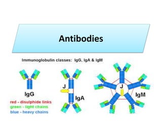
- 25. VI. CLASSES, SUBCLASSES, TYPES AND SUBTYPES A. Immunoglobulin classes • The immunoglobulins can be divided into five different classes, based on differences in the amino acid sequences in the constant region of the heavy chains. • All immunoglobulins within a given class will have very similar heavy chain constant regions. • These differences can be detected by sequence studies or more commonly by serological means (i.e. by the use of antibodies directed to these differences).
- 26. 1. IgG - Gamma heavy chains 2. IgM - Mu heavy chains 3. IgA - Alpha heavy chains 4. IgD - Delta heavy chains 5. IgE - Epsilon heavy chains CLASSES
- 27. B. Immunoglobulin Subclasses • The classes of immunoglobulins can be divided into subclasses based on small differences in the amino acid sequences in the constant region of the heavy chains. All immunoglobulins within a subclass will have very similar heavy chain constant region amino acid sequences. Again these differences are most commonly detected by serological means.
- 28. SUBCLASSES 1. IgG Subclasses • a) IgG1 - Gamma 1 heavy chains b) IgG2 - Gamma 2 heavy chains c) IgG3 - Gamma 3 heavy chains d) IgG4 - Gamma 4 heavy chains 2. IgA Subclasses • a) IgA1 - Alpha 1 heavy chains b) IgA2 - Alpha 2 heavy chains
- 29. C. IMMUNOGLOBULIN TYPES • Immunoglobulins can also be classified by the type of light chain that they have. • Light chain types are based on differences in the amino acid sequence in the constant region of the light chain. • These differences are detected by serological means. 1. Kappa light chains 2. Lambda light chains
- 30. D. IMMUNOGLOBULIN SUBTYPES • The light chains can also be divided into subtypes based on differences in the amino acid sequences in the constant region of the light chain. 1. Lambda subtypes a) Lambda 1 b) Lambda 2 c) Lambda 3 d) Lambda 4
- 31. E. Nomenclature • Immunoglobulins are named based on the class, or subclass of the heavy chain and type or subtype of light chain. • Unless it is stated precisely, you should assume that all subclass, types and subtypes are present. • IgG means that all subclasses and types are present.
- 32. F. Heterogeneity • Immunoglobulins considered as a population of molecules are normally very heterogeneous because they are composed of different classes and subclasses each of which has different types and subtypes of light chains. • In addition, different immunoglobulin molecules can have different antigen binding properties because of different VH and VL regions.
- 34. Properties and biological activities of Immunoglobulins Ig G Ig A Ig M Ig D Ig E 1. Structure Monomer Monomer in serum Dimer in secretion Pentamer Monomer Monomer 2. Heavy chain CH domain Gamma Three Alfa Three Mu Four Delta Three Epsilon Four 3. Mol. Wt. 1,50,000 1,60,000 9,00,000 1,80,000 1,90,000 4. Serum concentration (mg/ml) 12 2 1.2 0.03 0.00004 5. Present on membrane of mature B cell _ _ + + _ 5. Intravascular Distribution (%) 45 42 80 75 50 6. Crosses placenta + - - - - 7. Present in milk + + - - - 8. Selective secretion by seromucous glands - + - - - 9. Activation of complement Classical Alternate + - - + + - - - - - 10 Binds to FC receptor of phagocytes + - - - - 11 Induces mast cell degranulation - - - - +
- 35. VII. STRUCTURE AND SOME PROPERTIES OF IG CLASSES AND SUBCLASSES A. IgG • 1. Structure All IgG's are monomers (7S immunoglobulin). The subclasses differ in the number of disulfide bonds and length of the hinge region.
- 36. 2. Properties a) IgG is the major Ig in serum - 75% of serum Ig is IgG. b) IgG is the major Ig in extra vascular spaces c) Placental transfer - IgG is the only class of Ig that crosses the placenta. Transfer is mediated by a receptor on placental cells for the Fc region of IgG. Not all subclasses cross equally well; IgG2 does not cross well.
- 37. d) Fixes complement - Not all subclasses fix equally well; IgG4 does not fix complement e) Binding to cells - Macrophages, monocytes, PMNs and some lymphocytes have Fc receptors for the Fc region of IgG. Not all subclasses bind equally well; IgG2 and IgG4 do not bind to Fc receptors. A consequence of binding to the Fc receptors on PMNs, monocytes and macrophages is that the cell can now internalize the antigen better.
- 38. • The antibody has prepared the antigen for eating by the phagocytic cells. • The term opsonin is used to describe substances that enhance phagocytosis. IgG is a good opsonin. • Binding of IgG to Fc receptors on other types of cells results in the activation of other functions.
- 39. B. IgM • 1. Structure IgM normally exists as a pentamer (19S immunoglobulin) but it can also exist as a monomer. • In the pentameric form all heavy chains are identical and all light chains are identical. Thus, the valence is theoretically 10. • IgM has an extra domain on the mu chain (CH4) and it has another protein covalently bound via a S-S bond called the J chain. This chain functions in polymerization of the molecule into a pentamer.
- 41. 2. Properties • a) IgM is the third most common serum Ig. • b) IgM is the first Ig to be made by the fetus and the first Ig to be made by a virgin B cells when it is stimulated by antigen. • c) As a consequence of its pentameric structure, IgM is a good complement fixing Ig. Thus, IgM antibodies are very efficient in leading to the lysis of microorganisms.
- 42. d) As a consequence of its structure, IgM is also a good agglutinating Ig . Thus, IgM antibodies are very good in clumping microorganisms for eventual elimination from the body. e) IgM binds to some cells via Fc receptors.
- 43. C. IgA • 1. Structure Serum IgA is a monomer but IgA found in secretions is a dimer. • When IgA exits as a dimer, a J chain is associated with it. When IgA is found in secretions is also has another protein associated with it called the secretory piece or T piece; • sIgA is sometimes referred to as 11S immunoglobulin.
- 45. • Unlike the remainder of the IgA which is made in the plasma cell, the secretory piece is made in epithelial cells and is added to the IgA as it passes into the secretions. • The secretory piece helps IgA to be transported across mucosa and also protects it from degradation in the secretions.
- 46. 2. Properties • a) IgA is the 2nd most common serum Ig. b) IgA is the major class of Ig in secretions - tears, saliva, colostrum, mucus. Since it is found in secretions secretory IgA is important in local (mucosal) immunity. c) Normally IgA does not fix complement, unless aggregated. d) IgA can bind to some cells - PMN's and some lymphocytes.
- 47. D. IgD 1. Structure . IgD exists only as a monomer. 2. Properties a) IgD is found in low levels in serum; its role in serum uncertain.
- 48. b) IgD is primarily found on B cell surfaces where it functions as a receptor for antigen. IgD on the surface of B cells has extra amino acids at C-terminal end for anchoring to the membrane. It also associates with the Ig-alpha and Ig-beta chains. • c) IgD does not bind complement.
- 49. E. IgE • 1. Structure IgE exists as a monomer and has an extra domain in the constant region. • 2. Properties • a) IgE is the least common serum Ig since it binds very tightly to Fc receptors on basophils and mast cells even before interacting with antigen.
- 50. • b) Involved in allergic reactions - As a consequence of its binding to basophils an mast cells, IgE is involved in allergic reactions. Binding of the allergen to the IgE on the cells results in the release of various pharmacological mediators that result in allergic symptoms.
- 51. Take home message Role of different immunoglobulin classes IgG: Protects the body fluids. IgA: Protects the body surfaces. IgM: Protects the blood stream. IgE: Mediates type I hypersensitivity. IgD: Role not known.
- 52. SUMMARY • DEFINITION • STRUCTURE • CLASSIFICATION