Downloaded 1,805 times
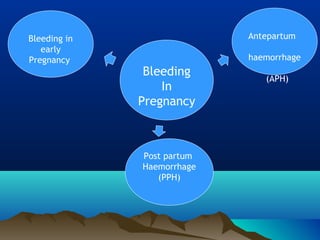


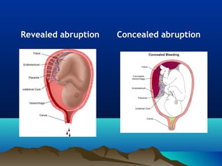
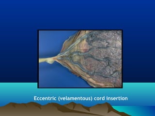

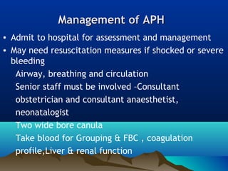
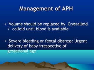


Antepartum haemorrhage (APH) refers to bleeding from the vagina during pregnancy after 24 weeks of gestation until delivery. The main causes of APH are placental abnormalities like placenta previa or abruption, as well as genital tract infections or trauma. Management involves resuscitation, monitoring for fetal distress, and delivery depending on the severity of bleeding and gestational age. Outcomes can be poor if APH is severe or left untreated, with risks of maternal and fetal death.