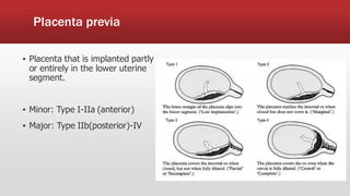
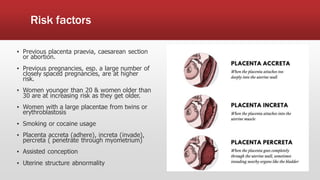
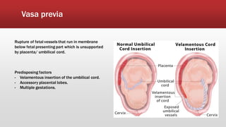
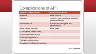
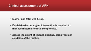
ANTEPARTUM
HEMORRHAGE
Definition
Bleeding from the genital tract in pregnancy before the onset of
labour at gestations of 22 weeks to delivery of the fetus.
Epidemiology
-Affects 3-3% of all pregnancies.
-3 times more common in multiparous than primiparous women.
Importance
▪ Obstetric emergency
▪ Attention should be sought immediately
▪ If left untreated can lead to death of the mother and/or foetus.
▪ Can lead to DVT
▪ Management reduce the risk of premature delivery and
maternal/perinatal morbidity/mortality.
Causes
▪ Placenta causes:
-Placenta Praevia
-Placenta Abruption
-Vasa Praevia
▪ Local causes:
-cervical polyps
-cervicitis
-vaginitis
-cervical cancer
▪ Indeterminate APH
Placenta previa
▪ Placenta that is implanted partly
or entirely in the lower uterine
segment.
▪ Minor: Type I-IIa (anterior)
▪ Major: Type IIb(posterior)-IV
Risk factors
▪ Previous placenta praevia, caesarean section
or abortion.
▪ Previous pregnancies, esp. a large number of
closely spaced pregnancies, are at higher
risk.
▪ Women younger than 20 & women older than
30 are at increasing risk as they get older.
▪ Women with a large placentae from twins or
erythroblastosis
▪ Smoking or cocaine usage
▪ Placenta accreta (adhere), increta (invade),
percreta ( penetrate through myometrium)
▪ Assisted conception
▪ Uterine structure abnormality
Clinical features
• Recurrent painless vaginal bleeding.
• Abdominal findings
- Uterus soft, relaxed and non tender
- Contraction may be palpated
- Presenting part is usually high
- Abnormal presentation
• Maternal cardiovascular compromise
• Fetal condition satisfactory until severe maternal compromise
• Vaginal examination- SHOULD NOT BE DONE.
Abruptio placenta
Seperation of normally located placenta after 22 weeks of gestation (>500g) and prior to delivery of fetus.
Risk factors
• Pre-eclampsia
• Abdominal trauma
• Abruptio in previous pregnancy (10 fold
increased risk)
• Multiparity- multiple gestation (over
distention of uterus)
• Cord traction
• Smoking
• Sudden decompression of the uterus
• Maternal Substance Abuse (Cocaine, alcohol)
• Maternal Tobacco abuse (2 fold increased
risk)
• Polyhydramnios
placenta praevia placenta abruptio
Pain painless painful
Uterus Soft, non tender
Tense, tender, irritable,
hard ly palpate fetal parts
Fetal position
Not engagement,
malpresentation
Normal, head maybe
engaged
Fetal heart Usually normal Absent or abnormal
A/w pre-eclampsia No Yes
Haemodynamic
signs
Proportional
Signs of hypovolaemic shock
with increase pulse rate,
hypotension, and peripheral
vasoconstriction.
Differencesbetweenplacentapraevia and placentaabruptio
Vasa previa
Rupture of fetal vessels that run in membrane
below fetal presenting part which is unsupported
by placenta/ umbilical cord.
Predisposing factors
- Velamentous insertion of the umbilical cord.
- Accessory placental lobes.
- Multiple gestations.
Velamentous insertion means that the cord is inserted on the amniotic membrane rather
than on the placenta with blood vessels stretching along the membrane between the
insertion point and the placenta.
•Occur when the fetal vessels run in the membranes below the presenting fetal part, unsupported
by placental tissue or umbilical cord at the cervical opening
•Spontaneous or artificial rupture of membranes often leads rupture of these vessels with likely
resultant fetal exsanguination(reported fetal mortality 33-100%).
•Must be suspected when APH occurs in a woman especially if,bleed is a bright red trickle .
•fetal heart shows sudden tachycardia or sudden deceleration (even persistent bradycardia!) and
the fetal distress appears disproportionate to the relatively ‘little’ bleed
•Occurs just after ARM
•Antenatal diagnosis can be made using transvaginal sonography in combination with color Doppler.
Clinical assessment of APH
• Mother and fetal well being.
• Establish whether urgent intervention is required to
manage maternal or fetal compromise.
• Assess the extent of vaginal bleeding, cardiovascular
condition of the mother.
Full history
Should be taken afterthe motheris stable.
• Associated with pain with the hemorrhage?
- Continuouspain: Placentalabruption
- Intermittent pain: Labour
• Risk factorsfor abruptionand placentapraeviashould be identified.
• Reduced fetal movement?
• If the APH is associated with spontaneousor iatrogenic ruptureof the fetal
membranes: rupturedvasa previa.
• Previous cervical smear history possibility of cervicalcancer. Symptomatic
pregnant women usually present with APH ( mostly postcoital)or vaginal
discharge.
Examination
• General: Pulse and BP
• Abdomen:
- Tense , tenderor woody feel to the uterusindicatesa significant abruption.
- Painlessbleeding, high fetal presentingpart- Placenta previa.
- Soft, non-tenderuterusmay suggest a lower genital tract cause or bleeding
form placentaor vasa previa.
• Speculum:
- identifycervical dilatation or visualisea lower genital tract cause.
• Digital vaginalexamination
- Should not be done until placenta praeviahas been excludedby USG.
Investigations
• FBC
• Coag
• GSH/GXM
• D-dimer: Abruptio placenta
• Ultrasound- TRP Placenta previa/IUD
• Colour doppler TVS - Vasa previa
• Fetal monitoring- CTG
Management
• Pad chart
• Minimise abdominal examination
• Appropriate investigations are done – FBC & GXM/GSH ( 2units)
• Monitor fetal well being
– Fetal kick chart(daily)
– CTG (weekly)
– U/S
• Steroid injection(> 24w, <36w)- IM dexamethasone12mg stat and
repeat the second dose after 12 hours.
- Placenta Praevia – mustdeliver by 38 weeks. If baby is dead, do not
perform Caesarean section, instead induce & augment labour
-Placenta abruptio- must deliver as soon as possible (within 2 hour)
Thank you

APH.pdf

  • 1.
  • 2.
    Definition Bleeding from thegenital tract in pregnancy before the onset of labour at gestations of 22 weeks to delivery of the fetus. Epidemiology -Affects 3-3% of all pregnancies. -3 times more common in multiparous than primiparous women.
  • 3.
    Importance ▪ Obstetric emergency ▪Attention should be sought immediately ▪ If left untreated can lead to death of the mother and/or foetus. ▪ Can lead to DVT ▪ Management reduce the risk of premature delivery and maternal/perinatal morbidity/mortality.
  • 4.
    Causes ▪ Placenta causes: -PlacentaPraevia -Placenta Abruption -Vasa Praevia ▪ Local causes: -cervical polyps -cervicitis -vaginitis -cervical cancer ▪ Indeterminate APH
  • 5.
    Placenta previa ▪ Placentathat is implanted partly or entirely in the lower uterine segment. ▪ Minor: Type I-IIa (anterior) ▪ Major: Type IIb(posterior)-IV
  • 6.
    Risk factors ▪ Previousplacenta praevia, caesarean section or abortion. ▪ Previous pregnancies, esp. a large number of closely spaced pregnancies, are at higher risk. ▪ Women younger than 20 & women older than 30 are at increasing risk as they get older. ▪ Women with a large placentae from twins or erythroblastosis ▪ Smoking or cocaine usage ▪ Placenta accreta (adhere), increta (invade), percreta ( penetrate through myometrium) ▪ Assisted conception ▪ Uterine structure abnormality
  • 7.
    Clinical features • Recurrentpainless vaginal bleeding. • Abdominal findings - Uterus soft, relaxed and non tender - Contraction may be palpated - Presenting part is usually high - Abnormal presentation • Maternal cardiovascular compromise • Fetal condition satisfactory until severe maternal compromise • Vaginal examination- SHOULD NOT BE DONE.
  • 8.
    Abruptio placenta Seperation ofnormally located placenta after 22 weeks of gestation (>500g) and prior to delivery of fetus.
  • 9.
    Risk factors • Pre-eclampsia •Abdominal trauma • Abruptio in previous pregnancy (10 fold increased risk) • Multiparity- multiple gestation (over distention of uterus) • Cord traction • Smoking • Sudden decompression of the uterus • Maternal Substance Abuse (Cocaine, alcohol) • Maternal Tobacco abuse (2 fold increased risk) • Polyhydramnios
  • 10.
    placenta praevia placentaabruptio Pain painless painful Uterus Soft, non tender Tense, tender, irritable, hard ly palpate fetal parts Fetal position Not engagement, malpresentation Normal, head maybe engaged Fetal heart Usually normal Absent or abnormal A/w pre-eclampsia No Yes Haemodynamic signs Proportional Signs of hypovolaemic shock with increase pulse rate, hypotension, and peripheral vasoconstriction. Differencesbetweenplacentapraevia and placentaabruptio
  • 11.
    Vasa previa Rupture offetal vessels that run in membrane below fetal presenting part which is unsupported by placenta/ umbilical cord. Predisposing factors - Velamentous insertion of the umbilical cord. - Accessory placental lobes. - Multiple gestations.
  • 12.
    Velamentous insertion meansthat the cord is inserted on the amniotic membrane rather than on the placenta with blood vessels stretching along the membrane between the insertion point and the placenta. •Occur when the fetal vessels run in the membranes below the presenting fetal part, unsupported by placental tissue or umbilical cord at the cervical opening •Spontaneous or artificial rupture of membranes often leads rupture of these vessels with likely resultant fetal exsanguination(reported fetal mortality 33-100%). •Must be suspected when APH occurs in a woman especially if,bleed is a bright red trickle . •fetal heart shows sudden tachycardia or sudden deceleration (even persistent bradycardia!) and the fetal distress appears disproportionate to the relatively ‘little’ bleed •Occurs just after ARM •Antenatal diagnosis can be made using transvaginal sonography in combination with color Doppler.
  • 14.
    Clinical assessment ofAPH • Mother and fetal well being. • Establish whether urgent intervention is required to manage maternal or fetal compromise. • Assess the extent of vaginal bleeding, cardiovascular condition of the mother.
  • 15.
    Full history Should betaken afterthe motheris stable. • Associated with pain with the hemorrhage? - Continuouspain: Placentalabruption - Intermittent pain: Labour • Risk factorsfor abruptionand placentapraeviashould be identified. • Reduced fetal movement? • If the APH is associated with spontaneousor iatrogenic ruptureof the fetal membranes: rupturedvasa previa. • Previous cervical smear history possibility of cervicalcancer. Symptomatic pregnant women usually present with APH ( mostly postcoital)or vaginal discharge.
  • 16.
    Examination • General: Pulseand BP • Abdomen: - Tense , tenderor woody feel to the uterusindicatesa significant abruption. - Painlessbleeding, high fetal presentingpart- Placenta previa. - Soft, non-tenderuterusmay suggest a lower genital tract cause or bleeding form placentaor vasa previa. • Speculum: - identifycervical dilatation or visualisea lower genital tract cause. • Digital vaginalexamination - Should not be done until placenta praeviahas been excludedby USG.
  • 17.
    Investigations • FBC • Coag •GSH/GXM • D-dimer: Abruptio placenta • Ultrasound- TRP Placenta previa/IUD • Colour doppler TVS - Vasa previa • Fetal monitoring- CTG
  • 18.
    Management • Pad chart •Minimise abdominal examination • Appropriate investigations are done – FBC & GXM/GSH ( 2units) • Monitor fetal well being – Fetal kick chart(daily) – CTG (weekly) – U/S • Steroid injection(> 24w, <36w)- IM dexamethasone12mg stat and repeat the second dose after 12 hours. - Placenta Praevia – mustdeliver by 38 weeks. If baby is dead, do not perform Caesarean section, instead induce & augment labour -Placenta abruptio- must deliver as soon as possible (within 2 hour)
  • 19.