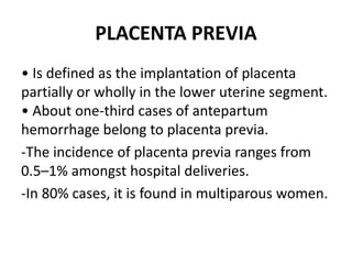
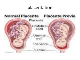
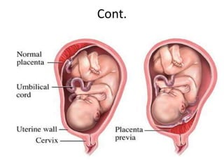
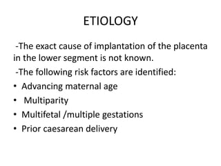
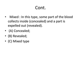
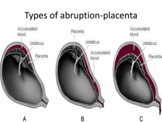
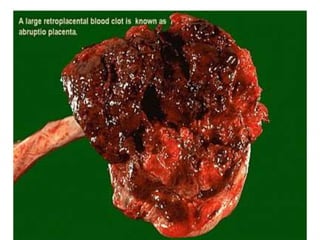
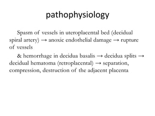
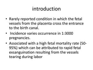
Antepartum heamorrhage
Dr Bilkisu Isa
25th -04-2022
Introduction/definition
• Death from hemorrhage still remains a leading
cause of maternal mortality.
• APH is defined as bleeding from the genital tract
in pregnancy from the age of viability(24 week’s
gestation –WHO but 28wks in Nigeria) and the
onset of labour.
• It affects 4% of all pregnancies. It is a medical
emergency.
• It is associated with increased risks of fetal and
maternal morbidity and mortality.
causes
• Placenta preavia
• Abruptio placenta
• Vasa preavia
• Excessive show
• Local causes ( bleeding from cervix, vagina
and vulva )
• Inderterminate APH
PLACENTA PREVIA
• Is defined as the implantation of placenta
partially or wholly in the lower uterine segment.
• About one-third cases of antepartum
hemorrhage belong to placenta previa.
-The incidence of placenta previa ranges from
0.5–1% amongst hospital deliveries.
-In 80% cases, it is found in multiparous women.
placentation
Cont.
Degrees of placenta previa with
findings on ultrasound examination
Cont.
ETIOLOGY
-The exact cause of implantation of the placenta
in the lower segment is not known.
-The following risk factors are identified:
• Advancing maternal age
• Multiparity
• Multifetal /multiple gestations
• Prior caesarean delivery
Cont.
• Smoking
• Prior placenta previa
• Uterine structural anomaly
• Assisted conception
TYPES OR DEGREES
• Type—I (Low-lying): the placental edge is in
the lower uterine segment but does not reach
the internal os
• Type—II (Marginal): The placenta reaches the
margin of the internal os but does not cover it.
Divided into anterior and posterior.
Cont.
• Type—III (Incomplete or partial central): The
placenta covers the internal os partially
(covers the internal os when closed but does
not entirely do so when fully dilated).
• Type—IV (Central or total): The placenta
completely covers the internal os even after it
is fully dilated.
CAUSE OF BLEEDING
• Bleeding results from small disruptions in the
placental attachment during normal development
and thinning of the lower uterine segment
• As the placental growth slows down in later
months and the lower segment progressively
dilates, the inelastic placenta is sheared off the
wall of the lower segment.
• This leads to opening up of uteroplacental vessels
and leads to an episode of bleeding.
CLINICAL FEATURES
• Bleeding: usually mild but it could be severe;
recurrent, painless and causeless.
• Soft and non-tender uterus
• Normal fetal heart rate (unless there is severe
bleeding or associated abruption).
• High presenting part.
Cont.
• Fetal malpresentation
(breech/transverse/oblique).
• General condition and anemia are
proportionate to the visible blood loss
Abdominal examination
• The size of the uterus is proportionate to the
period of gestation
• The uterus feels relaxed, soft and elastic
without any localized area of tenderness.
• Persistence of malpresentation. There is also
increased frequency of twin pregnancy.
• The head is floating in contrast to the period
of gestation. The head cannot be pushed
down into the pelvis.
Cont.
• Fetal heart sound is usually present.
• Vulval inspection: the blood is bright red as
the bleeding occurs from the separated utero-
placental sinuses close to the cervical opening
and escapes out immediately
• Vaginal examination is contraindicated
DIAGNOSIS
• Painless and recurrent vaginal bleeding in the
second half of pregnancy should be taken as
placenta previa unless proved otherwise.
• Ultrasonography is the initial procedure either
to confirm or to rule out the diagnosis
• Localization of placenta
Cont.
• Sonography –– Transabdominal ultrasound
(TAS) –– Transvaginal ultrasound (TVS) ––
Transperineal ultrasound –– Color Doppler
flow study
• Magnetic resonance imaging (MRI)
• Clinical –– By internal examination (double set
up examination) –– Direct visualization during
caesarean section –– Examination of the
placenta following vaginal delivery
MANAGEMENT
• PREVENTION:
• — Adequate antenatal care
• — Antenatal diagnosis at 20th week
• — Significance of “warning hemorrhage”
should not be ignored
TREATMENT ON ADMISSION
• IMMEDIATE ATTENTION: Overall assessment
of the case is quickly made as regards :
- Amount of the blood loss — by noting the
general condition, pallor, pulse rate and blood
pressure;
- Blood samples are taken for group, cross
matching and estimation of hemoglobin;
Cont.
• A large-bore IV cannula is sited and an
infusion of normal saline is started and
compatible cross matched blood transfusion
should be arranged;
• Gentle abdominal palpation to ascertain any
uterine tenderness and auscultation to note
the fetal heart rate
• Inspection of the vulva to note the presence
of any active bleeding
FORMULATION OF THE LINE OF
TREATMENT
The definitive treatment depends:
• upon the duration of pregnancy,
• fetal and maternal status
• and extent of the hemorrhage
Expectant management
• The aim is to continue pregnancy for fetal
maturity without compromising the maternal
health. Conduct of expectant treatment:
• Strict Bed rest;
• Investigations—like hemoglobin estimation,
blood grouping and urine for protein are done;
• Periodic inspection of the vulval pads and fetal
surveillance with USG at interval of 2–3 weeks;
Cont.
• Supplementary hematinics should be given
and the blood loss is replaced by adequate
cross matched blood transfusion, if the patient
is anemic;
• Steroid for lung maturation if gestational age
is less than 34 weeks
Cont.
• Use of tocolysis (magnesium sulfate) can be
done if vaginal bleeding is associated with
uterine contractions;
• Rh immunoglobin should be given to all Rh
negative (unsensitized) women.
Active Management
Delivery :
• 1. Bleeding occurs at or after 37 weeks of
pregnancy
• 2. Patient is in labor
• 3. Fetal distress
• 4. Torrential Bleeding
• 5. Congenital anomaly not compatible with
life
Cont.
• 6. Intrauterine fetal death
• Cesarean delivery is done for all women with
sonographic evidence of placenta previa
where placental edge is within 2 cm from the
internal os.
• It is especially indicated if it is posterior or
thick
Clinical classification
Minor : Deliver vaginally
• Type 1 (anterior/posterior)
• Type 2 anterior
Major: Caesarean section
• Type 2 posterior
• Type 3
• Type 4
Complications
• MATERNAL • During pregnancy: Antepartum
haemorrhage, Malpresentation, Preterm
labour.
• During labour: PROM, Cord prolapse,
Intrapartum hemorrhage, Increased incidence
of operative interference, Postpartum
hemorrhage, Retained placenta, Shock.
• Puerperium: Sepsis, Subinvolution, Embolism
FETAL
• Low birth weight,
• Asphyxia, Intrauterine fetal death,
• Birth injuries.
ABRUPTIO PLACENTAE
definition
• It is one form of antepartum hemorrhage
where the bleeding occurs due to premature
separation of normally situated placenta after
the age of viability.
• Occurs in 1-2% of all pregnancies
• Perinatal mortality rate associated with
placental abruption was 119 per 1000 births
compared with 8.2 per 1000 for all others.
Types of abruption placenta
• Revealed : Following separation of the
placenta, the blood comes out of the cervical
canal to be visible externally.
• Concealed : The blood collects behind the
separated placenta or collected in between
the membranes and decidua
Cont.
• Mixed : In this type, some part of the blood
collects inside (concealed) and a part is
expelled out (revealed).
• (A) Concealed;
• (B) Revealed;
• (C) Mixed type
Types of abruption-placenta
pathophysiology
--- Spasm of vessels in uteroplacental bed (decidual
spiral artery) → anoxic endothelial damage → rupture
of vessels
& hemorrhage in decidua basalis → decidua splits →
decidual hematoma (retroplacental) → separation,
compression, destruction of the adjacent placenta
adjacent placenta
CLINICAL CLASSIFICATION
• Grade—0: Clinical features may be absent.
The diagnosis is made after inspection of
placenta following delivery.
• Grade—1 (40%): (i) Vaginal bleeding is slight
(ii) Uterus: irritable, tenderness may be
minimal or absent (iii) Maternal BP and
fibrinogen levels unaffected (iv) FHS is good.
Cont.
• Grade—2 (45%): (i) Vaginal bleeding mild to
moderate (ii) Uterine tenderness is always
present (iii) Maternal pulse ↑, BP is
maintained (iv) Fibrinogen level may be
decreased (v) Shock is absent (vi) Fetal distress
or even fetal death occurs.
Cont.
• Grade—3 (15%): (i) Bleeding is moderate to
severe or may be concealed (ii) Uterine
tenderness is marked (iii) Shock is pronounced
(iv) Fetal death is the rule (v) Associated
coagulation defect or anuria may complicate
Risk factors
• The primary cause of placental abruption is
unknown, but there are several associated
conditions.
• Increased age and parity
• Preeclampsia, Chronic hypertension
• Preterm ruptured membranes
Cont.
• Multifetal gestation, Hydramnios
• Cigarette smoking, Folic acid deficiency
• Thrombophilias
• Cocaine use
• Prior abruption
• Uterine leiomyoma
• External trauma
Clinical Presentation
• Bleeding: revealed/concealed, so clinical
picture is important.
• Pain on the uterus and this increases in
severity.
• Signs of shock (hypovolaemia): fainting and
collapse.
• Woody hard tender uterus ( uterine tetany)
• Couvelaire uterus (Bluish uterus).
Cont.
• Difficult to palpate the fetal parts and to hear
the fetal heart.
• Normal fetal lie and presentation
• Ultrasonography: is done to confirm fetal
viability, assess fetal growth & normality,
measure liquor
MANAGEMENT
• Treatment for placental abruption varies
depending on gestational age and the status
of the mother and fetus.
• Admit patient, take History & examination
• Assess blood loss, Nearly always more than
revealed
• IV access, X match, DIC screen, Assess fetal
well-being
• Placental localization
Principle of management
• Early delivery (50% of abruption present in
labour).
• Adequate blood transfusion.
• Adequate analgesia.
• Detailed maternal and fetal monitoring.
• Coagulation profile (30% develop DIC).
Cont.
indication for caesarean section:
• Fetal distess, severe bleeding,
• alive baby and not in advanced labour.
• Perinatal mortality rate is 15-20%.
Vaginal delivery:
• very low gestation
• dead baby
Cont.
Conservative management
• small abruption,
• well mother and fetus,
• if the gestational age < 34, give steroids
COMPLICATIONS
MATERNAL
• Shock, Blood coagulation disorders,
• Oliguria and anuria, Postpartum
haemorrhage,
• Puerperal sepsis,
• Acute renal failure: acute tubular or cortical
necrosis.
Fetal complications
• Intrauterine growth restriction
• Anaemia
• Premature delivery
• Fetal distress and death
VASA PRAEVIA
introduction
• Rarely reported condition in which the fetal
vessels from the placenta cross the entrance
to the birth canal.
• Incidence varies occurrence in 1:3000
pregnancies.
• Associated with a high fetal mortality rate (50-
95%) which can be attributed to rapid fetal
exsanguination resulting from the vessels
tearing during labor
vasapreavia
Causes
• There are three causes typically noted for vasa
praevia:
• Bi-lobed placenta
• Velamentous insertion of the umbilical cord
• Succenturiate (Accessory) lobe
Risk factors
• Bilobed and succenturiate placentas
• Velamentous insertion of the cord
• Low-lying placenta
• Multiple gestation
• Pregnancies resulting from in vitro fertilization
• Palpable vessel on vaginal exam
Symptoms
• Usually asymptomatic
• Sudden onset of painless bleeding in second
or third trimester or at rupture of membranes
• No sign or symptom of placenta praevia or
abruption
• IUGR , Congenital malformation
• Abnormal fetal heart rate.
MANAGEMENT
• Detection of nucleated red blood cells
(Singer’s alkali denaturation test) or fetal
hemoglobin is diagnostic.
• Management depends on fetal gestational
age, severity, persistence or recurrence of
bleeding, and the presumed cause of bleeding
Cont.
• Pregnancy > 37 weeks and bleeding recurrent
— delivery is recommended.
• The mode of delivery depends on the state of
the fetus,
• and other associated factors (cervix).
Cont,
• Expectant management can be done in
selected cases for fetal maturity similar to
placenta previa.
• Fetal monitoring must be carefully done.
Intrapartum diagnosis of vasa previa, needs
expeditious delivery.
• Neonatal blood transfusion may be needed.
THANK YOU
THANK YOU

Antepartum heamorrhage.pptx

  • 1.
  • 2.
    Introduction/definition • Death fromhemorrhage still remains a leading cause of maternal mortality. • APH is defined as bleeding from the genital tract in pregnancy from the age of viability(24 week’s gestation –WHO but 28wks in Nigeria) and the onset of labour. • It affects 4% of all pregnancies. It is a medical emergency. • It is associated with increased risks of fetal and maternal morbidity and mortality.
  • 3.
    causes • Placenta preavia •Abruptio placenta • Vasa preavia • Excessive show • Local causes ( bleeding from cervix, vagina and vulva ) • Inderterminate APH
  • 4.
    PLACENTA PREVIA • Isdefined as the implantation of placenta partially or wholly in the lower uterine segment. • About one-third cases of antepartum hemorrhage belong to placenta previa. -The incidence of placenta previa ranges from 0.5–1% amongst hospital deliveries. -In 80% cases, it is found in multiparous women.
  • 5.
  • 6.
  • 7.
    Degrees of placentaprevia with findings on ultrasound examination
  • 8.
  • 9.
    ETIOLOGY -The exact causeof implantation of the placenta in the lower segment is not known. -The following risk factors are identified: • Advancing maternal age • Multiparity • Multifetal /multiple gestations • Prior caesarean delivery
  • 10.
    Cont. • Smoking • Priorplacenta previa • Uterine structural anomaly • Assisted conception
  • 11.
    TYPES OR DEGREES •Type—I (Low-lying): the placental edge is in the lower uterine segment but does not reach the internal os • Type—II (Marginal): The placenta reaches the margin of the internal os but does not cover it. Divided into anterior and posterior.
  • 12.
    Cont. • Type—III (Incompleteor partial central): The placenta covers the internal os partially (covers the internal os when closed but does not entirely do so when fully dilated). • Type—IV (Central or total): The placenta completely covers the internal os even after it is fully dilated.
  • 13.
    CAUSE OF BLEEDING •Bleeding results from small disruptions in the placental attachment during normal development and thinning of the lower uterine segment • As the placental growth slows down in later months and the lower segment progressively dilates, the inelastic placenta is sheared off the wall of the lower segment. • This leads to opening up of uteroplacental vessels and leads to an episode of bleeding.
  • 14.
    CLINICAL FEATURES • Bleeding:usually mild but it could be severe; recurrent, painless and causeless. • Soft and non-tender uterus • Normal fetal heart rate (unless there is severe bleeding or associated abruption). • High presenting part.
  • 15.
    Cont. • Fetal malpresentation (breech/transverse/oblique). •General condition and anemia are proportionate to the visible blood loss
  • 16.
    Abdominal examination • Thesize of the uterus is proportionate to the period of gestation • The uterus feels relaxed, soft and elastic without any localized area of tenderness. • Persistence of malpresentation. There is also increased frequency of twin pregnancy. • The head is floating in contrast to the period of gestation. The head cannot be pushed down into the pelvis.
  • 17.
    Cont. • Fetal heartsound is usually present. • Vulval inspection: the blood is bright red as the bleeding occurs from the separated utero- placental sinuses close to the cervical opening and escapes out immediately • Vaginal examination is contraindicated
  • 18.
    DIAGNOSIS • Painless andrecurrent vaginal bleeding in the second half of pregnancy should be taken as placenta previa unless proved otherwise. • Ultrasonography is the initial procedure either to confirm or to rule out the diagnosis • Localization of placenta
  • 19.
    Cont. • Sonography ––Transabdominal ultrasound (TAS) –– Transvaginal ultrasound (TVS) –– Transperineal ultrasound –– Color Doppler flow study • Magnetic resonance imaging (MRI) • Clinical –– By internal examination (double set up examination) –– Direct visualization during caesarean section –– Examination of the placenta following vaginal delivery
  • 20.
    MANAGEMENT • PREVENTION: • —Adequate antenatal care • — Antenatal diagnosis at 20th week • — Significance of “warning hemorrhage” should not be ignored
  • 21.
    TREATMENT ON ADMISSION •IMMEDIATE ATTENTION: Overall assessment of the case is quickly made as regards : - Amount of the blood loss — by noting the general condition, pallor, pulse rate and blood pressure; - Blood samples are taken for group, cross matching and estimation of hemoglobin;
  • 22.
    Cont. • A large-boreIV cannula is sited and an infusion of normal saline is started and compatible cross matched blood transfusion should be arranged; • Gentle abdominal palpation to ascertain any uterine tenderness and auscultation to note the fetal heart rate • Inspection of the vulva to note the presence of any active bleeding
  • 23.
    FORMULATION OF THELINE OF TREATMENT The definitive treatment depends: • upon the duration of pregnancy, • fetal and maternal status • and extent of the hemorrhage
  • 24.
    Expectant management • Theaim is to continue pregnancy for fetal maturity without compromising the maternal health. Conduct of expectant treatment: • Strict Bed rest; • Investigations—like hemoglobin estimation, blood grouping and urine for protein are done; • Periodic inspection of the vulval pads and fetal surveillance with USG at interval of 2–3 weeks;
  • 25.
    Cont. • Supplementary hematinicsshould be given and the blood loss is replaced by adequate cross matched blood transfusion, if the patient is anemic; • Steroid for lung maturation if gestational age is less than 34 weeks
  • 26.
    Cont. • Use oftocolysis (magnesium sulfate) can be done if vaginal bleeding is associated with uterine contractions; • Rh immunoglobin should be given to all Rh negative (unsensitized) women.
  • 27.
    Active Management Delivery : •1. Bleeding occurs at or after 37 weeks of pregnancy • 2. Patient is in labor • 3. Fetal distress • 4. Torrential Bleeding • 5. Congenital anomaly not compatible with life
  • 28.
    Cont. • 6. Intrauterinefetal death • Cesarean delivery is done for all women with sonographic evidence of placenta previa where placental edge is within 2 cm from the internal os. • It is especially indicated if it is posterior or thick
  • 29.
    Clinical classification Minor :Deliver vaginally • Type 1 (anterior/posterior) • Type 2 anterior Major: Caesarean section • Type 2 posterior • Type 3 • Type 4
  • 30.
    Complications • MATERNAL •During pregnancy: Antepartum haemorrhage, Malpresentation, Preterm labour. • During labour: PROM, Cord prolapse, Intrapartum hemorrhage, Increased incidence of operative interference, Postpartum hemorrhage, Retained placenta, Shock. • Puerperium: Sepsis, Subinvolution, Embolism
  • 31.
    FETAL • Low birthweight, • Asphyxia, Intrauterine fetal death, • Birth injuries.
  • 32.
  • 33.
    definition • It isone form of antepartum hemorrhage where the bleeding occurs due to premature separation of normally situated placenta after the age of viability. • Occurs in 1-2% of all pregnancies • Perinatal mortality rate associated with placental abruption was 119 per 1000 births compared with 8.2 per 1000 for all others.
  • 34.
    Types of abruptionplacenta • Revealed : Following separation of the placenta, the blood comes out of the cervical canal to be visible externally. • Concealed : The blood collects behind the separated placenta or collected in between the membranes and decidua
  • 35.
    Cont. • Mixed :In this type, some part of the blood collects inside (concealed) and a part is expelled out (revealed). • (A) Concealed; • (B) Revealed; • (C) Mixed type
  • 36.
  • 38.
    pathophysiology --- Spasm ofvessels in uteroplacental bed (decidual spiral artery) → anoxic endothelial damage → rupture of vessels & hemorrhage in decidua basalis → decidua splits → decidual hematoma (retroplacental) → separation, compression, destruction of the adjacent placenta adjacent placenta
  • 39.
    CLINICAL CLASSIFICATION • Grade—0:Clinical features may be absent. The diagnosis is made after inspection of placenta following delivery. • Grade—1 (40%): (i) Vaginal bleeding is slight (ii) Uterus: irritable, tenderness may be minimal or absent (iii) Maternal BP and fibrinogen levels unaffected (iv) FHS is good.
  • 40.
    Cont. • Grade—2 (45%):(i) Vaginal bleeding mild to moderate (ii) Uterine tenderness is always present (iii) Maternal pulse ↑, BP is maintained (iv) Fibrinogen level may be decreased (v) Shock is absent (vi) Fetal distress or even fetal death occurs.
  • 41.
    Cont. • Grade—3 (15%):(i) Bleeding is moderate to severe or may be concealed (ii) Uterine tenderness is marked (iii) Shock is pronounced (iv) Fetal death is the rule (v) Associated coagulation defect or anuria may complicate
  • 42.
    Risk factors • Theprimary cause of placental abruption is unknown, but there are several associated conditions. • Increased age and parity • Preeclampsia, Chronic hypertension • Preterm ruptured membranes
  • 43.
    Cont. • Multifetal gestation,Hydramnios • Cigarette smoking, Folic acid deficiency • Thrombophilias • Cocaine use • Prior abruption • Uterine leiomyoma • External trauma
  • 44.
    Clinical Presentation • Bleeding:revealed/concealed, so clinical picture is important. • Pain on the uterus and this increases in severity. • Signs of shock (hypovolaemia): fainting and collapse. • Woody hard tender uterus ( uterine tetany) • Couvelaire uterus (Bluish uterus).
  • 45.
    Cont. • Difficult topalpate the fetal parts and to hear the fetal heart. • Normal fetal lie and presentation • Ultrasonography: is done to confirm fetal viability, assess fetal growth & normality, measure liquor
  • 46.
    MANAGEMENT • Treatment forplacental abruption varies depending on gestational age and the status of the mother and fetus. • Admit patient, take History & examination • Assess blood loss, Nearly always more than revealed • IV access, X match, DIC screen, Assess fetal well-being • Placental localization
  • 47.
    Principle of management •Early delivery (50% of abruption present in labour). • Adequate blood transfusion. • Adequate analgesia. • Detailed maternal and fetal monitoring. • Coagulation profile (30% develop DIC).
  • 48.
    Cont. indication for caesareansection: • Fetal distess, severe bleeding, • alive baby and not in advanced labour. • Perinatal mortality rate is 15-20%. Vaginal delivery: • very low gestation • dead baby
  • 49.
    Cont. Conservative management • smallabruption, • well mother and fetus, • if the gestational age < 34, give steroids
  • 50.
    COMPLICATIONS MATERNAL • Shock, Bloodcoagulation disorders, • Oliguria and anuria, Postpartum haemorrhage, • Puerperal sepsis, • Acute renal failure: acute tubular or cortical necrosis.
  • 51.
    Fetal complications • Intrauterinegrowth restriction • Anaemia • Premature delivery • Fetal distress and death
  • 52.
  • 53.
    introduction • Rarely reportedcondition in which the fetal vessels from the placenta cross the entrance to the birth canal. • Incidence varies occurrence in 1:3000 pregnancies. • Associated with a high fetal mortality rate (50- 95%) which can be attributed to rapid fetal exsanguination resulting from the vessels tearing during labor
  • 54.
  • 55.
    Causes • There arethree causes typically noted for vasa praevia: • Bi-lobed placenta • Velamentous insertion of the umbilical cord • Succenturiate (Accessory) lobe
  • 56.
    Risk factors • Bilobedand succenturiate placentas • Velamentous insertion of the cord • Low-lying placenta • Multiple gestation • Pregnancies resulting from in vitro fertilization • Palpable vessel on vaginal exam
  • 57.
    Symptoms • Usually asymptomatic •Sudden onset of painless bleeding in second or third trimester or at rupture of membranes • No sign or symptom of placenta praevia or abruption • IUGR , Congenital malformation • Abnormal fetal heart rate.
  • 58.
    MANAGEMENT • Detection ofnucleated red blood cells (Singer’s alkali denaturation test) or fetal hemoglobin is diagnostic. • Management depends on fetal gestational age, severity, persistence or recurrence of bleeding, and the presumed cause of bleeding
  • 59.
    Cont. • Pregnancy >37 weeks and bleeding recurrent — delivery is recommended. • The mode of delivery depends on the state of the fetus, • and other associated factors (cervix).
  • 60.
    Cont, • Expectant managementcan be done in selected cases for fetal maturity similar to placenta previa. • Fetal monitoring must be carefully done. Intrapartum diagnosis of vasa previa, needs expeditious delivery. • Neonatal blood transfusion may be needed.
  • 61.