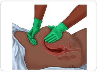
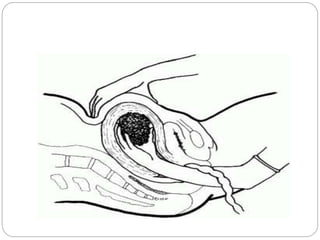
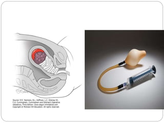
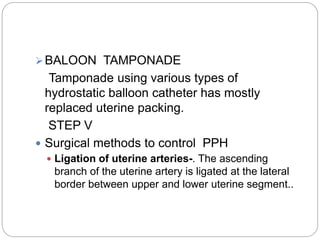
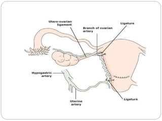
POST PARTUM
HAEMORRHAGE
INTRODUCTION-
PPH is one of the most alarming and serious
emergencies a midwife may face and is
especially terrifying if it occurs immediately
following a birth.
DEFINITION
๏‚— The amount of blood loses in excess of 500
ml following birth of the baby (WHO).
๏‚— Clinical definition โ€œAny amount of bleeding
from or into genital tract following birth of the
baby up to the end of the puerperium, which
adversely affects the general condition of the
patient evidence by rise in pulse rate and
falling blood pressure, is called post partum
hemorrhage.โ€
๏‚— The incidence is about 4-6% of all deliveries.
TYPES
๏‚— Primary โ€“ hemorrhage occurs within 24 hours
following the birth of the baby.
Third stage hemorrhage โ€“ bleeding occurs
before expulsion of placenta.
True stage hemorrhage โ€“ bleeding occurs
subsequent to expulsion of placenta
(majority).
๏‚— Secondary- hemorrhage occurs beyond 24
hours within puerperium.
PRIMARY PPH-
CAUSES-
1.ATONIC UTERUSES (80%)
Atonicity of the uterus is the commonest cause of
post partum hemorrhage. This is a failure of the
myometrium at the placental site to contract and
retract and to compress blood vessel and control
blood loss by living ligature action. Following are
the conditions
๏‚— Grand multipara โ€“ inadequate retraction and
frequent adherent placenta.
๏‚— Over-distension of the uterus- as in multiple
pregnancy, hydramnious and large baby.
Imperfect retraction.
๏‚— Malnutrition and anemia โ€“ even slight blood
loss can develop PPH.
๏‚— APH
๏‚— Prolonged labour โ€“ poor retraction, infection
(amnionitis), dehydration are important factors.
๏‚— Anesthesia โ€“ depth of anesthesia and the
analgesic may cause atonicity.
๏‚— Initiation or augmentation of delivery by oxytocin.
๏‚— Malformation of the uterus โ€“ implantation of the
placenta in the uterine septum of a septate
uterus.
๏‚— Uterine fibroid
๏‚— Mismanaged third stage of labour.
๏‚— Precipitate labour.
2.Traumatic (20%)
Trauma to the genital tract usually occurs
following operative delivery, even after
spontaneous delivery. Trauma involves
usually the cervix, vagina, perineum
(episiotomy wound and lacerations) and Para
urethral region.
3.Retained tissues-Bits of placenta, blood
clots cause PPH due to imperfect uterine
retraction.
4.Drugs
Use of Tocolytic drugs (ritodrine), MgSO4,
Nifedipine
5.Blood coagulation disorders, acquired or
congenital
Blood coagulation disorder is less common
cause of PPH.
CLINICAL FEATURE-
MANAGEMENT OF PPH-
๏‚— Management of 3rd stage bleeding
๏‚— Actual management
๏ถ MANAGEMENT OF 3RD STAGE BLEEDING
Steps of management
1. Placental site bleeding-
๏‚— To palpate the fundus and massage the uterus to
make it hard. The massage is to be done by
placing four fingers behind the uterus and thumb
in front.
๏‚— To start crystalloid solution (NS or RL) with
oxytocin (1L with 20 units) at 60 drops per minute
and to arrange for blood transfusion if necessary.
๏‚— Oxytocin 10 unit IM or methergine 0.2 mg is given
intravenously.
๏‚— To catheterize the bladder.
๏‚— To give antibiotics (Ampicillin 2gm and
Metronidazole 500mg IV)
2. Management of traumatic bleed
๏‚— The uterovaginal canal is to be explored under
general anesthesia after the placenta is expelled
and haemostatic sutures are placed on the
offending sites.
STEPS OF MANUAL REMOVAL OF PLACENTA
๏‚— The patient is placed in lithotomy position. With
all aseptic measures, the bladder is catheterized.
๏‚— One hand is introduced into the uterus in cone
shaped manner following the cord. While
introducing the hand, the labia are separated by
the fingers at the other hand.
๏‚— Counter pressure on the uterine fundus is applied
by the hand placed over the abdomens. The
abdominal hand should steady the fundus and
guide the movement of the fingers inside the
uterine cavity till the placenta is completely
separated.
๏‚— As soon as the placenta margin is reached, the
fingers are inserted between the placenta and the
uterine wall with the back of the hand in contact
with the uterine wall.
๏‚— When the placenta is completely separated, it is
extracted by traction of the cord by the other
hand. The uterine hand is still inside the uterus for
exploration of the cavity to be sure that nothing is
left behind.
MANAGEMENT OF TRUE PPH
๏‚— Immediate measures are to be taken by the
attending House officer (Doctor/ Midwife).
๏‚— Call for extra help.
๏‚— Send blood for group, cross matching, diagnostic
test
๏‚— Infuse rapidly 2 liters of normal saline.
๏‚— Give O2 by mask 10-15 L/min.
๏‚— Start 20 units of oxytocin in 1 L of NS IV at the
rate of 60drop per min.
ACTUAL MANAGEMENT
The first step is to control the fundus and to note
the feel of the uterus. If the uterus is flabby, the
bleeding is likely to be from the atonic uterus. If
the uterus is firm and contracted, the bleeding is
likely to traumatic origin.
๏ƒ˜ ATONIC UTERUS
STEP I
๏‚— Massage the uterus to make it hard and express
the blood clot.
๏‚— Inject oxytocin drip is started (10 units in 500 ml
of NS) at the rate of 40-60 drops/ min.
๏‚— FC to keep bladder empty and to monitor urine
output.
๏‚— To examine the expelled placenta and
membranes for evidence of missing cotyledon or
piece of membranes.
๏‚— If the uterus fails to contract, proceed to next
STEP II
๏‚— Simultaneous inspection of the cervix and vagina
is to be done.
๏‚— T. Misoprostol 1000 mg per rectum is effective.
๏‚— When uterus atony is due to Tocolytic drug,
calcium gluconate (1g IV slowly) should be given
to neutralize the calcium blocking effect of the
drug.
๏‚— STEP III
๏ƒ˜ UTERINE MASSAGE AND BIMANUAL
COMPRESION
๏‚— The whole hand is introduced into the vaginal in
cone shaped fashion after separating the labia with
the finger of the other hand.
๏‚— The vaginal hand is clenched into a fist with the
back at the hand directed posteriosly and the
knuckles in the anterior fornix.
๏‚— The other hand is placed over the abdomen behind
the uterus to make it anteverted.
๏‚— The uterus is firmly squeezed between the two
hands.
๏‚— It may be necessary to continue the compression
for a prolonged period until the tone of the uterus is
๏‚— STEP IV
๏ƒ˜ UTERINE TAMPONADE
๏‚— Tight intrauterine packing is done
๏‚— A 5 meter long strip of gauze, 8cm wide folded twice
is required.
๏‚— The gauge is soaked in antiseptic cream before
introduction. The gauge is placed high up and
packed into the fundal area first while the uterus is
steadied by the external hand.
๏‚— Gradually the rest of the cavity is packed so that no
empty space is left behind.
๏‚— Antibiotic should be given and the plug should be
removed after 24 hours.
๏ƒ˜BALOON TAMPONADE
Tamponade using various types of
hydrostatic balloon catheter has mostly
replaced uterine packing.
STEP V
๏‚— Surgical methods to control PPH
๏‚— Ligation of uterine arteries-. The ascending
branch of the uterine artery is ligated at the lateral
border between upper and lower uterine segment..
๏‚— Ligation of the ovarian and uterine artery
anastomosis- If bleeding continue is done just
below the ovarian ligament.
๏‚— Ligation of anterior division of internal iliac
artery โ€“ reduced distal blood flow.
STEP VI
๏‚— Hysterectomy โ€“ rarely uterus fails to contract and
bleeding continues in spite of the above
measures.
๏‚— MANAGEMENT OF TRAUMATIC PPH
The trauma to the perineum, vagina and the
cervix is to be searched under good light by
speculum examination and haemostasis is
achieved by appropriate catgut sutures.
SECONDARY PPH
CAUSES
๏‚— The bleeding usually occurs 8th โ€“ 14th day of
delivery.
๏‚— Retained bits of cotyledon or membrane.
๏‚— Infection and separation of slough over a deep
cervico โ€“viganal laceration.
๏‚— Endometritis and subinvolutionof the placental
site due to delayed healing process.
DIAGNOSIS
๏‚— The bleeding is bright red in color and of
varying amount.
๏‚— Degree of anemia and evidence of sepsis.
๏‚— Sub involution.
๏‚— USG
MANAGEMENT
๏‚— SUPPORTIVE THERAPY
๏‚— Blood transfusion
๏‚— Administer antibiotic as routine.
๏‚— CONSERVATIVE
๏‚— If the bleeding is slight and no apparent
cause is detected, a careful watch for a
period of 24 hrs or so is done in the hospital.
๏‚— ACTIVE MANAGEMENT
๏‚— The retained bits are removed by ovum
forceps.
๏‚— Gentle curettage is done by using flushing
curette.
๏‚— Methargin 0.2 mg is given IM.
PREVENTION-
1.Antenatal
๏‚— Improvement of the health status.
๏‚— High risk patients who are likely developing
PPH are to be screened and delivered in a
well equipped hospital.
๏‚— Blood grouping should be done for all women
so that no time is wasted during emergency.
๏‚— High risk cases should be delivered by a
senior obstetrician.
2. Intranatal
๏‚— Active management of the third stage for all
women in labour should be a routine as it reduces
PPH by 60%.
๏‚— Cases with induced or augmented labour by
oxytocin, the infusion should be continued for at
least one hour after the delivery.
๏‚— Woman delivered by caesarean sections,
oxytocin 5IU slow IV is to be given to reduce
blood loss.
๏‚— Exploration of the utero-vaginal canal for
evidence of trauma following difficult labour or
instrumental delivery.
๏‚— Observation for about two hours of the delivery to
make sure that the uterus is hard and well
contracted before sending her .
๏‚— Expert- obstetric anesthetist is needed when
the delivery is conducted under general
anesthesia.
๏‚— Examination of the placenta and membranes
should be a routine so as to detect at the
earliest any missing part.
NURSING MANAGEMENT-
ASSESSMENT
1.Determine that normal third stage progress is
occurring.
๏‚— Rhythmic contraction until the placenta is born.
๏‚— Birth of placenta occurs 5 -30 min after birth of
the baby.
๏‚— Signs of placental separation is seen
๏‚— Fundus rises slightly in abdomen.
๏‚ง Umbilical cord lengthens.
๏‚— Slight gush of blood noted.
๏‚— Placenta separation
๏‚— The mother may experience chills or shivering.
๏‚— Assess maternal blood pressure following birth of
baby.
๏‚— Assess the status of the uterus โ€“ contraction will
continue until birth of the placenta.
๏‚— Examine placenta to document that all cotyledons
and membrane are present.
NURSING DIAGNOSIS
๏‚— Anxiety related to knowledge deficit.
๏‚— Risk of infection
๏‚— Pain
๏‚— Fatigue
๏‚— Impaired skin integrity.
PLANNING
๏‚— Monitor maternal and newborn status.
๏‚— Provide support in parental newborn
interactions.
๏‚— Provide support and comfort measure during
third stage.
IMPLEMENTATION
๏‚— Observe and record birth of placenta.
๏‚— Monitor maternal blood pressure.
๏‚— Dry the baby completely.
๏‚— Administer oxytocics drugs as per physicianโ€™s
order.
EVALUATION
๏‚— Be sure that mother and newborn maintain
normal physical parameter.
๏‚— Monitor mother โ€“ baby attachment/ bonding.
๏‚— Make sure that mother feels comfortable and
supported during the third stage.
POST PARTUM HEMORRHAGE(PPH)

POST PARTUM HEMORRHAGE(PPH)

  • 2.
  • 3.
    INTRODUCTION- PPH is oneof the most alarming and serious emergencies a midwife may face and is especially terrifying if it occurs immediately following a birth.
  • 4.
    DEFINITION ๏‚— The amountof blood loses in excess of 500 ml following birth of the baby (WHO). ๏‚— Clinical definition โ€œAny amount of bleeding from or into genital tract following birth of the baby up to the end of the puerperium, which adversely affects the general condition of the patient evidence by rise in pulse rate and falling blood pressure, is called post partum hemorrhage.โ€
  • 5.
    ๏‚— The incidenceis about 4-6% of all deliveries. TYPES ๏‚— Primary โ€“ hemorrhage occurs within 24 hours following the birth of the baby. Third stage hemorrhage โ€“ bleeding occurs before expulsion of placenta. True stage hemorrhage โ€“ bleeding occurs subsequent to expulsion of placenta (majority). ๏‚— Secondary- hemorrhage occurs beyond 24 hours within puerperium.
  • 6.
    PRIMARY PPH- CAUSES- 1.ATONIC UTERUSES(80%) Atonicity of the uterus is the commonest cause of post partum hemorrhage. This is a failure of the myometrium at the placental site to contract and retract and to compress blood vessel and control blood loss by living ligature action. Following are the conditions ๏‚— Grand multipara โ€“ inadequate retraction and frequent adherent placenta. ๏‚— Over-distension of the uterus- as in multiple pregnancy, hydramnious and large baby. Imperfect retraction. ๏‚— Malnutrition and anemia โ€“ even slight blood loss can develop PPH.
  • 7.
    ๏‚— APH ๏‚— Prolongedlabour โ€“ poor retraction, infection (amnionitis), dehydration are important factors. ๏‚— Anesthesia โ€“ depth of anesthesia and the analgesic may cause atonicity. ๏‚— Initiation or augmentation of delivery by oxytocin. ๏‚— Malformation of the uterus โ€“ implantation of the placenta in the uterine septum of a septate uterus. ๏‚— Uterine fibroid ๏‚— Mismanaged third stage of labour. ๏‚— Precipitate labour.
  • 8.
    2.Traumatic (20%) Trauma tothe genital tract usually occurs following operative delivery, even after spontaneous delivery. Trauma involves usually the cervix, vagina, perineum (episiotomy wound and lacerations) and Para urethral region. 3.Retained tissues-Bits of placenta, blood clots cause PPH due to imperfect uterine retraction.
  • 9.
    4.Drugs Use of Tocolyticdrugs (ritodrine), MgSO4, Nifedipine 5.Blood coagulation disorders, acquired or congenital Blood coagulation disorder is less common cause of PPH.
  • 10.
  • 11.
    MANAGEMENT OF PPH- ๏‚—Management of 3rd stage bleeding ๏‚— Actual management ๏ถ MANAGEMENT OF 3RD STAGE BLEEDING Steps of management 1. Placental site bleeding- ๏‚— To palpate the fundus and massage the uterus to make it hard. The massage is to be done by placing four fingers behind the uterus and thumb in front. ๏‚— To start crystalloid solution (NS or RL) with oxytocin (1L with 20 units) at 60 drops per minute and to arrange for blood transfusion if necessary.
  • 13.
    ๏‚— Oxytocin 10unit IM or methergine 0.2 mg is given intravenously. ๏‚— To catheterize the bladder. ๏‚— To give antibiotics (Ampicillin 2gm and Metronidazole 500mg IV) 2. Management of traumatic bleed ๏‚— The uterovaginal canal is to be explored under general anesthesia after the placenta is expelled and haemostatic sutures are placed on the offending sites.
  • 14.
    STEPS OF MANUALREMOVAL OF PLACENTA ๏‚— The patient is placed in lithotomy position. With all aseptic measures, the bladder is catheterized. ๏‚— One hand is introduced into the uterus in cone shaped manner following the cord. While introducing the hand, the labia are separated by the fingers at the other hand. ๏‚— Counter pressure on the uterine fundus is applied by the hand placed over the abdomens. The abdominal hand should steady the fundus and guide the movement of the fingers inside the uterine cavity till the placenta is completely separated.
  • 15.
    ๏‚— As soonas the placenta margin is reached, the fingers are inserted between the placenta and the uterine wall with the back of the hand in contact with the uterine wall. ๏‚— When the placenta is completely separated, it is extracted by traction of the cord by the other hand. The uterine hand is still inside the uterus for exploration of the cavity to be sure that nothing is left behind.
  • 17.
    MANAGEMENT OF TRUEPPH ๏‚— Immediate measures are to be taken by the attending House officer (Doctor/ Midwife). ๏‚— Call for extra help. ๏‚— Send blood for group, cross matching, diagnostic test ๏‚— Infuse rapidly 2 liters of normal saline. ๏‚— Give O2 by mask 10-15 L/min. ๏‚— Start 20 units of oxytocin in 1 L of NS IV at the rate of 60drop per min.
  • 18.
    ACTUAL MANAGEMENT The firststep is to control the fundus and to note the feel of the uterus. If the uterus is flabby, the bleeding is likely to be from the atonic uterus. If the uterus is firm and contracted, the bleeding is likely to traumatic origin.
  • 19.
    ๏ƒ˜ ATONIC UTERUS STEPI ๏‚— Massage the uterus to make it hard and express the blood clot. ๏‚— Inject oxytocin drip is started (10 units in 500 ml of NS) at the rate of 40-60 drops/ min. ๏‚— FC to keep bladder empty and to monitor urine output. ๏‚— To examine the expelled placenta and membranes for evidence of missing cotyledon or piece of membranes. ๏‚— If the uterus fails to contract, proceed to next
  • 20.
    STEP II ๏‚— Simultaneousinspection of the cervix and vagina is to be done. ๏‚— T. Misoprostol 1000 mg per rectum is effective. ๏‚— When uterus atony is due to Tocolytic drug, calcium gluconate (1g IV slowly) should be given to neutralize the calcium blocking effect of the drug.
  • 21.
    ๏‚— STEP III ๏ƒ˜UTERINE MASSAGE AND BIMANUAL COMPRESION ๏‚— The whole hand is introduced into the vaginal in cone shaped fashion after separating the labia with the finger of the other hand. ๏‚— The vaginal hand is clenched into a fist with the back at the hand directed posteriosly and the knuckles in the anterior fornix. ๏‚— The other hand is placed over the abdomen behind the uterus to make it anteverted. ๏‚— The uterus is firmly squeezed between the two hands. ๏‚— It may be necessary to continue the compression for a prolonged period until the tone of the uterus is
  • 23.
    ๏‚— STEP IV ๏ƒ˜UTERINE TAMPONADE ๏‚— Tight intrauterine packing is done ๏‚— A 5 meter long strip of gauze, 8cm wide folded twice is required. ๏‚— The gauge is soaked in antiseptic cream before introduction. The gauge is placed high up and packed into the fundal area first while the uterus is steadied by the external hand. ๏‚— Gradually the rest of the cavity is packed so that no empty space is left behind. ๏‚— Antibiotic should be given and the plug should be removed after 24 hours.
  • 25.
    ๏ƒ˜BALOON TAMPONADE Tamponade usingvarious types of hydrostatic balloon catheter has mostly replaced uterine packing. STEP V ๏‚— Surgical methods to control PPH ๏‚— Ligation of uterine arteries-. The ascending branch of the uterine artery is ligated at the lateral border between upper and lower uterine segment..
  • 26.
    ๏‚— Ligation ofthe ovarian and uterine artery anastomosis- If bleeding continue is done just below the ovarian ligament. ๏‚— Ligation of anterior division of internal iliac artery โ€“ reduced distal blood flow. STEP VI ๏‚— Hysterectomy โ€“ rarely uterus fails to contract and bleeding continues in spite of the above measures.
  • 29.
    ๏‚— MANAGEMENT OFTRAUMATIC PPH The trauma to the perineum, vagina and the cervix is to be searched under good light by speculum examination and haemostasis is achieved by appropriate catgut sutures.
  • 30.
    SECONDARY PPH CAUSES ๏‚— Thebleeding usually occurs 8th โ€“ 14th day of delivery. ๏‚— Retained bits of cotyledon or membrane. ๏‚— Infection and separation of slough over a deep cervico โ€“viganal laceration. ๏‚— Endometritis and subinvolutionof the placental site due to delayed healing process.
  • 31.
    DIAGNOSIS ๏‚— The bleedingis bright red in color and of varying amount. ๏‚— Degree of anemia and evidence of sepsis. ๏‚— Sub involution. ๏‚— USG MANAGEMENT ๏‚— SUPPORTIVE THERAPY ๏‚— Blood transfusion ๏‚— Administer antibiotic as routine.
  • 32.
    ๏‚— CONSERVATIVE ๏‚— Ifthe bleeding is slight and no apparent cause is detected, a careful watch for a period of 24 hrs or so is done in the hospital. ๏‚— ACTIVE MANAGEMENT ๏‚— The retained bits are removed by ovum forceps. ๏‚— Gentle curettage is done by using flushing curette. ๏‚— Methargin 0.2 mg is given IM.
  • 33.
    PREVENTION- 1.Antenatal ๏‚— Improvement ofthe health status. ๏‚— High risk patients who are likely developing PPH are to be screened and delivered in a well equipped hospital. ๏‚— Blood grouping should be done for all women so that no time is wasted during emergency. ๏‚— High risk cases should be delivered by a senior obstetrician.
  • 34.
    2. Intranatal ๏‚— Activemanagement of the third stage for all women in labour should be a routine as it reduces PPH by 60%. ๏‚— Cases with induced or augmented labour by oxytocin, the infusion should be continued for at least one hour after the delivery. ๏‚— Woman delivered by caesarean sections, oxytocin 5IU slow IV is to be given to reduce blood loss. ๏‚— Exploration of the utero-vaginal canal for evidence of trauma following difficult labour or instrumental delivery. ๏‚— Observation for about two hours of the delivery to make sure that the uterus is hard and well contracted before sending her .
  • 35.
    ๏‚— Expert- obstetricanesthetist is needed when the delivery is conducted under general anesthesia. ๏‚— Examination of the placenta and membranes should be a routine so as to detect at the earliest any missing part.
  • 36.
    NURSING MANAGEMENT- ASSESSMENT 1.Determine thatnormal third stage progress is occurring. ๏‚— Rhythmic contraction until the placenta is born. ๏‚— Birth of placenta occurs 5 -30 min after birth of the baby. ๏‚— Signs of placental separation is seen ๏‚— Fundus rises slightly in abdomen. ๏‚ง Umbilical cord lengthens. ๏‚— Slight gush of blood noted. ๏‚— Placenta separation ๏‚— The mother may experience chills or shivering.
  • 37.
    ๏‚— Assess maternalblood pressure following birth of baby. ๏‚— Assess the status of the uterus โ€“ contraction will continue until birth of the placenta. ๏‚— Examine placenta to document that all cotyledons and membrane are present. NURSING DIAGNOSIS ๏‚— Anxiety related to knowledge deficit. ๏‚— Risk of infection ๏‚— Pain ๏‚— Fatigue ๏‚— Impaired skin integrity.
  • 38.
    PLANNING ๏‚— Monitor maternaland newborn status. ๏‚— Provide support in parental newborn interactions. ๏‚— Provide support and comfort measure during third stage. IMPLEMENTATION ๏‚— Observe and record birth of placenta. ๏‚— Monitor maternal blood pressure.
  • 39.
    ๏‚— Dry thebaby completely. ๏‚— Administer oxytocics drugs as per physicianโ€™s order. EVALUATION ๏‚— Be sure that mother and newborn maintain normal physical parameter. ๏‚— Monitor mother โ€“ baby attachment/ bonding. ๏‚— Make sure that mother feels comfortable and supported during the third stage.